Visual Testin g Lab Anatomy Special Senses Test

Visual Testin g Lab Anatomy – Special Senses

Test 1 – Visual Acuity Snellen Eye Chart � Stand on the “X” marked with teal masking tape � � 4 locations Cover one eye, have partner point to letters and check accuracy Cover other eye, repeat Numbers out to the side of the letter rows… these indicate your vision at the distance If wearing glasses, complete one trial with and one without the glasses. Highlight the trial with glasses What’s normal vision?

Test 2 – Near Point Accommodation How well do your ciliary body muscles change the shape of your lens to allow you to see objects close up? Meter or half meter stick… hold it at a right angle to the face; grab an index card with print on it Have test subject close one eye, start at the very back of the meter stick and slowly bring the card toward their open eye Have them indicate when the writing on the card is no longer in sharp focus – blurry

Near Point of Accommodation Indicate how far away the card is from their face (cm) Record this number and compare it to the chart on page 3 of your lab to figure out the age correlation of vs. accommodation Tablewith 2. 1 your near point. Age Average Near Point (cm) 10 7 40 20 20 10 50 45 30 13 60 90 Hole’s Human Anatomy Lab Manual

Test 3 – Color Vision Testing Hole’s Anatomy and Physiology Lab Manual � Page 288 – 3 already set up for you! Images are numbered 1 -4 � Show these to your lab partner one at a time � Indicate on your lab document what number they see in appropriate column Table 3 Color Vision Testing Name 1 2 3 4

Test 4 – Eye Tracking Have the test subject sit in a chair and sit directly in front of them Have the test subject track an object as your move it back and forth � You can use your finger, pen, ruler, etc. Indicate if their movements are smooth or jerky � If it’s jerky – you will recognize this… I promise.

Word to the Wise… Keep your phones up… if I see phones out when you should be participating with your group the whole group will get a 0 Do NOT start the questions on the back page until all the data is collected � You should be working as a group to answer the questions. Not just one or two people doing all the work. � Read the background info provided to help with the questions! Group sizes – 2 to 5 people Submit lab when finished! Notes will start promptly at 9: 20

Label the eye on your notes when you get finished with the lab!

The Eye Special Senses – Day 4

Are the squares inside the blue and yellow squares all the same color?

Bezold effect The smaller squares inside the blue and yellow squares are all the same color. They seem different (magenta and orange) because a color is perceived differently depending on its relation to adjacent colors (here blue or yellow depending on the outer square).

Are the horizontal lines straight or crooked?

Does Lincoln’s face look normal?
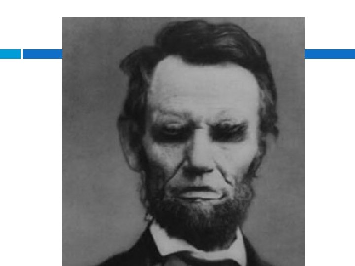

Illusory Contour: a form of visual illusion where contours are perceived without a luminance or color change across the contour

Can you see a baby?


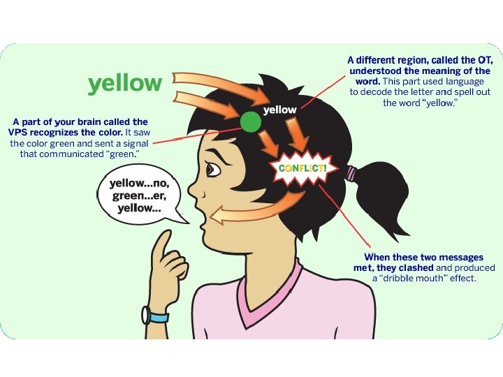
How quickly can you say the color of the words below?

Eye Fun Facts Your eye muscle that functions to dilate and constrict the pupil is the fastest reacting muscle in the body Week four of pregnancy is when cells from the developing brain tissue begin developing the optic nerves An ostrich's eye is bigger than their brain “Red eye” in photographs occurs when light from a camera's flash reflects off of the retina's blood vessels 70% of your sensory cells in the body are located in

Eye and Sense of Sight Vision system �Eyes �Optic nerves �Vision centers in the brain �Accessory structures Eye Structure �Processes light to produce images �Three layers �Two chambers �Various Specialized parts

Outer Eye Anatomy – Fibrous Layer Connective Tissue Sclera � White of the eye � Protects the eye � Muscles attach here! Cornea � Front of eye � “Window” that allows light into eye � Bends light as it enters 35 -23

Middle Eye Anatomy ~ Vascular Layer � Choroid: vascular layer containing connective tissue, between the retina and the sclera Brings oxygen/nutrients to the eye � Iris: colored portion of eye that regulates light entering Iris color is due to variable amounts of eumelanin (brown/black melanins) and pheomelanin (red/yelow melanins) produced by melanocytes � Pupil: central opening in the iris, it regulates the amount of light that enters the eye Dilator muscle (smooth muscle) that contracts and relaxes to open or close pupil

Middle Eye Anatomy � Ciliary body: muscles; control the shape of the lens to allow for focusing; anchors lens; produces aqueous humor � Lens: transparent structure inside the eye; bends and refracts light rays, focuses light on nerve cells of retina

Chambers of the Eye �Anterior chamber Between the iris and the cornea's innermost surface Filled with aqueous humor – nourishes and bathes anterior eye �Posterior Behind chamber lens Contains vitreous humor – maintains shape of eyeball and holds retina in place

Inner Eye Anatomy – Nervous Layer Retina: light-sensitive tissue lining the back of eye. � Light rays are focused onto the retina through the cornea, pupil and lens. � The retina converts the light rays into impulses that travel through the optic nerve to our brain, where they are interpreted as the images Retina is layered with nerve cells and photoreceptors known as rods and cones Optic disc: Raised disk on the retina at the point of entry of the optic nerve lacking visual receptors Fovea Centralis: small depression in the retina of the eye where visual acuity is highest. The center of the field of vision is focused in this region, where retinal cones are particularly concentrated

Visual Receptors of the Eye Rods: scotopic vision � Very sensitive to light � Contains rhodopsin: biological pigment in rod cells Will function in dim light – “limited” night vision Animals that have good vision in the dark have a higher percentage of rods in comparison to humans Do not provide sharp image or detect color � Many more rod cells in comparison to cones Cones: photopic vision � Function in bright light � Contains photopsin: allows for perception of color Sensitive to color and provide sharp images *Fovea Centralis contains highest concentration of cones � The light levels where both are operational are called mesopic


Special Senses – The Eye Day 2

Accessory Structures of the Eye orbits � Eye sockets � Form a protective shell around the eyes � Eyebrows protect eyes Eyelids � Skin, muscle, and connective tissue � Blinking Prevents surface from drying out Keeps foreign material out of eye
 Conjunctivas � Mucus membranes")
Eye and Sense of Sight: Visual Accessory Organs (cont. ) Conjunctivas � Mucus membranes � Line inner surfaces of eyelids Lacrimal apparatus � Lacrimal glands Lateral edge of eyeballs Produce tears � Nasolacrimal Medial ducts aspect of eyeballs Drain tears into nose
: lateral movement Medial")
35 -34 Eye Muscles 6 muscles per eye Lateral Rectus (abducens): lateral movement Medial Rectus (oculomotor): medial movement Superior rectus (oculomotor): superior and medial movement Inferior rectus (oculomotor): inferior and medial movement Inferior oblique (oculomotor): superior and lateral movement Superior oblique (trochlear): inferior and lateral movement

The Art of Seeing… There are major functional differences between the rods and cones. � Rods are extremely sensitive, and can be triggered by as few as 6 photons. At very low light levels, visual experience is based solely on the rod signal. This explains why colors cannot be seen at low light levels: only one type of photoreceptor cell is active � Cones require significantly brighter light (i. e. , a larger numbers of photons) in order to produce a signal. In humans, there are three different types of cone cells, distinguished by their pattern of response to different wavelengths of light

Types of Cones Three types, each with different pigment: � S-cones, M-cones and L- cones � Each cone is therefore sensitive to visible wavelengths of light that correspond to short-wavelength medium-wavelength long-wavelength light

Structure…
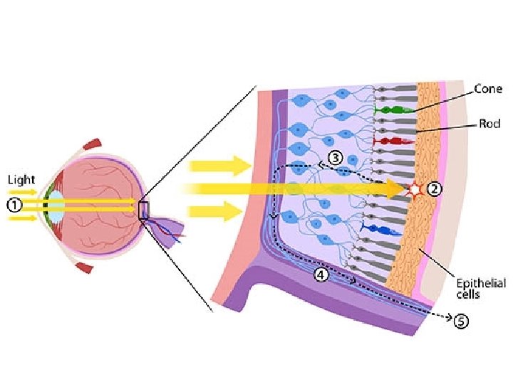

Organization of Rod and Cone Cells The discs containing rhodopsin or photopsin are constantly recycled to keep your visual system healthy. � RPE can absorb scattered light vision is a lot clearer! � By having the discs right next to the epithelial cells (retinal pigmented epithelium: RPE) at the back of the eye, parts of the old discs can be carried away by cells in the RPE. Light can also have damaging effects, so this set up also helps protect your rods and cones from unnecessary damage. Rods and cones are constantly sending signals, which requires the movement of lots of molecules, which they need to replenish to keep working. � Because the RPE is right next to the discs, it can easily help reload photoreceptor cells and discs with the molecules they need to keep sending

35 -40 Visual Pathway Eye works like a camera � Light enters the eye through the lens � Refraction – cornea, lens, and fluids bend light to focus it on the retina Image upside down on retina Retina converts light to nerve impulse Optic nerve Optic chiasm Occipital lobe of cerebrum Image turned rightside up

Why are images upside down on the retina? Images on your retina are reversed; retina “sees” everything backwards brain reorients image Retinal image reversal is an adaptive advantage providing us with tremendous peripheral vision and the ability to view objects much larger our eyes

35 -42 Vision Testing Professionals include � Ophthalmologist – medical doctor who is an eye specialist � Optometrist – provides vision screening and diagnostic testing � Opticians – fills vision prescriptions for glasses and contacts

Both the cornea and the lens of the eye have convex surfaces and help to focus light rays onto the retina
 – impairment of distance vision Hyperopia (farsighted) – impairment of")
Vision Impairments Myopia (nearsighted) – impairment of distance vision Hyperopia (farsighted) – impairment of near vision Objects near are seen � Distant objects clear, clearly, but objects those close are blurry farther away are blurry. � Eyeball is shorter � Eyeball is too long � Light focused posterior � Light focuses anterior to to retina � Presbyopia Impairment due to aging Loss of lens elasticity

How to decipher your contact prescription OD = Oculus dexter, Right Eye OS = Oculus sinister, Left Eye BC / Base Curve (a number between 8. 0 and 10. 0) DIA / Diameter (a number between 13. 0 and 15. 0) Power / Sphere / Rx (a number between -20 and +20) � � (-) indicates near-sightedness (+) indicates far-sightedness For astigmatic patients, they will also need: � � Cylinder (a number between 4. 00 and +4. 00) Axis (a number between 0 and 180) Cylinder and Axis are usually separated by "X" and read as "times"

Astigmatism Imperfection in the curvature of your cornea or lens causing distortion of images � Vision for both near and far objects appears blurry or distorted

35 -47 Common Diseases and Disorders Disorder / Disease Description Amblyopia Lazy eye; one eye is not used regularly; poor depth perception; often concurrent with strabismus Strabismus Convergent Misalignment of eyes Crossed eyes; one or both eyes turn inward Divergent Wall eye; one or both eyes turn outward Conjunctivitis Pink eye; highly contagious bacterial infection Cataracts Opaque structures in lens prevent light from passing through; vision fuzzy
 35 -48 Disorder / Disease Description Entropion Inversion")
Common Diseases and Disorders (cont. ) 35 -48 Disorder / Disease Description Entropion Inversion of lower eyelid Glaucoma Increase in intraocular pressure due to a buildup of aqueous humor in anterior chamber Macular degeneration Progressive disease; inadequate blood supply to retina; most common cause of vision loss; affects people over 50 years Nystagmus Rapid, involuntary eye movements
- Slides: 49