Vision Lab Review eye anatomy especially Path of

Vision Lab Review eye anatomy especially: Path of light through eyeball Cellular layers of retina Intrinsic eye muscles Blind spot and fovea centralis

Vision Process can be Divided into Three Steps 1. Light enters eye, is focused by lens onto retina 2. Photoreceptors transduce light energy into electrical signal

3. Electrical signals sent along neural pathways are processed in visual cortex

Pupils are dilated in response to the contraction of the A. Radial ciliary muscle B. Circular ciliary muscle C. Radial iris muscle D. Circular iris muscle E. Both C) and D) apply

Light Modification Pre-Retina • Amount of light is changed by altering pupil aperture from 1. 5 – 8 mm • • • Pupillary constriction due to ? Dilation ? Pupillary reflex is consensual

Fig. 10. 28 Postganglionic sympathetic axon In dim light In normal light From superior cervical ganglion Radially arranged smooth muscle fibers of the iris Circularly arranged smooth muscle fibers of the iris Pupil Ciliary ganglion In bright light Postganglionic parasympathetic axon From oculomotor nerve

The Lens … … focuses light by changing its shape Refraction: Light waves are bent, or refracted when they pass from one medium into another, due to different densities.

Copyright © Mc. Graw-Hill Education. Permission required for reproduction or display. Fig. 10. 34 Ciliary muscle fibers relaxed Suspensory ligament taut Lens thin and focused for distant vision (a) Ciliary muscle fibers contracted Suspensory ligament relaxed Lens thick and focused for close vision Fig 10. 34 (b)
 by changing lens shape Lens")
Accommodation: Light is focused (to keep objects in focus) by changing lens shape Lens attached to ciliary muscle via suspensory ligament (= zonulas) Ciliary muscle contracts. . . Lens bulges up

Vision Problems Accommodation Ability of eye to focus differentially on objects of near vision (< 6 m or 20 feet) Fig 10 -31

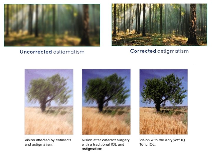
More Vision Problems § Astigmatism: asymmetry of cornea / lens § Presbyopia: loss of accommodation

Testing for Astigmatism

Determine Near-Point of Accomodation

Testing Visual Acuity Snellen chart

Fig. 10. 35 Copyright © Mc. Graw-Hill Education. Permission required for reproduction or display. Emmetropia (normal vision) Rays focus on retina (a) No correction necessary Hyperopia (farsightedness) Rays focus behind retina (c) Convex lens corrects farsightedness Myopia (nearsightedness) Rays focus in front of retina (b) Concave lens corrects nearsightedness Astigmatism Rays do not focus (d) Uneven lens corrects astigmatism

Visual Field and Binocular Vision 3 vs. 2 dimensional view
- Slides: 17