Viruses Chapter outline 5 1 General Properties of


, and plant")
 vary widely in size and shape. Viruses are smaller than cells,")


 of 2. Penetration (injection) of 4.")


 as an example (1) penetration by the")


- Slides: 51
Viruses
Chapter outline 5. 1 General Properties of Viruses 5. 2 General Features of Virus Reproduction 5. 3 Overview of Bacterial Viruses 5. 4 Temperate Bacteriophages: Lysogeny and Lambda 5. 5 Overview of Animal Viruses 5. 6 Pox Viruses 5. 7 Adcnoviruses 5. 8 Retroviruses 5. 9 Viroids and Prions
5. 1 General Properties of Viruses viruses differ from living cells in at least three ways: (1) their simple, acellular organization (2) the absence of both DNA and RNA in the same virion, (3) their inability to reproduce independently of cells and carry out cell division as prokaryotes and eukaryotes do.
Viruses can exist in two phases extracellular and intracellular Virion, the extracellular phase, posses few if any enzymes and can not reproduce independently of living cells. In the intracellular phase, viruses exist primarily as replicating nucleic acids that induce host metabolism to synthesize virion components; eventually complete virus particles or virions are released.
Hosts and size Three main classes - animal viruses, bacterial viruses (bacteriophages), and plant viruses. The particular host range of a virus is determined by the virus's requirements for its specific attachment to the host cell and the availability within the potential host of cellular factors required for viral multiplication.
Virus particles (virions) vary widely in size and shape. Viruses are smaller than cells, ranging in size from 0. 02 to 0. 3 μm. A common unit of measure for viruses is the nanometer, which is 1000 times smaller than 1 um and 1 million times smaller than 1 mm. Smallpox virus, one of the largest viruses, is about 200 nm in diameter (a bit smaller than the size of the smallest bacteria); poliovirus, one of the smallest, is only 28 nm in diameter (about the size of a ribosome).
Size Viruses vary considerably in size. Although most are quite a bit smaller than bacteria, some of the larger viruses (such as the smallpox virus) are about the same size as some very small bacteria (such as the mycoplasmas, rickettsias, and chlamydias). Viruses range from 20 to 300 nm in diameter
Genome in virion Viral genomes. The genomes of viruses can be composed of either DNA or RNA, and some use both as their genomic material at different stages in their life cycle. However , only one type of nucleic acid is found in the virion of any particular type of virus. This can be singlestranded (ss), double-stranded (ds), or in the case of the hepadnaviruses, partially double-stranded.
The comparative sizes of several viruses and bacteria:
Structure of viruses • Most viruses are too small to be seen under light microscope. • All viruses consists of an RNA or DNA core genome surrounded by a protein coat capsid. • The combined viral genome and capsid is called the nucleocapsid.
The nucleic acid of a virus is surrounded by a protein coat called the capsid Each capsid is composed of protein subunits called capsomeres. In some viruses, the capsid is covered by an envelope, which usually consists of some combination of lipids, proteins, and carbohydrates. Depending on the virus, envelopes may or may not be covered by spikes, which are carbohydrateprotein complexes that project from the surface of the envelope.
Structure of viruses • Most viruses are too small to be seen under light microscope. • All viruses consists of an RNA or DNA core genome surrounded by a protein coat capsid. • The combined viral genome and capsid is called the nucleocapsid.
Complex viruses Some viruses, particularly bacterial viruses, have very complicated structures and are called complex viruses. Examples of complex viruses are poxviruses, which do not contain clearly identifiable capsids but have several coats around the nucleic acid. Certain bacteriophages have capsids to which additional structures are attached.
A virus can have either DNA or RNA but never both !!
A virus can have either DNA or RNA but never both !!
GENERAL MORPHOLOGY Viruses may be classified into several morphological types on the basis of their capsid architecture as revealed by electron microscopy and a technique called x-ray crystallography.
A, Some viruses, such as tobacco mosaic virus, have a helical symmetry with the capsid surrounding an RNA genome. B, Many viruses that infect bacteria, such as the T-even bacteriophage, have a complex capsid with DNA contained within the head structure. C, Some animal viruses, such as adenovirus, have isometric symmetry and a DNA genome. D, others, such as coronavirus(冠状病毒), have complex capsids and an envelope with protruding proteins surrounding an RNA genome.
Helical viruses resemble long rods that may be rigid or flexible. Surrounding the nucleic acid, their capsid is a hollow cylinder with a helical structure. An example of a helical virus that is a rigid rod is the tobacco mosaic virus
Polyhedral viruses The capsid of most polyhedral viruses is in the shape of a regular polyhedron with 20 triangular faces and 12 corners. The capsomeres of each face form an equilateral triangle. An example of a polyhedral virus in the shape of an icosahedron is the adenovirus. Another icosahedral virus is the poliovirus.
Enveloped viruses The capsid of some viruses is covered by an envelope. Enveloped viruses are roughly spherical but highly variable in shape because the envelope is not rigid. When helical or polyhedral viruses are enclosed by envelopes, they are called enveloped helical and enveloped polyhedral viruses. An example of an enveloped helical virus is the influenza virus.
Complex viruses Some viruses, particularly bacterial viruses, have very complicated structures and are called complex viruses. Examples of complex viruses are poxviruses, which do not contain clearly identifiable capsids but have several coats around the nucleic acid. Certain bacteriophages have capsids to which additional structures are attached.
Viral Multiplication For a virus to multiply, it must invade a host cell and take over the host's metabolic machinery. A single virus can give rise to several or even thousands of similar viruses in a single host cell. This process drastically changes the host cell and often causes its death.
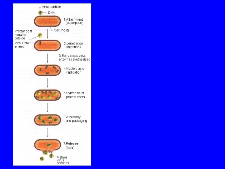
MULTIPLICATION OF BACTERIOPHAGES The multiplication cycle of bacteriophages, like that of all viruses, can be divided into several distinct stagesattachment penetration biosynthesis of viral components Maturation Release
7. Release of mature virions 1. Attachment (adsorption) of 2. Penetration (injection) of 4. Replication of the virus 5. Synthesis of proteins used 6. Assembly of structural 3. Early steps in replication the virion to a susceptible host the virion or its nucleic acid. as structural subunits of the subunits (and membrane from the cell during which the host cell. into the cell. virus coat. components in enveloped biosynthetic machinery is altered viruses) and packaging of as a prelude to virus nucleic acid into new virus synthesis. Virus-specific particles. enzymes are typically made.
Attachment of phage to host cell : After a chance collision between phage particles and bacteria, an attachment site on the virus attaches to a complementary receptor site on the bacterial cell.
Penetration: During the process of penetration, the bacteriophage's tail releases an enzyme, phage lysozyme, which breaks down a portion of the bacterial cell wall. then the bacteriophage injects its DNA (nucleic acid) into the bacterium.
Biosynthesis of viral components: Any RNA transcribed in the cell is m. RNA transcribed from phage DNA for biosynthesis of phage enzymes and capsid protein. The host cell's ribosomes, enzymes, and amino acids are used for translation
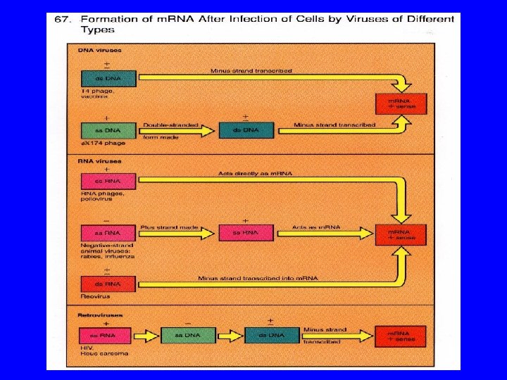
Formation of m. RNA after infection of cells by viruses of different types. The chemical sense of the m. RNA is considered as plus (+). The sense of the various virus nucleic acids are indicated as + if the same as m. RNA, as – if opposite, or as +- if double-stranded. Examples are indicated next to the virus nucleic acid. Although examples of viruses containing SS DNA of the - sense are known, none are discussed in the text.
Maturation and release: The phage heads and tails are separately assembled from protein subunits, the head is packaged with phage DNA, and the tail is attached
Release: Lysozyme, whose code is provided by a phage gene, is synthesized within the cell. This enzyme causes a breakdown of the bacterial cell wall, and the newly produced bactedophages are released from the host cell. The number of newly synthesized phage particles released from a single cell usually ranges from about 50 to 200.
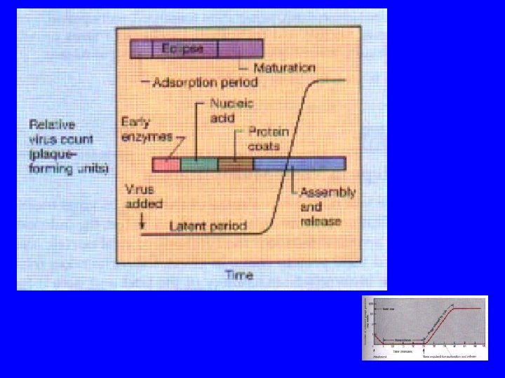
eclipse period There are genetic controls that regulate when different regions of phage DNA are transcribed into m. RNA during the multiplication cycle. • There are early messages that are translated into early phage proteins, the enzymes used in the synthesis of phage DNA. • There are late messages that are translated into late phage proteins for the synthesis of capsid proteins. This control mechanism is mediated by RNA polymerase.
Time course of events in phage T 4 infection Latent period
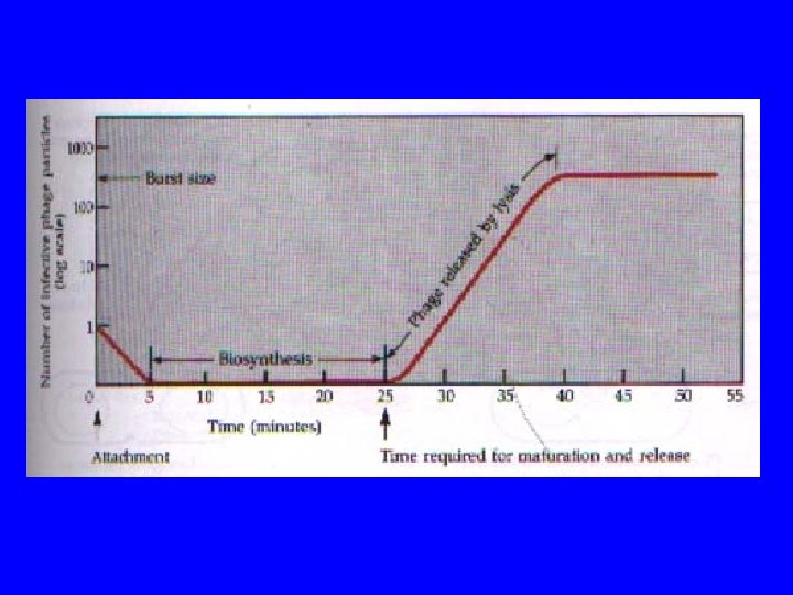
one-step growth curve
Lysogeny Some phages can incorporate their DNA into the host cell's DNA. , The phage remains latent and does not cause lysis of the host cell. Such a state is called lysogeny. Such phages are called lysogenic phages or temperate phages. The participating bacterial host cells are known as lysogenic cells.
Lysogenic cycle of bacteriophage lambda in E. coli
The consequences of infection by a temperate bacteriophage. The alternatives on infection are integration of the virus DNA into the host DNA (lysogenization) or replication and release of mature virus (lysis). The lysogenic cell can also be induced to produce mature virus and lyse.
The consequences of infection by a temperate bacteriophage. The alternatives on infection are integration of the virus DNA into the host DNA (lysogenization) or replication and release of mature virus (lysis). The lysogenic cell can also be induced to produce mature virus and lyse.
Think and answer following two questions !! 1. What are the two pathways available to temperate virus? 2. Describe how a single protein like the lambda represor can act both as an activator and a represor.
Some important characteristics for viral classification 1. Nature of the host-animal, plant, bacterial, insect, fungal 2. Nucleic acid characteristics-DNA or RNA, single or double stranded, molecular weight 3. Capsid symmetry-icosahedral, helical 4. Presence of an envelope and ether sensitivity 5. Diameter of the virion or nucleocapsid 6. Number of capsomers in icosahedral viruses 7. Immunologic properties 8. Intracellular location of viral replication
Recently, the International Committee for Taxonomy of Viruses has developed a uniform classification system and divided viruses into 50 families. The committee places greatest weight on three properties: (1)nucleic acid type (2) nucleic acid strandedness (3) presence or absence of an envelope
Schematic representations of the main types of bacterial viruses Tailed bacteriophage : Genome: DNA, double-stranded. Virion: complex shape, binary symmetry, variable number of capsomers. The tails of the phage are long and contractile in group A , long and noncontractile in group B, and very short in group C. Example: T-even coliphages.
Cubic bacteriophage: Group 1 Genome: DNA, single-stranded. Virion: icosahedral, cubic symmetry, 12 capsomers. Example: X 174 Group 2 Genome: DNA, double-stranded. Virion: cubic symmetry, enveloped. Example: PM-2. Group 3 Genome: RNA, single-stranded. Virion: icosahedral, cubic symmetry, 32 capsomers. Group 4 Genome: RNA, double-stranded. Virion: cubic symmetry, enveloped. Example: 06.
Filamentous bacteriophage: Genome: DNA, single-stranded. Virion: rod-shaped, helical symmetry. Example: fd.
Plant Viruses : Tobacco mosaic virus (TMV) as an example (1) penetration by the virus of a susceptible plant cellgenerally through abrasions or insect bites, (2) tincoating of the viral nucleic acid within the plant cell, (3) assumption by the viral genome of control of the synthetic activities of the host cell, (4) expression of the viral genome so that viral nucleic acid and capsid components are synthesized, (5) assembly of the viral particles within the host cell, and, (6) release of the complete viral particles from the host plant cell.
Animal Viruses:
Viroids and Prions: Viroids are small, circular, single-stranded RNA molecules that are the smallest known pathogens. The extracellular form of the viroid is naked RNA-there is no capsid of any kind. Prions have a distinct extracellular form, but the extracellular form seems to be entirely protein. It apparently does not contain any nucleic acid, or if it does, the molecule is not long enough to encode the single kind of protein of which the prion is composed
Maturation and release: The phage heads and tails are separately assembled from protein subunits, the head is packaged with phage DNA, and the tail is attached