Viruses and Prokaryotes Biology II Viruses Replicate only

Viruses and Prokaryotes Biology II

Viruses • Replicate only by infecting living cells • Differ widely in size and structure • One thing in common: enter living cells and use the machinery of the infected cell to produce more viruses
 made up of: –")
What is a Virus? • A particle (not a cell!) made up of: – Nucleic acid • DNA or RNA – Protein • DNA or RNA is surrounded by a protein coat, or capsid – Sometimes lipids

Capsid • Capsid proteins bind to the surface of a cell • Proteins “trick” the cell into allowing it inside • Viral genes take over the cell • Cell transcribes the viral genes, putting the genetic program of the virus into effect • Often times the virus destroys the cell

Viral Infection: Lytic or Lysogenic • Viruses must bind precisely to proteins on the cell surface and then use the host’s genetic system • Most viruses are highly specific to the cells they infect • Bacteriophage: a virus that infects bacteria
 • A virus enters a cell, makes a copy of")
Lytic Infection (HOSTAGE SITUATION) • A virus enters a cell, makes a copy of itself, and causes the cell to burst • Virus injects its DNA into the host • Usually the host cannot tell that the DNA is foreign and ends up copying the viral DNA! • The infected cell lyses, or bursts, and releases hundreds of virus particles that may go on to infect other cells
 - Repiratory and then systemic. (Last known \"natural\"")
Human Lytic Viruses • Smallpox (Variola) - Repiratory and then systemic. (Last known "natural" case in Somalia 1977. There was a laboratory acquired case later). • Respiratory Syncytial Virus (RSV) - Severe lung infections in infants. Infects respiratory tract but spread by contact. Handwashing is important. • Adenovirus - Mild to severe infections of the respiratory tract, Eye infections and gastrointestinal infections. • Rhinovirus - "Common cold virus" infects the upper respiratory tract. • Norwalk Virus - Gastroenteritis in older children and adults. Fecal-oral transmission. • Rotavirus - Gastroenteritis in infants and young children. Fecal-oral transmission. • Measles (rubeola) - Repiratory infection first and then systemic • Rubella (german measles) - Respiratory first and then systemic. Congenital infections are a serious concern. • Influenza Virus - Severe respiratory infections. Three serotypes: A, B and C. Serotype A also infects birds and other mammals where it trades genetic information with bird and mammal strains of influenza virus. Giving aspirin to children with influenza and chickenpox may lead to Reye's syndrome. • Polio Virus - Infects the GI tract first then through the blood to the central nervous system. Spread by oral-fecal route. • Rabies Virus - Spread by the saliva in the bite of an infected animal. Infects muscle cells at bite site then moves to the central nervous system and the salivary glands. • Hemorrhagic fevers- A number of viruses cause damage to the small blood vessels causing hemorrhage throughout the body: Ebola - spread by blood and body fluids, reservoir unknown; Lassa Fever - spread by rodent urine and feces and also by blood and body fluids of infected individuals; Dengue Fever (breakbone fever)- spread by mosquitoes that have bitten infected people; Yellow Fever - spread by mosquitoes that have bitten infected people or infected monkeys. • Hepatitis A and E - Transmitted through fecal-oral route. Infects the intestine and then through the blood to the liver.
 • A virus embeds its DNA into the DNA of")
Lysogenic Infection (BRAINWASHED CELL) • A virus embeds its DNA into the DNA of the host cell and is replicated along with the host cell’s DNA • Prophage: the DNA that is embedded in the host’s DNA • Infection that causes the host cell to make copies of the virus indefinitely • Do not lyse the host cell right away • Eventually the viral DNA will remove itself from the host DNA and direct the synthesis of new virus particles Laundry example… these must be my socks…I’ll wash them

Human Lysogenic Viruses • Herpes Viruses: • Herpes simplex - infects epithelium of mouth or genitals then travels up nerve axons to become latent in ganglia of the spinal column. Neonatal infections are very severe. Spread by secretions from active lesions. • Varicella-Zoster Virus - Causes both chickenpox and shingles (herpes zoster). Shingles is the manifestation of a latent infection. Spread by the respiratory route. • Epstein-Barr Virus (EB virus) - Causes mononucleosis which is spread by contact with infected saliva. Initially infects the oral or genital mucosa but then spreads to B-lymphocytes. This virus is associated with two malignancies: Burkitt's lymphoma and nasopharyngeal carcinoma. • Cytomegalovirus (CMV) - Infection from contact with infected saliva and other secretions. The virus infects mucous epithelium and white blood cells. Many children and adults harbor latent infections which are totally assymptomatic. Immunocompromised people may suffer devastating lung, central nervous system and other systemic infections. • Papilloma Virus - These viruses cause a variety of warts and they are spread by close contact. Three types (16, 18 and 31) have been associated with cervical and penile cancers. • Hepatitis B (HBV) - Spread through saliva and all other secretions and also vertically (from mother to child during birth or rarely in utero). Severe infection of the liver. Some people may develop chronic hepatitis and become life long carriers of the virus. This virus is strongly associated with liver cancer (hepatocellular carcinoma). • Retrovirus - Both Human Immunodeficiency Virus (HIV) and Human T-cell Lymphotropic Virus-1 (HTLV-1) belong to this group. These viruses infect T-cells and are spread by blood and body fluids. HTLV-1 is notable for two reasons: (1) it is transmitted to infants through mother's milk and (2) it causes adult onset T-cell leukemia.

Viruses and Disease • Polio, measles, AIDS, mumps, influenza, rabies, common cold • In most viral infections, viruses attack and destroy certain cells in the body, causing the symptoms of the disease • Vaccines – A weakened or killed virus or viral proteins that stimulates the immune system to produce immunity to the disease – Used to prevent infection – Provide protection only if used before an infection begins

Viruses and Cancer • Oncogenic viruses cause cancer in animals • Carry genes that disrupt the normal controls over cell growth and division • By studying such viruses, scientists have identified many of the genes that regulate cell growth in eukaryotes

Retroviruses • Viruses that contain RNA as their genetic information • “Retrovirus” – Genetic information is copied backward – From RNA to DNA • Mistakes can be made • This DNA is inserted in the host DNA • Responsible for some types of cancer in animals • HIV, the seasonal flu

Prions • Infectious particle made up of a misfolded protein rather than RNA or DNA • Prions accumulate, especially in the nervous system, that cells become damaged or destroyed • Prions are resistant to heat and digestive enzymes, so they are not destroyed by cooking infected meat An extremely simplified and highly unrealistic depiction of the normal prion protein (A) and the misfolded prion protein (B).

Mad Cow Disease • In 1985, veterinarians in Great Britain found cows suffering from a disease that, like scrapie in sheep, attacked and destroyed parts of the brain • BSE (for "bovine spongiform encephalopathy"), but the erratic behavior of infected cattle led to the common name of "mad cow disease" • When evidence emerged that mad cow disease and Creutzfeldt. Jakob disease (CJD), a similar disease in humans, might be caused by prions, people began to worry. • Was it possible that more than 100 people in Britain had died from CJD caused by prion-infected beef?

Mad Cow Disease • In 1996, British authorities concluded that the practice of using tissue from sheep and cows to prepare cattle feed had made it possible for BSE to spread rapidly to cattle and then to humans who ate contaminated beef. • They banned the use of cattle tissue in feed, and a similar ban was put in force in the United States the very next year. • In 2002, however, BSE was discovered in cows in Canada, and near the end of 2003, tissue from a Washington state cow with BSE was discovered after its meat had been processed.

H 1 N 1 What is H 1 N 1? • A new influenza virus that was causing illness in people • First detected in people in the United States in April 2009 • June 11, 2009, the World Health Organization (WHO) signaled that a pandemic of 2009 H 1 N 1 flu was underway Why was it called “swine flu”? • Laboratory testing showed that many of the genes in this new virus were very similar to influenza viruses that normally occur in pigs (swine) in North America • Further study has shown that it is very different from what normally circulates in North American pigs • It has two genes from flu viruses that normally circulate in pigs in Europe and Asia and bird (avian) genes and human genes • 2 surface proteins: hemagglutinin (H) & neuraminidase (N)

H 1 N 1 How did H 1 N 1 virus spread? What were the signs & symptoms? • The same way that seasonal flu spreads • • • Person to person – coughing or sneezing – touching something (such as a surface or object) with flu viruses on it and then touching the mouth or nose Fever Cough Sore throat Runny or stuffy nose Body aches Headache Chills Fatigue Diarrhea Vomiting Severe illnesses and death

H 1 N 1 How long can an infected person spread this virus to others? • • 1 day before getting sick to 5 to 7 days after. This can be longer in some people, especially children and people with weakened immune systems What can I do to protect myself from getting sick? • Vaccination BEFORE sickness • Cover your nose and mouth with a tissue when you cough or sneeze. Throw the tissue in the trash after you use it. • Wash your hands often with soap and water, especially after you cough or sneeze. • Avoid touching your eyes, nose or mouth. • Try to avoid close contact with sick people. • If you are sick with flu-like illness, stay home for at least 24 hours after your fever is gone except to get medical care or for other necessities.

Are Viruses Alive? • FOR: Viruses share the genetic code with living things and affect living things • AGAINST: They do not have all the characteristics of life (chapter 1) • VERDICT: NO!!!

Prokaryotes • Single-celled organisms that lack a nucleus • Circular DNA • Range in size from 1 -5 micrometers • (Eukaryotic cells range in size from 10 -100 micrometers)

Classifying Prokaryotes Current Classification Domain Bacteria Domain Archaea Kingdom Eubacteria Kingdom Archaebacteria (Former Kingdom Monera) Domain Eukarya Kingdom Protista Kingdom Plantae Kingdom Fungi Kingdom Animalia

Domain Bacteria: Kingdom Eubacteria • What we think about when we say “bacteria” • Cell well contains peptidoglycan (murein) – Made of sugars and amino acids • Larger of the two prokaryotic domains • Live almost anywhere

Domain Bacteria: Examples • Cyanobacteria: photosynthetic, like plants, which means that they use the sun’s energy to make food for themselves. • Spirochetes: are gram-negative spiral-shaped, and heterotrophic. Some of them live in the presence of oxygen, others don’t. • Gram-positive bacteria: includes the strain of streptococcus bacteria that causes strep throat. It also includes the bacteria that produces yogurt, by growing and fermenting in milk (producing lactic acid). These bacteria also produce many of our antibiotics. • Proteobacteria: is one of the largest phyla of all the bacteria. Many are gramnegative. They are divided into several subgroups, such as enteric bacteria, chemoautotrophs, and nitrogen-fixing bacteria. The enteric bacteria live mainly in intestinal tracts, like E. coli.

Domain Archaea: Kingdom Archaebacteria • Under a microscope look similar to eubacteria • Lack peptidoglycan (murein) in cell walls • Membrane lipids are quite different • Found in very harsh conditions • Early Earth’s atmosphere was filled with poisonous gases and was very hot – nothing could survive, except the archaebacteria. • Obligate anaerobes – bottom of the sea – volcanic vents – They cannot live in the presence of oxygen

Domain Archaea: Examples • Methanogens: are characterized by their ability to harvest energy by converting H 2 and CO 2 into methane gas; found in marshes and in the intestinal tracts of humans and some animals • Halophiles: salt-loving; found in the Dead Sea, the Great Salt Lake, and other areas with a high salt content • Thermoacidophiles: found in extremely acidic conditions and in areas with very high temperatures. They can survive in areas with temperatures as high as 230ₒF and with p. Hs below 2; locations include volcanic vents and hydrothermal vents (cracks in the ocean floor where scalding water leaks out)

Archaea to Eukarya • DNA of archaebacteria genes are more like those of eukaryotes than those of eubacteria • Scientists believe they may be the ancestors of eukaryotes

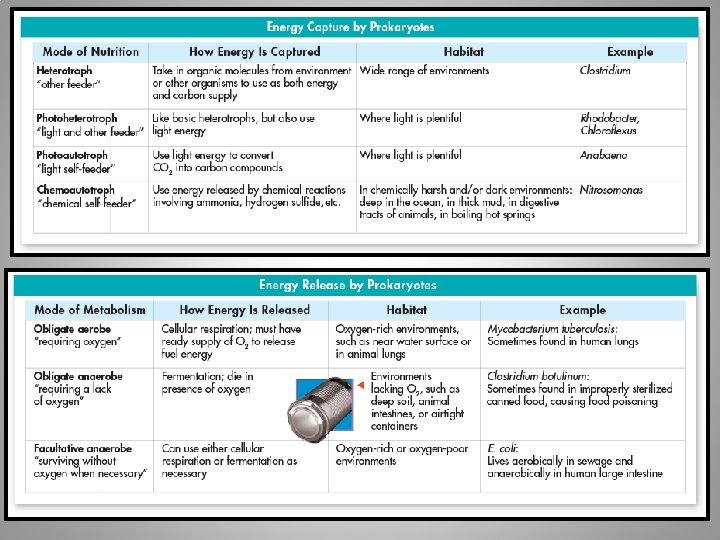
Identifying Prokaryotes • Shape • Cell wall chemical structure • Movement • Energy obtainment

Prokaryote Shape • Bacilli – Rod-shaped • Cocci – Spherical-shaped • Spirilla – Spiral and corkscrew-shaped

Cell Wall Chemical Structure: Gram Staining in Eubacteria • Determines: – Resistance to antibiotics – Identify an unknown bacteria • Staining a group of bacteria with four different liquids

Gram Positive: Purple • Cell wall containing many peptidoglycan, which absorbs the gram stain • More susceptible to antibiotics than gram-negative bacteria – Why? • They are not selectively permeable • They let everything into the cell…including antibiotics – Good for us, bad for them!

Gram Negative: Red • Cell wall containing a second, outer layer of lipids & carbohydrates • Selectively permeable and gram stain cannot pass through – Antibiotics cannot pass through either – Good for them, bad for us!

Prokaryote Movement • Flagella • Lash • Snake • Spiral • Move on a slime trail • No movement at all

Growth and Reproduction • When conditions are favorable, prokaryotes can grow and divide at astonishing rates • As often as every 20 minutes!

Reproduction: Binary Fission • Asexual reproduction • Prokaryote replicates its DNA • Divides in half, produces two identical daughter cells

Reproduction: Conjugation • Sexual reproduction • Paramecia and some prokaryotes • Exchange genetic information through a hollow bridge • (Think “conjunction-junction” song)

Reproduction: Endospore • Formed when conditions become unfavorable for reproduction • Bacterium produces a thick internal wall • Wall encloses its DNA and a portion of its cytoplasm to remain dormant until good conditions arise

Bacteria in Nature • Bacteria are vital to maintaining the living world • Some producers that capture energy by photosynthesis • Other help to break down the nutrients in dead matter and the atmosphere, allowing other organisms to use the nutrients

Decomposers • Help the ecosystem recycle nutrients • When a tree dies and falls to the forest floor, armies of bacteria attack and digest the dead tissue • The bacteria break the dead matter into simpler substances, which are released into the soil and taken up by the plants (picture: food in human digestive tract)
")
Nitrogen Fixers • Plants and animals depend on bacteria for nitrogen (amino acids proteins) • 80% of the atmosphere is nitrogen gas • Plants need nitrogen in the form of ammonia • Certain bacteria are able to convert nitrogen • Nitrogen Fixation: process of converting nitrogen gas into ammonia

Bacteria and Disease • Cause disease by destroying living cells or by releasing chemicals that upset homeostasis • Damaging host tissue • Releasing toxins

Antibiotics • Compounds that block the growth and production of bacteria • Can be used to cure many bacterial diseases

Human Uses of Bacteria: Foods • Cheese • Pickles • Yogurt • Sauerkraut • Buttermilk • Vinegar from wine • Sour cream
")
Human Uses of Bacteria: Industry • Digest petroleum (helpful in cleaning up oil spills) • Remove waste products and poisons from water • Mine minerals from the ground • Synthesize drugs and chemicals through genetic engineering

Controlling Bacteria • • • Physical removal – Soap and running water that dislodge bacteria and viruses Disinfectants – Chemical solutions that kill bacteria Food storage – Low temperatures slow bacteria growth Food processing – Boiling, frying, or steaming can kill bacteria Sterilization by Heat – Use of heat on objects such as medical instruments can prevent the growth of potentially dangerous bacteria
- Slides: 45