VIRUS 2 ND PROF PHARM D Definition Viruses


, introduced the term virus in microbiology. Edward Jenner")


, which")

 derived from: 1.")


 DNA viruses are in class 1 • The")


 • Class V(negative")

 sense RNA. They")

 strand RNA viruses that")
 sense")
 • • Picornaviridae Togaviridae Flaviviridae Retroviridae")
 • • • Paramyxoviridae Rhabdoviridae Orthomyxoviridae Filoviridae Bunyaviridae Reoviridae (double-stranded)")


- Slides: 76
VIRUS 2 ND PROF. PHARM. D
Definition Viruses are non cellular particles made up of genetic material and protein that can invade living cells.
Discovery of viruses Edward Jenner (1798), introduced the term virus in microbiology. Edward Jenner noticed that milk maids who were infected with cowpox, develop immunity against small pox. He inoculated a boy with the vesicle fluid taken from the hand of infected maid. The boy developed immunity against small pox. Edward Jenner assumed that the vesicle fluid that has been taken from the hand of the milk maid contained the poison (virus), that was responsible for immunity.
Discovery of viruses In 1884 C. Chamber land, in Pasteur's lab, discovered that if you passed a liquid containing bacteria through an unglazed PORCELAIN tube, the bacteria were COMPLETELY RETAINED and the solution that passed through (the FILTRATE) was sterile. In 1892 D. IWANOWSKI applied this test to a filtrate of plants suffering from TOBACCO MOSAIC DISEASE with shocking results; the filtrate the fully capable of producing the original disease in the new host.
Discovery of viruses Nothing could be seen in the filtrates using the most powerful microscopes, nor could anything be cultivated from the filtrates. Iwanowski concluded that the bacteria was so small / or they made a filterable toxin.
Discovery of Viruses Filtration of a mixture of bacteria and viruses. If a mixture of viruses and bacteria are filtered through a bacterial-proof filter (red), the viruses will pass through into the filtrate in the flask. Filtered beer is produced by a similar process.
Discovery of Viruses
Are Viruses living or non-living? Biologists consider viruses to be non-living because: -Are not cells -Do not grow or respond to their surroundings -Cannot make food, take in food, or produce wastes -Viruses do not respond to stimuli. They can only multiply if in another living cell
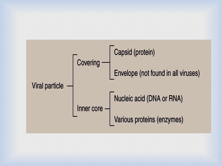
General characteristics of viruses Viruses are smaller than bacteria, they range in size between 20 -300 nanometer (nm). Viruses contain only one type of nucleic acid, either DNA or RNA, but never both. Viruses consist of nucleic acid surrounded by a protein coat. Some viruses have additional lipoprotein envelope.
General characteristics of viruses Viruses are obligate cellular parasites. They replicate only inside living cells. Viruses replicate through replication of their nucleic acid and synthesis of the viral protein. Viruses do not multiply in chemically defined media. Viruses do not undergo binary fission.
Virus and Living organisms Viruses must infect a living cell in order to grow and reproduce. They also take advantage of the host’s respiration, nutrition and all the other functions that occur in living things. Therefore, viruses are considered to be parasites.
Virion – Complete virus particle : nucleic acid + protein coat, which may be surrounded by an envelope – It is the form in which the virus moves between cells or hosts Viral Genome – EITHER RNA or DNA genome surrounded by a protective virus-coded protein coat (Capsid)
Viroids & Prions • Viroids – ss RNA genome and the smallest known pathogens. • – Affects plants Prions – – – Infectious particles that are entirely protein. No nucleic acid Highly heat resistant Animal disease that affects nervous tissue Affects nervous tissue and results in • Bovine spongiform encephalitis (BSE) “mad cow disease”, • Scrapie in sheep • kuru & Creutzfeldt-Jakob Disease (CJD) in humans 13
Naming Viruses • International Committee on Taxonomy of Viruses names them based on three characteristics: • Type of nucleic acid (DNA or RNA) • Is the nucleic acid double or single stranded • Presence or absence of nuclear envelope
Prokaryotes Vs. Eukaryotes Vs. Viruses • No membrane bound nucleus • Has a cell wall • Only a few organelles or none at all. • Has a capsule surrounding it • Three main types. • Nucleus with membrane • Only plants have cell wall • Contains many organelles • Has a lipid bilayer membrane surrounding it. • Specialized by thousands of different sizes and shapes. • • No nucleus No membranes No organelles Cannot reproduce on its own • Generally not considered alive by most standards
Origin of virus According to a hypothesis, viruses are bits of nucleic acid that ‘escaped’ from cellular organism. Some traces are from and bacterial cells. animal cells, plant cells Their multiple origins species-specific. explain why viruses are However, some other have broader range of host cells
REACTION TO PHYSICAL AND CHEMICAL AGENTS
Heat and Cold • Viral infectivity is generally destroyed by heating at 50– 60°C for 30 minutes • Viruses can be preserved by storage at subfreezing temperatures and can thus be preserved in the dry state at 4°C. p. H • Viruses are usually stable between p. H values of 5. 0 and 9. 0. • Some viruses (eg, enteroviruses) are resistant to acidic conditions. • All viruses are destroyed by alkaline conditions.
Radiation • Ultraviolet, x-ray, and high-energy particles inactivate viruses. Formaldehyde • Formaldehyde destroys viral infectivity by reacting with nucleic acid. • Viruses with single-stranded genomes are inactivated much more readily than those with double-stranded genomes. Ether Susceptibility • Ether susceptibility can be used to distinguish viruses that possess an envelope from those that do not
Configuration of Virus • The DNA or RNA genome may be : – ss – single stranded or – ds – double stranded • Genomes may be either: – (+) sense: Positive-sense viral RNA is identical to viral m. RNA and thus can be immediately translated into protein by the host cell. OR – (-) sense: Negative-sense viral RNA is complementary to m. RNA and thus must be converted to positive-sense RNA by an RNA polymerase before translation. 2 0
The nucleic acid genome plus the protective protein coat is called the nucleocapsid which may have icosahedral, helical or complex symmetry.
T h e Envelope • Enveloped viruses obtain their envelope by budding through a host cell membrane • In some cases, the virus buds through the plasma membrane but in other cases the envelope may be derived from internal cell membranes such as those of the Golgi body or the nucleus
T h e Envelope • Enveloped viruses do not necessarily have to kill their host cell in order to be released, since they can bud out of the cell - a process that is not necessarily lethal to the cell - hence some budding viruses can set up persistent infections
T h e Envelope • Enveloped viruses are readily infectious only if the envelope is intact (since the viral attachment proteins which recognize the host cell receptors are in the viral envelope) • This means that agents that damage the envelope, such as alcohols and detergents, reduce infectivity
General Morphology Capsid Structure determines shape: • Helical Viruses = nucleic acid is inside a hollow cylindrical capsid with a helical structure • Rabies, Ebola viruses, Tobacco Mosaic Virus • Polyhedral viruses = many sided; icosahedron is common with 20 equilateral triangles as sides and 12 vertices • Poliovirus, Adenovirus, herpes, • Complex structures • Pox virus & bacteriophage
ICOSAHEDRAL CAPSID • Icosahedral morphology is characteristic of the nucleocapsids of many “spherical” viruses • The icosahedral capsid structure of adenovirus is made up of three proteins, hexon, penton base, and fiber • Some proteins are associated with viral DNA, whereas others are associated with hexon and are involved in the formation of the capsid
HELICAL CAPSID • The icosahedral capsid structure of adenovirus is made up of three proteins, hexon, penton base, and fiber • Helical morphology is seen in nucleocapsids of many filamentous and pleomorphic viruses • Helical nucleocapsids are characterized by length, width, pitch of the helix, and number of protomers per helical turn
Bacteriophage • Viruses that infect bacterial cells are called bacteriophages (phages for short), which means ‘bacteria eaters’ • These are large, complex viruses, with a characteristic head and tail structure • The double-stranded, linear DNA genome contains over 100 genes, and is contained within the icosahedral head
Bacteriophage Much of the knowledge comes from studying bacteriophage, because they can be cultured easily within living bacteria. Their tail fibers are the base used to attach themselves to bacterial host cell The tail is the channel for their genetic material to be injected to the host cell.
Nomenclature of Viruses Various approaches, (do not obey the binomial nomenclature) derived from: 1. Named after the diseases eg. Measles virus, smallpox virus 2. Name after the places where the disease first reported eg. Newcastle disease virus, Ebola virus, Norwalk virus, Bunyaviridae 3. Host and signs of disease eg. Tobacco mosaic virus, cauliflower mosaic virus brome mosaic virus
4. Latin and Greek words eg. Coronaviridae Parvoviridae – “small” – “crown” 5. Virus discovers eg. Epstein-Barr virus 6. How they were originally thought to be contracted eg. dengue virus (“evil spirit”), influenza virus (the “influence” of bad air) 7. Combinations of Rous Sarcoma virus the above eg.
Reasons beyond classification Classification of virus been determined bythe structural and chemical composition of virus Are apply to all plant viruses, animal viruses and bacterial viruses Virus is acellular cell – cannot be categorised using taxonomic classification It used International Committee on Taxonomy of Viruses (ICTV) to classify the viruses
Before discovery • • Dermotropic – infected skin cell Neurotrophic – infected nerve cell Viscerotropic – infect organ of digestive tract Pneumotropic – infected respiratory system
After discovery • Type and structure of their nucleic acids • Methods of replication • Host range • Chemical and physical characteristics
Classification � Viruses are not classified as members of the kingdoms � Do not obey the biological taxonomy � Generally based on: 1. Classical - eg. animal, plant, bacterial virus system - eg. naked or enveloped virus 2. Genomic - Baltimore classification 3. Serology - classification based on Diagnostic virology - eg. Infectious bronchitis virus (IBV) of chickens (a coronavirus) – 3 different types present, these types have significant antigenic differences, but perhaps very little genetic or biological difference between these viruses.
Classification of Viruses 1. 2. 3. 4. The following criteria are used to classify viruses: Morphology – structure of capsid – presence or absence of envelope Size of the virion Type of host/host structures the virus infected - Bacteriophages: infect bacterial cells - Plant viruses infect plant cells -Animal viruses are subgrouped by the tissues they attack: a) Dermotrophic: if they infect the skin b) Neurotrophic: if they infect nerve tissue Genome composition – DNA / RNA – ds/ss DNA and ds/ss RNA
Classification of Viruses 1. 2. 3. 4. Taxonomic groups – family, subfamily, genus and species The names of virus families (family) are italicized - End in Latin suffix –viridae The genera (genus) end in the suffix – virus The species – English common name
• Viruses are divided into three groups, based on the morphology of the nucleocapsid and the arrangement of capsomeres. Symmetry of viruses • • Arrangement of capsomers in the virus. Two primary shapes of virus is rod and spherical. Rod shaped virus-helical symmetry Spherical virus-icosahedron
Baltimore Classification of viruses • The division of the viruses into classes based on genome type and mode of replication and transcription • Suggested by David Baltimore – Seven Baltimore classes. • Major groups of viruses are distinguished first by their nucleic acid content as either DNA or RNA • RNA and DNA viruses can be single-stranded (ss. RNA, ss. DNA) or double-stranded (ds. RNA, ss. DNA)
7 class of Baltimore classification Class Description of genome and replication strategy I Double stranded DNA genome II Example of bacterial virus Example of animal virus Lamda, T 4 Herpesvirus, poxvirus Single stranded DNA genome ØX 174 Chicken anemia virus III Double stranded RNA genome Ø 6 Reovirus IV Single stranded RNA genome plus MS 2 sense Poliovirus V Single stranded RNA genome minus sense Influenza virus, Rabies virus VI Single stranded RNA genome that replicated with DNA intermediate Retrovirus VII Double stranded DNA genome that replicates with RNA intermediate Hepatitis B virus
Class I • Double-stranded (ds) DNA viruses are in class 1 • The production of m. RNA and genome replication in such viruses occurs as it would from the host genome. Class II • Single-stranded(ss) DNA viruses. • These viruses form a double stranded DNA intermediate during replication and this intermediate used for transcription. • RNA polymerase requires double-stranded DNA as template.
Positive and Negative strand RNA viruses • The production of m. RNA and genome replication is much different with RNA viruses (Class III-VI). • m. RNA is the complementary base sequence to the template strand of DNA. • In virology, m. RNA is said to be plus(+) configuration. • While its complement is said to be the minus (-) configuration. • How does these viruses replicate? • Cellular RNA polymerases do not catalyze formation of RNA from an RNA template but from DNA template. • RNA viruses whether plus, negative or double stranded require a specific RNA-dependant RNA polymerase.
Class IV • Positive-strand of RNA viruses • Viral genome is of the plus configuration and hence serve directly as m. RNA. • The viruses required other protein, therefore m. RNA encodes a virus specific and RNA dependent RNA polymerase. • Once synthesized, this polymerase makes complementary minus strands of RNA and then use as template to make more plus strand.
Class III and Class V • Class III (double-stranded RNA viruses) • Class V(negative strand RNA virus) • m. RNA must be first synthesized, however cells does not have RNA polymerase. • To circumvent , these viruses contain enzyme in the virion, enters cell along with the genomic RNA. • Therefore, in this case complementary plus strand is synthesized by RNA dependant RNA polymerase and used as m. RNA. • Plus strand used as template to make more negativestrand genome.
Class VI • Single-stranded RNA genome that replicates with DNA intermediate. • This RNA virus require reverse transcriptase to copy the information found in RNA to DNA. Class VII • Double-stranded DNA genome that replicates with RNA intermediate. • Required reverse transcriptase • Mechanism producing m. RNA is similar in virus Class I
Retroviruses: are enveloped viruses that have two complete copies of (+) sense RNA. They also contain the enzyme reverse transcriptase, which uses the viral RNA to form a complementary strand of DNA, which is then replicated to form a ds. DNA retro, latin for “backward” (Class IV) in Baltimore Classification
Ambisense genome • A virus genome composed of ss. DNA or ss. RNA that is partly (+) sense and partly (-) sense. • Example: - Bunyaviridae ((-) sense RNA) and Arenavirus ((-) sense RNA)
Baltimore Classification - Advantages 1. Can classify between the (+) strand RNA viruses that do (Class VI) and do not (Class IV) undergo reverse transcription 2. Can classify between the ds. DNA viruses that do (Class VII) and do not (Class I) carry out reverse transcription
General Properties of RNA Viruses � Many ss. RNA viruses contain positive (+) sense RNA, and during an infection acts like m. RNA and can be translated by host’s ribosomes. � Other ss. RNA viruses have negative (-) sense RNA and the RNA acts as a template during transcription to make a complementary (+) sense m. RNA. � Negative (-) sense RNA must carry a RNA polymerase RNA dependent RNA polymerase – within the virion. Class III, IV and V � RNA viruses must either carry enzymes or have genes for those enzymes in order to copy RNA genomes after infecting a host cell
RNA viruses (+ve sense) • • Picornaviridae Togaviridae Flaviviridae Retroviridae
RNA viruses (-ve sense) • • • Paramyxoviridae Rhabdoviridae Orthomyxoviridae Filoviridae Bunyaviridae Reoviridae (double-stranded)
DNA viruses • Double – stranded • Adenoviridae • Herpesviridae • Poxviridae • Papovaviridae • Hepadnaviridae • Single – stranded • Parvoridae
Classification of major viral families based on genome structure and virion morphology. A, DNA viruses. L, linear genome; C, circular genome. B, RNA viruses. S, segmented genome.
Important viruses of humans VIRUS DISEASE Genome Type Adenovirus Respiratory infections ds. DNA Epstein barr virus Infectious mononucleosis Nasopharyngeal carcinoma Burkitt's lymphoma ds. DNA Herpes simplex Type 1 Cold sores ds. DNA Herpes simplex Type 2 Genital warts ds. DNA HIV AIDS (+)ss. RNA HPV Warts ds. DNA Influenza virus Influenza (-)ss. RNA
VIRUS Ebola virus DISEASE Haemorrhagi c fever Paramyxo virus Mumps Polio virus Poliomyelitis Rabies Virus Rabies Rubella virus German measles Small pox Vericella Zoster Chicken pox, shingles Genome Type (-)ss. RNA (+)ss. RNA ds. DNA
In visualizing the structure of a virus X-RAY CRYSTALLOGRAPHY • It is a means of determining the physical structure, dimensions of the individual proteins and components of the virus. Electron microscopy • It is used to generate information about the overall shape of the virus; it is also used with diagnostic purposes through detection of virus
Viruses In Dental Diseases • HSV Virus • Varicella Zoster Virus • Small Pox Virus • Paramyxovirus • HPV Virus • Measles Virus • Coxsackie Virus • HIV Virus • HHV-8 Virus
Viral Reproduction • Steps of Lytic Cycle • Attachment • Entry • Replication • Assembly • Lysis/Release (lyses the cell)
How do viruses replicate? 2 methods of replication: 1. Lytic Cycle – the virus enters the cell, replicates itself hundreds of times, and then bursts out of the cell, destroying it. 2. Lysogenic Cycle – the virus DNA integrates with the host DNA and the host’s cell helps create more virus DNA. An environmental change may cause the virus to enter the Lytic Cycle.
In the lytic cycle, the virus reproduces itself using the host cell's chemical machinery. The red spiral lines in the drawing indicate the virus's genetic material. The orange portion is the outer shell that protects it.
In the lysogenic cycle, the virus reproduces by first injecting its genetic material, indicated by the red line, into the host cell's genetic instructions.
1. ATTACHMENT • The first step in viral infection is attachment, interaction of a virion with a specific receptor site on the surface of a cell. • Receptor molecules differ for different viruses but are generally glycoproteins. • human immunodeficiency virus binds to the CD 4 receptor on cells of the immune system, • Rhinoviruses bind intercellular adhesion molecule 1 (ICAM -1), • Epstein-Barr virus recognizes the CD 21 receptor on B cells. • The presence or absence of receptors plays an important determining role in viral pathogenesis. • The attachment step may initiate irreversible structural changes in the virion.
2. Penetration • After binding, the virus particle is taken up inside the cell. This step is referred to as penetration
3. UNCOATING • Uncoating occurs shortly after penetration. • Uncoating is the physical separation of the viral nucleic acid from the outer structural components of the virion so that it can function. • The genome may be released as free nucleic acid (picornaviruses) or as a nucleocapsid (reoviruses). • The nucleocapsids usually contain polymerases. • Uncoating may require acidic p. H in the endosome. 4. TRANSCRIPTION • Virus m. RNA is produced using either cellular enzymes or virus-coded enzymes.
5. GENOME REPLICATION • This stage can take place in either the cytoplasm or nucleus of the infected cell. • Depending on the size of the virus genome the enzymes involved in genome replication may be encoded by either the virus itself or the host cell 6. TRANSLATION • This stage uses the host cell machinery – ribosomes and enzymes etc. • Various proteins are synthesised - structural - only in virion -and non-structural- detected only in the virus infected cell.
7. VIRION ASSEMBLY • The newly formed virus proteins and genomic nucleic acid assemble to produce the new virus particles. 8. VIRION RELEASE • Various strategies are available for the release of the progeny virus from the infected cell depending on the particular virus group. • The virus may bud through the cell membrane OR • The virus may simply cause lysis of the cell resulting in cell death and the release of progeny virus particles.
• Surface of virions consists of possibly 240 subunits comprising of 3 different polypeptides termed Large, . middle, small surface(HBs) proteins • The lipid in the outer protein shell or the HBs particles is derived from an intracellular compartment & not the plasma membrane
Viruses and cancer • 10% of cancer is caused by viruses • An oncovirus is a virus that can cause cancer • An oncogene is a cancer causing gene • Oncogenic viruses • DNA viruses • Human papillomavirus – cervical cancer • Hepatitis B causes liver cancer
Major Human Oncogenic Virus DNA VIRUSES - Small DNA tumor viruses Human Papilloma virus (HPV) -Adenovirus Herpesviruses (large) - Epstein Barr virus (EBV) - Kaposi’s Sarcoma Herpesvirus (KSHV) Other - Hepatitis virus B RNA viruses Human T-cell Leukemia Virus 1 (HTLV 1) Hepatitis virus C
Viruses Associated With The Development Of Human Neoplasia VIRUSES NEOPLASMS DNA VIRUSES Human papilloma virus Herpes simplex virus II Epstein-Barr virus Human Herpes virus 8 Hepatitis B virus Herpes simplex virus 6 (HBLV) Cervical Ca, warts, ano genital carcinoma Cervical carcinoma NPCa, African Burkitt’s Kaposi’s sarcoma Hepatocellular Ca Certain B cell lymphomas 58
Viruses Associated With The Development Of Human Neoplasia VIRUSES RNA VIRUSES Human T-cell leukemia virus II NEOPLASMS Some T-cell leukemia, Lymphoma Some cases of Human immunodeficiency virus hairy cell leukemia Lymphoma; Kaposi’s sarcoma 59
Transmission of Viruses • • • Respiratory transmission • Influenza A virus Faecal-oral transmission • Enterovirus Blood-borne transmission • Hepatitis B virus Sexual Transmission • HIV Animal or insect vectors • Rabies virus
Common Methods of Inactivating Viruses for Various Purposes • Sterilize laboratory supplies and equipment, • Disinfect surfaces or skin, • Sterilization may be accomplished by 1. steam under pressure, 2. dry heat, 3. ethylene oxide, 4. γ-irradiation. • Surface disinfectants include 1. sodium hypochlorite, 2. glutaraldehyde, 3. formaldehyde. • Skin disinfectants include • chlorhexidine, 70% ethanol, and iodophores.