Viral diseases NEWCASTLE DISEASE ND Def viral disease
 Def. : viral disease affect many species of birds")


 • Def. : An acute, highly contagious virus-caused disease of chickens")
 Serous, catarrhal, or caseous exudates in the trachea")

 (Gomboro) Def. : an acute, contagious viral disease of young")


 Def. : An acute viral disease of chickens & pheasants characterized")


 • is a slow spreading viral disease of chickens, turkey")


 § eosinophilic/red intracytoplasmic inclusions (Bollinger bodies).")


 • an infectious disease of birds on which E.")
 Airsacculitis: • Lesions may follow vaccination or infection with Mycoplasma,")
 Omphalitis: (Navel infection) In recently hatched chickens the navel is swollen, inflamed and")


 o Is a respiratory disease of chickens")


- Slides: 47
Viral diseases NEWCASTLE DISEASE (ND) Def. : viral disease affect many species of birds characterized by marked variation in PM. Lesions due to variation of pathotypes. Lentogenic (Low pathogenic) low mortality Mesogenic (moderate pathogenic). Velogenic most pathogenic(Vicerotropic or Neurotropic)
q. The disease is usually characterized by a short course, marked respiratory sings, diarrhea, and paralysis followed by death.
Depression Diarrhea Mortality
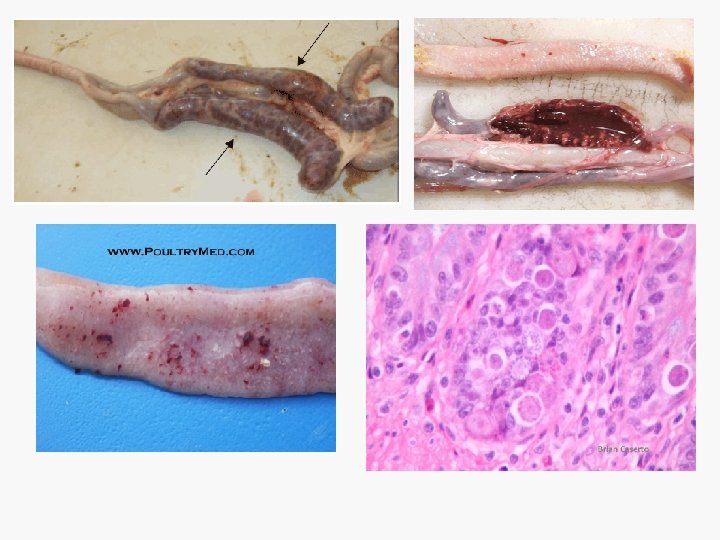
Gross lesions of velogenic Newcastle disease. • Severe inflammation of trachea and air sacs. • Hemorrhagic or necrotic focal lesions in the intestinal mucosa appear as circumscribed bluish red raised necrotic tissue easily to enucleated. • Hemorrhagic necrosis of cecal tonsils. • Hemorrhages on the mucosal surface of the proventriculus or in the gizzard. • Hemorrhages on the serous membranes, and the mucosa of the esophagus.
Microscopical examination • Neuoronal degeneration. • perivascular cuffing with lymphocytic cells. • endothelial hypertrophy.
Infectious bronchitis (IB) • Def. : An acute, highly contagious virus-caused disease of chickens characterized by: • respiratory signs • severe renal disease • marked decrease in egg production and bad egg quality (misshaped egg and watery albumin). • Cause: Coronavirus
PM. Lesions • Respiratory Lesions: 1) Serous, catarrhal, or caseous exudates in the trachea and bronchi lumen. 1) Plugs of yellow caseous material obstructing the bronchi and lower parts of trachea 1) There may or may not air saculitis.
Uremic form: • The kidneys are pale, swollen and the ureters distended with uric acid crystals In layers v The oviducts may be hypoplastic atrophied or cystic v The birds deposit yolk or fully formed eggs in the abdominal cavity (internal layers).
Infectious bursal disease (IBD) (Gomboro) Def. : an acute, contagious viral disease of young chickens characterized by: Ø diarrhea, vent picking, trembling, incoordination Ø Large, oedematous bursa followed by atrophy of the bursa of Fabricius. Ø immunosuppression.
Clinical symptoms • Closed eyes and death. • Ruffled feathers • Depression and trembling • Watery and whitish diarrhoea
Gross lesions • Enlargement of bursa fabricius to almost double its normal size • Haemorrhage on the internal and serosal surfaces of the bursa of fabricius. • Hemorrhage in the Proventriculus and Gizzard junction.
• haemorrhages on the leg, thigh and pectoral muscles. • kidneys, may be swollen and the ureters contain urates.
Microscopical lesions: ü In bursa there is lymphoid follicle depletion, cystic formation and destruction followed by atrophy. ü The spleen, Thymus and cecal tonsils showed similar changes like bursa. ü The variant strain induce rapid bursal atrophy and severe immunosuppression.
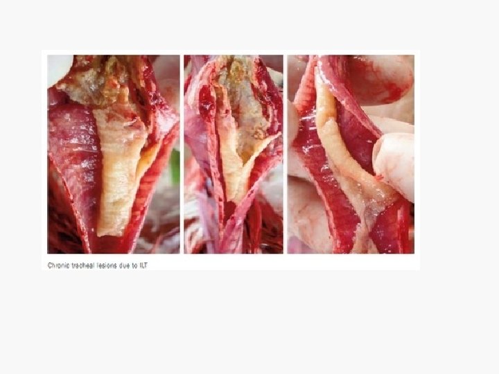
Infectious laryngotracheitis (ILT) Def. : An acute viral disease of chickens & pheasants characterized by: § marked dyspnea § coughing § gasping § expectoration of bloody exudate. Cause: Herps virus.
PM. Lesions • Severe laryngotracheitis, with bloody exudate in the trachea. • an occluding pseudomembrane or cheesy plugs in the trachea, death having occurred from suffocation. • bloody peak or blood on the face, head or feathers. • In less pathogenic outbreaks, mild conjunctivitis and sinusitis
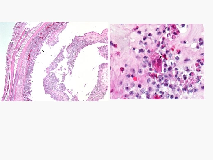
Microscopical lesions ü at the first few days of the disease reveal intranuclear inclusion bodies in epithelial lining the trachea. ü Desquamate necrotizing tracheitis.
Fowl pox (Avian pox) • is a slow spreading viral disease of chickens, turkey and other birds characterized by: • cutaneous lesions on unfeathered skin of the head, neck, legs and feet and/or by • diphtheritic membrane in the upper digestive and respiratory tract.
Gross lesions Cutaneous lesions: q The reaction against poxvirus is mainly proliferative rather than exudative. Characterized by appearance of wart like papillae or nodules, on face and unfeathered areas, comb and wattles, which soon become dark brown and hard.
Diphtheritic form: Lesions are raised, buff to yellow plaques on mucous membranes. The lesions appear in the upper respiratory and upper digestive tracts.
§ keratinocytes have marked cytoplasmic vacuolation (ballooning degeneration) § eosinophilic/red intracytoplasmic inclusions (Bollinger bodies). § The dermis is moderately infiltrated by lymphocytes, macrophages and heterophils.
Avian viral tumours Feature Age Symptoms Gross pathology Neural enlargement Bursa of fabricucs Tumors in skin muscle and proventriculus Microscopic lesions Neural involvement Liver tumours Spleen Bursa Centeral nervous system Lymphoid proliferation on skin and feather follicle. Cytology of tumour Category of neoplastic lymphoid cells Marek's disease 6 weeks or older Lymphoid leucosis Not less than 16 weeks Frequently paralysis Non-specific. Frequent atrophy May be present Absent nodular tumor usually absent Yes Often perivascular Diffuse Interfollicular tumour and/or atrophy of the follicle Yes No focal or diffuse often focal Pleomorphic lymphoid cells including lymphoblasts, small medium and large lymphocytes and reticular cells. Lymphoblasts T cells B cells. Interfollicular tumour. No No
Bacterial diseases
Avian colibacillosis (E. Coli infection) • an infectious disease of birds on which E. coli is the primary or secondary pathogen. Characterized by coligranuloma, peritonitis, salpingitis, synnovitis, omphalitis, and airsacculitis. • Caused by E. coli.
Gross Lesions • 1) Airsacculitis: • Lesions may follow vaccination or infection with Mycoplasma, 1 B, NDV, and ILT. • Lesions in this type are characterized by thickened air sacs which instead of being thin and glistening become thick, dull, and opaque. In severe cases caseous material in the air sacs are present.
2) Omphalitis: (Navel infection) In recently hatched chickens the navel is swollen, inflamed and . Abnormal yolk material and peritonitis are seen. 3) Acute septicemic picture ( Coli septicemia). 4) Enteritis 5) Salpingitis 6) Coligranuloma. (Hjarre's disease) 7) Synovitis and arthritis 8) Panophthalmitis 9) Pericarditis
Avian salmonellosis 1 - Pullorum disease. White Diarrhea; Bacillary white diarrhea; BWD is an infectious, egg transmitted disease of poultry, characterized by: v white diarrhea and v high mortality in young birds v asymptomatic adult carrier. Cause : Salmonella pullorum.
Gross pathology In Adults: § Occasionally there is nodular myocarditis, pericarditis or abnormal gonads. § Ovary may be hemorrhagic, atrophic or discolored follicles. § Oviduct impaction § peritonitis and ascites § atrophic testes.
In young birds Birds died after short septicemic course. Many birds have pasted white feces in the vent area. Classically there are gray nodules in one or more of the following sites: Lungs, liver, gizzard wall, heart, intestinal or cecal wall, spleen and peritoneum. Petechial hemorrhage or foci of necrosis in the liver.
Joints may be swollen in some birds. When the intestine is opened white plaques may be found in the intestine or ceca. Spleen frequently enlarged Ureters frequently distended with urats. Unabsorbed yolk sac
2 - Fowl Typhoid It is an infectious disease, primarily of chickens and turkeys with many of the lesions that occur with pullorum disease. Cause: Salmonella gallinarum.
Gross pathology 1 - In chickens and young poults • Lesions of fowl typhoid as in pullorum 2 - Lesions of acute fowl typhoid in older birds • Bile stained ("Bronzed") enlarged liver with or without small necrotic foci. • Enlarged spleen and kidneys. • Enteritis often with ulceration. • 3 - Lesions in older birds (Chronic fowl typhoid), . Resembles those seen in pullorum disease.
Fowl cholera Pasteurellosis Is an infectious disease of poultry appears as: Ø an acute septicemic disease with high morbidity and mortality. Ø A chronic form (Localized) form occurs follow an acute form or independently. Cause: Pasteurella multocida.
Gross lesions 1 - In peracute form: ü Lesions may be absent ü petechial and ecchymotic hemorrhages on heart, under serous membranes and mucous membranes of gizzard and or abdominal fat. ü Duodenitis.
2 - In acute form: Ø Lesions appear as disseminating intravascular coagulation of blood (Thrombi). Ø In layers and breeders hens, free yolk in the peritoneal cavity. Acute oophoritis with regressing follicles and acute diffuse peritonitis. Ø Diffuse streak hemorrhages of the liver with or without small necrotic foci in the liver (Corn meal liver).
3 - In chronic form: v Localized chronic inflammatory lesions involve joints and tendon sheath v wattle edema v Abscess in conjunctival sac, infraorbital sinus, and the nasal turbinate. v Lung sequestration mainly in turkeys.
Mycoplasma Gallisepticum Infection Chronic respiratory disease (CRD) o Is a respiratory disease of chickens and turkeys, characterized by respiratory signs and lesions o Cause: Mycoplasma galisepticum. o The organism is often associated with one or more of the following agents and these associations enhance pathogenisity. Infectious bronchitis, Newcastle disease, E. coli, pastuerella multocida and hemophilus para gallinarrum.
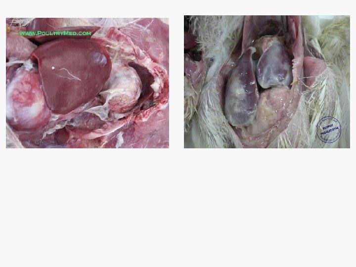
Gross Lesions Ø Poor physical condition and loss of weight. Ø Catarrhal inflammation of the nasal passages, sinuses, trachea, and bronchi. Ø Air sacs often are thickened, opaque and it often contains slimy or cheesy exudate. Ø The classic lesions of the disease are Airsacculitis, . Fibrinous perihepatilis and Adhesive pericarditis.
Mycotoxicosis • Mycotoxicosis is a disease caused by toxic fungal metabolite which produced by fungi that colonize and invade grains and feeds. • The environmental factors as high humidity play role in aggravating toxin production
Aflatoxicosis Aflatoxins are mostly Hepatotoxic. Gross pathology q Jaundice and generalized edema. q Hemorrhages on the liver. q Tan yellow discoloration of liver (fatty liver). q Swelling of kidneys and pale in colour.
Ochratoxicosis Gross pathology Ø Ochratoxins is mostly nephrotoxic Ø Reduction in weight gain. Ø Nepherotoxicity in kidneys (pale swollen kidneys). Ø Visceral gout and dehydration. Ø Immunosuppression.
Coccidiosis is a protozoal disease of poultry characterized by diarrhea and enteritis caused by different species of Eimeria. 1 - Those affect the anterior one third of the intestine Cause: Eimeria acervulina v Enteritis may be mild or severe and can lead to thickening of the mucosa. v white to gray striation (plaques) are often visible in the mucosa.
2 - Those affect the middle one third Cause Eimeria necatrix and Eimeria maxima Gross pathology Ø Congestion, hemorrhages, necrosis and white to yellow foci (very large schizonts), and petechial hemorrhages may be seen through the serosa. Ø Thickening of the intestinal wall and Bloody intestinal content.
3 Those affect the posterior one third Cause: Eimeria burntti and Eimeria tenella Gross pathology: q Presence of caseous or cheesy cores in the cecum. q Marked typhlitis. q Blood appear in ceca and feces in early cases.