Violation of blood circulation hyperaemia anaemia stasis bleeding

Violation of blood circulation: hyperaemia, anaemia, stasis, bleeding, thromboses, embolisms, infarcts As. -prof. Volodymyr D. Voloshyn (According to prof. Yaroslav Ya. Bodnar et al. , V. Serov et al. )

A pathological process is the appropriate reaction of organism as a reply to a damage factor. Alteration (from a lat. alteratio is change) or damage – there are the changes of cells structure, intercellular matter, tissues and organs which are expressed in violation of their function. . dystrophy >>>>> necrosi

n Violation of blood circulation causes the damage of structures of cells and tissues, that is expressed in the change of tissue and cellular metabolism, and in development of different types of dystrophy up to necrosis. They can develop, both in separate and in all organs and to stipulate the decline of their function. Above all things these disorders are inherent by all disease of the cardiac-vessels system. Knowledge about these gives us the possibility right to estimate the dynamics of motion and foresee the consequences of illnesses.

Violation of blood circulation Hyperaemia Thromboses Anaemia Embolisms Infarcts Bleeding Stasis Plasmorrhagy

Arterial hyperaemia is an intensive increased fill up by blood of organ or tissues through the surplus wave of arterial blood

ARTERIAL HYPERAEMIA Physiological Acute Pathological Chronic TOTAL LOCAL Angioneurotic Plethora Erytremia Collateral Postanaemia Vacate Inflammatic At arterio-venous canales


Venous hyperaemia is increase of blood supplying of organ or tissue through the deceleration ( ) of outflow of blood, influxes of blood here not changed or diminished. затрудненням

General venous hyperaemia arises up at pathology of heart, which conduces to insufficiency of cardiac activity
. There are")
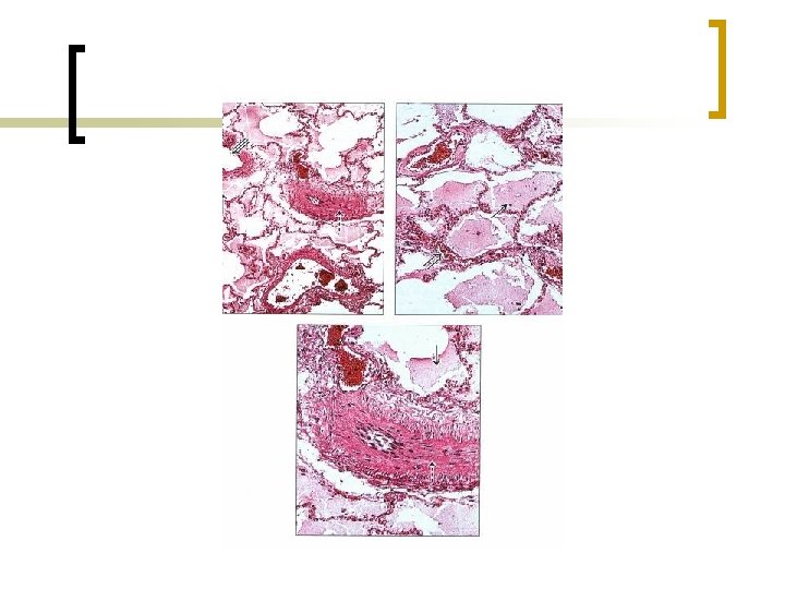
n At acute cardiac insufficiency (infarct of myocardium, acute decompensation of heart). There are the results of hypoxy and increase of capillary permeability lead to the plasmatic impregnation (plasmorrhage), swollen, spot diapedetic hemorrhages, dystrophic and necrotizing changes in the parenchimatic elements, for example, in lungs at the infarct of the left ventricle of heart.

Acute venous hyperaemia
 insufficiency which are developed at the congenital and acquired")
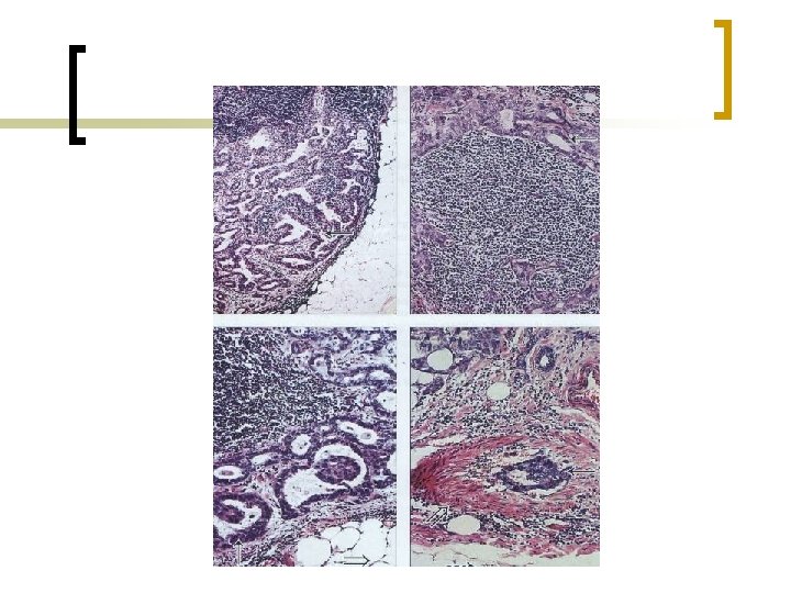
n At chronic cardiac (cardiac-vessels) insufficiency which are developed at the congenital and acquired defects of heart disease, miocarditis, cardiosclerosis there is a chronic venous hyperaemia. The latter Chronic hypoxia comes, and which conduces to development not only plasmorrhagy, edema and spot hemorrhages but also atrophy and sclerosis of tissues and organs.

Morphological signs of right ventricle cardiac insufficiency Cyanosis Skin Liver Lungs Atrophy Atrophic ulcers Nutmeg liver Sclerosis Dilatation of vein canal Cirrhosis Portal hypertension Brown induration Kidneys Cyanotic induration Lien Cyanotic induration Gastrointestinal Swelling Atrophic catarrh
 liver")
Nutmeg (Muscat) liver
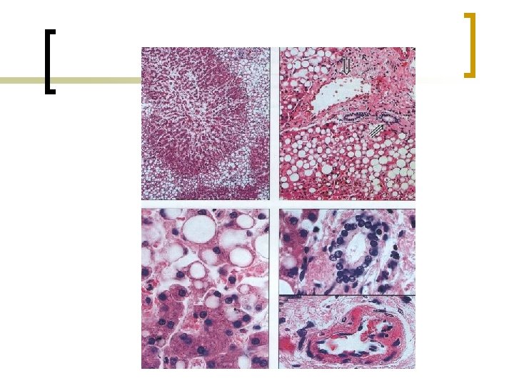

n The sclerotic changes are predefined by that the hypoxia stimulates the synthesis of collogen by fibroblasts; at the same time there is atrophy of parenchimatic elements. Thus the parenchima is replaced by connecting tissue, organs and tissues became dense – there is induration.

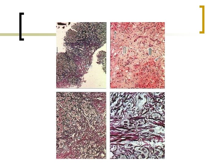
Brown induration of lungs

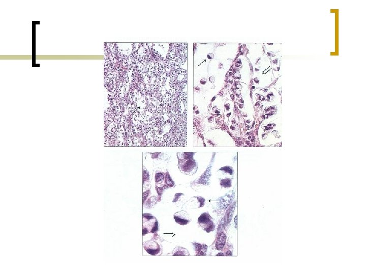
The local venous hyperemia develops at the difficult tide off blood from organs or from parts of body because there is the corking of the vein by thrombi, emboli or vein press by tumor or increased next (neighbouring) organ.
 is diminishing")
Anemia n Anaemia or ischemia (from a lat. ischo – to detain) is diminishing of blood supplying of organ, tissue, part of body as a result of insufficient of blood tide. Acute Chronic
 Spastic Ischemia from at the redistribution of blood")
Types of anaemia (depending on reason) Spastic Ischemia from at the redistribution of blood Obturatic Compressible

Depending on reasons and terms of origin distinguish such types of anaemia: n n Spastic (reflex) is spasm of arteries, for example at the pain irritation, at negative emotions. Obturatic is partial or absolute closing of artery by a tromb, clot (embol), disattached atherosclerotic plate, connecting tissue which overgrew at artery wall inflammation. Compressible is compression of artery by a tumour, liquid, ligature, bandage. Ischemia from at the redistribution of blood. At emitting of ascytic liquid blood comes to the vessels of abdominal region and a cerebral ischemia develops. In the cases of stand up quickly the blood comes into the low areas of body, and cerebral ischemia and dizziness come, orthostatic shock develops, the consciousness loss.

Obturatic myocardial ischemia. Infarct
 is stopping of blood motion")
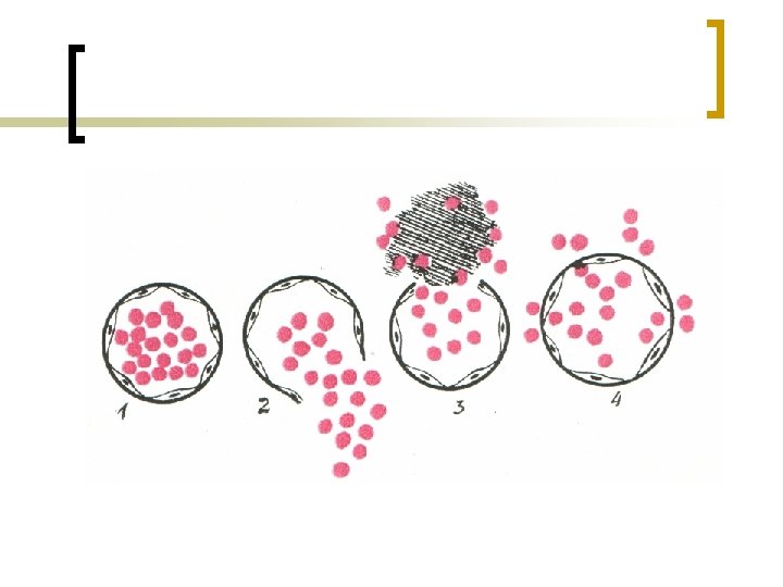
Stasis n Stasis (from a lat. stasis is stop) is stopping of blood motion in the microcirculatory vessels, mainly in capillaries.

 is the exit of blood from the road clearance of")
Bleeding n Bleeding (haemorrhagia) is the exit of blood from the road clearance of vessels or heart in an environment (external) or in the cavity of the body (internal).

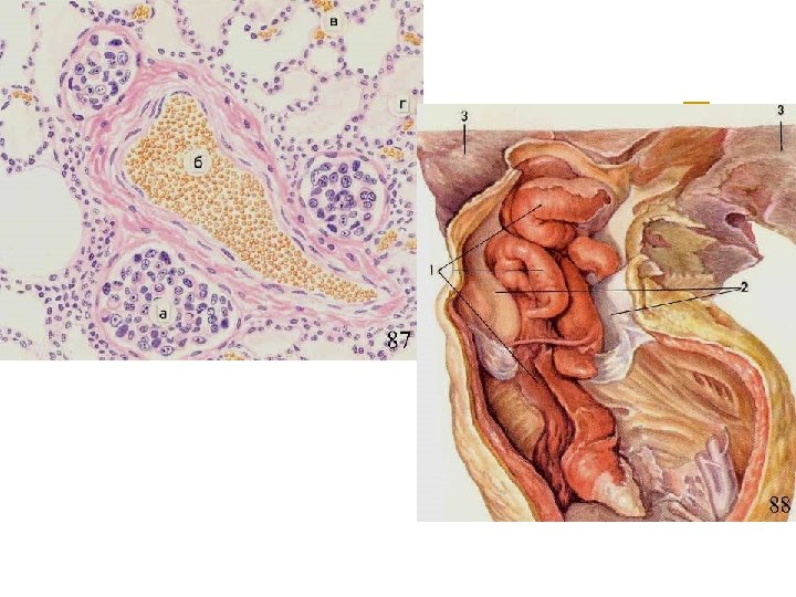
Pipe pregnancy is broken

n Hemorrhages are the blood accumulation in tissues and which follows from vessels
 External Petechiae")
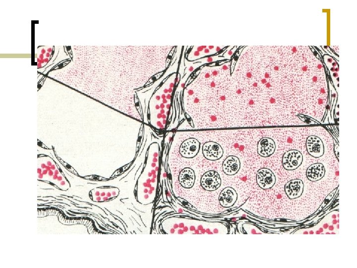
Morphological signs of blood outing from vessel canal Bleeding Hemorrhages (in tissue) External Petechiae Internal Ecchymoses Owing to rupture Bruise синець Owing to corrode Haematoma Owing to increasing of vessels permeability Haemorrhagic infiltration

Petechial hemorrhage

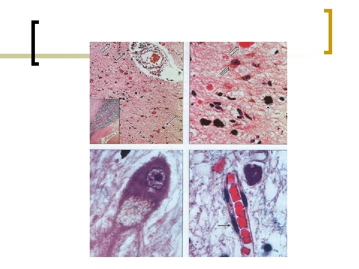
Haematoma of cerebellum and hemorragic infiltration of pia

Thrombosis n A thrombosis is an aggregation of blood in the road clearance of vessels or heart in alive. Blood clot, which appeared is named a tromb. The intravessel clot of lymph is also named a tromb

n n To the local factors of trombformation belong: damage of endothelia, deceleration and violation of laminarity of blood flow. To the general: unbalance between the convolutional and anticonvolutional systems of blood and the change of its composition.

Classification of the thrombus According to structure According to vessel’s canal Spheroid Red White Mixed Hyaline Specific forms Ordinary (atwall) Obstructive (Obturative) Horseman thrombus Delatic Progressive Marantic Migratory thrombus

Red Obstructive thrombus of vein

Consequences of thrombosis

Embolism n Embolism is circulation in the blood or in the lymph of particles which does not meet in a norm in them.

Classification of Embolism According to direction of move According to structure Typical Thrombemboli Atypical Lipid Gas Retrograde Aerial Paradoxical Cell Bacterial By foreign bodies

Bacterial embolism

86

Infarct n A infarct is the hearth of necrosis, which arises up as a result of the stopping of blood supplying. Other name is ischemia.
 White Triangular Red Incorrect")
Classification of infarcts According to view According to form (shape) White Triangular Red Incorrect form White infarct with red border

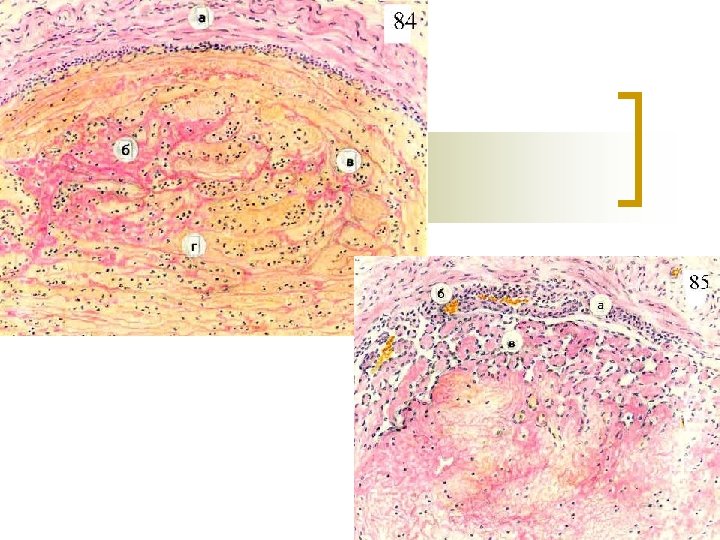
White infarct n White infarct – it is good marked off from surrounding tissue area of necrosis of whiteyellow color. Mainly arises up in organs with insufficient development of collaterals (spleen).

Ischemic stroke
 n White infarct with the hemorragic rim")
White infarct with the hemorragic rim (border) n White infarct with the hemorragic rim is the area of necrosis of white-yellow color which is off from surrounding tissue by expanded collateral vessels and diapedetic hemorrhages. Framing (border) is the result of transition of spasm in paretic expansion of vessels and increasing their vascular permeability.

Infarct of myocardium

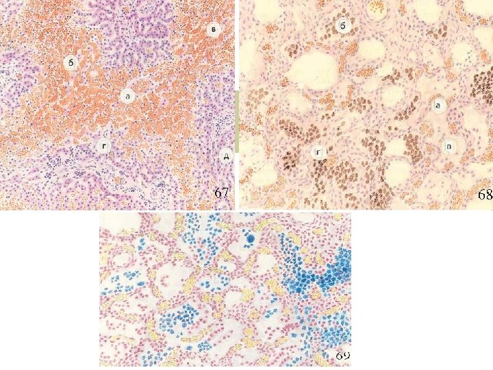
Hemorragic infarct n Hemorragic infarct is the area of necrosis which is saturated with blood. The development of him is predefined to the angiaarchitectinic of organ – double blood supplying with the presence of anastomoses (inosculations). .

A hemorragic infarct of lungs

Organ Kind of infarct Kind of necrosis HEART White infarct with red border Coagulative necrosis with secondary Colikvation LUNGS Red Coagulative KIDNEY White infarct with red border Coagulative White & red Colikvative White Coagulative Red Colikvative BRAIN SPLEEN INTESTINE

Thank you for attention!
- Slides: 59