VESTIBULOCOCHLEAR NERVE AND AUDITORY PATHWAY VESTIBULOCOCHLEAR NERVE It

VESTIBULOCOCHLEAR NERVE AND AUDITORY PATHWAY

VESTIBULOCOCHLEAR NERVE. It is the 8 th cranial nerve. . It is entirely sensory. . It is also called as auditory nerve/stato-acoustic nerve. . Developmentally it is derived from the embryonic otic placode.

• Vestibulocochlear nerve is attached to the surface of the brainstem at the lower border of the pons , posterolateral to the attachment of the facial nerve. Facial nerve Vestibulocochle ar Nerve

• It consists of two anatomically and functionally distinct parts vestibular nerve, distributed to the organ of equilibrium cochlear nerve, distributed to the hearing organ.

Medial /dorsal/chief vestibular nucleus: located in medulla")
VESTIBULAR NUCLEI. Are four in no. : 1)Medial /dorsal/chief vestibular nucleus: located in medulla 2)Lateral vestibular nucleus/nucleus of deiters: located in upper medulla 3)Inferior vestibular nucleus: located in lower medulla 4)Superior vestibular nucleus: located in pons
Ventral cochlear nucleus: located on the anterolateral aspect of")
COCHLEAR NUCLEI. Two in number: 1)Ventral cochlear nucleus: located on the anterolateral aspect of inferior cerebellar peduncle. 2)Dorsal cochlear nucleus: located on the post. aspect of the inferior cerebellar peduncle lateral to the floor of 4 th ventricle.

FUNCTIONAL COMPONENT • Both the components are SPECIAL SOMATIC AFFERENT: They carry sensory information necessary for the maintainance of equilibrium & hearing from the membranous labyrinth of the internal ear. • The fibres carrying sensory information for equillibrium terminate in the vestibular nuclei within the brainstem. • The fibres carrying sensory information for hearing terminate in dorsal & ventral cochlear nuclei located on inferior cerebeller peduncle.

VESTIBULAR NERVE • It contains about 19, 000 nerve fibres. • It originates in the internal acoustic meatus, which is a channel in the petrous part of temporal bone, through which facial nerve , auditory nerve and some vessels pass.

. The nerve fibers of the vestibular nerve are the central processes of the nerve cells located in the vestibular ganglion(scarpa’s ganglion)which is situated in the trunks of the vestibular nerve at the bottom of internal acoustic meatus. . These central processes form the trunk of the vestibular nerve, which pass medially in a spiral fashion to occupy ventromedial part of cohlear nerve.

• Then it enters the ant surface of brainstem in a groove b/w lower border of pons & the upper part of medulla oblongata.

• On reaching the lower border of pons the vestibular nerve passes medial to inferior cerebeller peduncle, divides into ascending & descending branches & makes synapsis with four groups of vestibular nuclei , located beneath the floor of fourth ventricle. • A few fibres reach directly the flocculonodular lobe of the cerebellem through juxtarestiform body.

• Some efferent fibres extend from the vestibular to the cochlear nerves by the vestibulo-cochlear anastomosis(Oort’s bundle).

• Peripheral processes of the bipolar cells at the bottom of the internal acoustic meatus divide into upper, lower and posterior branches.
 of the internal acoustic meatus • Transverse")
Features at the blind end ( floor) of the internal acoustic meatus • Transverse crest • AREA ABOVE CREST shows: • 1) opening into facial canal anteriorly • 2) superior vestibular area posteriorly{ perforated} • AREA BELOW CREST: • 1) Tractus spiralis foraminosus • 2)inferior vestibular area • 3) foramen singulare

Upper branch • Foramina in superior vestibular area • Macula of utricle and ampullae of the anterior and lateral semicircular ducts.

Lower branch • Foramina in the inferior vestibular area. • Macula of the saccule

Posterior branch • Foramen singulare • Ampulla of the posterior semicircular duct.
 • The vestibular pathway transmits impulses from vestibular receptors")
VESTIBULAR PATHWAY(neural pathway for balance) • The vestibular pathway transmits impulses from vestibular receptors in the internal ear to the vestibular nuclei in the brainstem, which in turn project to widespread area of the central nervous system. • It is responsible for maintainance of bodybalance(static & kinetic).

VESTIBULAR RECEPTORS • The receptors for static balance are maculae in the wall of utricle & saccule of vestibule(static labyrinth). The static labyrinth detects the position of head with respect to gravity& it also responds to linear acceleration/deceleration. • Macula consists of: 1)Supporting cells 2)Hair cells The hair cells are embedded into a gelatinous mass containing irregularly shaped concretions(otoliths). This gelatinous mass moves in response to gravity, bending the hair cells and initiating action potentials in the associated neurons.
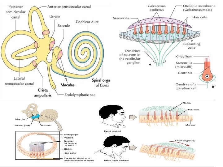

• The receptor for kinetic balance is crista ampularis (present in the expanded base of each semicircular canal). The kinetic labyrinth is invoved in evaluating head movements & angular acceleration. • Each crista consists of: 1) Supporting cells 2) Hair cells 3) Cupula(crest of epithelium with curved gelatinous mass without otoliths)
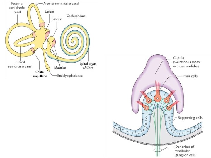

• The endolymph movement within each semicircular canal moves the cupula, bends the hair & initiates the action potential in the associated neurons.
sensory neurons form vestibular ganglion •")
PATHWAY • The cell bodies of first order (bipolar)sensory neurons form vestibular ganglion • Peripheral processes of these cells end on hair cells of maculae of utricle and saccule and on hair cells of cristae of semicircular ducts of internal ear.

• Central processes of bipolar cells form vestibular nerve. These relay in vestibular nuclei and flocculonodul ar lobe of cerebellum
Flocculonodular lobe (vestibulocerebellar")
• Second order sensory neurons of vestibular nuclei project to: a)Flocculonodular lobe (vestibulocerebellar tract) b)Motor nuclei of 111 rd, 1 Vth&V 1 th cranial nerves( Medial longitudinal fasciculus) c)Anterior horn cells of spinal cord(vestibulospinal tract). . The vestibular nuclei also project to posteroventral nucleus of thalamus.

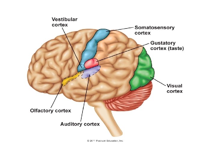
• Third order sensory neurons from thalamus project to vestibular area of the cerebral cortex in postcentral gyrus.

FUNCTIONS OF VESTIBULAR SYSTEM. The vestibular connections contribute to the coordination of muscle contraction in maintenance of upright posture. . Through connections of the medial longitudinal fasciculus of the same & opp: side, the vestibular system controls some vestibulo-ocular & postural reflexes (vestibulospinal &vestibulocollic reflex), eg conjugate eye movements & movements of the trunk & neck in response to vestibular stimulation. . The vestibulothalamocortical connections give a sense of balance.

VESTIBULO-OCULAR REFLEX • The VOR is a reflex, where activation of vestibular system causes eye movement. • This reflex functions to stablize images on retina during head movements by producing eye movements in the direction opp: to head movement, thus preserving image on the center of visual field. • Since slight head movement is present all the time, the VOR is very important for stablizing vision.

CIRCUIT OF VESTIBULO-OCULAR REFLEX HEAD ROTATION activates SEMICIRCULAR CANALS sends impulses via VESTIBULAR NERVE impulses pass to VESTIBULAR NUCLEI IN BRAINSTEM
: fibres pass directly to lateral rectus fibres")
fibres ativate C/L ABDUCENT NUCLEUS(but inhibit ipsilateral): fibres pass directly to lateral rectus fibres pass via MLF to C/L occulomotor nucleus , which activates medial rectus Hence causing conjugate eye movements

The dysfunction of vestibulothalamocortical connections is associated with the symptoms of motion sickness. The most common causes of damage to vestibular nerve are; Exposure to ototoxic antibiotics, Ménière's disease Encephalitis Typically, patients with a damaged nerve suffer from acute attacks of vertigo accompanied by nausea/vomiting, inability to maintain posture and horizontal nystagmus.

COCHLEAR NERVE. Cochlear nerve conducts the nerve impulses concerned with sound from the organ of corti in the cochlea. . It contains about 23, 500 nerve fibres.

• The fibres of cochlear nerve are the central processes of the bipolar spiral ganglion cells located in the spiral ganglion of the cochlea(present in the modiolus of internal ear). • These fibres pass through the tractus spiralis foraminosus at the bottom of the internal acoustic meatus & unite to form the trunk of cochlear nerve.

The trunk enters the anterior surface of the brainstem at the lower border of pons on the lateral side of the emerging facial nerve & is separated from it by the vestibular nerve.

• On entering the pons the nerve fibres divide, with one branch entering the posterior cochlear nucleus & the other branch entering the anterior cochlear nucleus(which are situated on the surface of inferior cerebeller peduncle).

• The peripheral fibres end in terminals around the bases of the inner and outer hair cells of the organ of Corti( peripheral receptor for sound)

In mammals, cochlear nerve fibers are classified as: . Type I neurons 9095% innervate the inner hair cells. They have relatively large diameters, are bipolar, and are myelinated. Each type I axon innervates only a single inner hair cell, but each inner hair cell is innervated by up to 30 such nerve fibers, depending on species and location within the cochlea. • Type II neurons 5 -10% innervate the outer hair cells. They have relatively small diameters are unipolar, and are unmyelinated

Hearing is the second in importance among the special senses in the humans after vision.
 • It is highly specialized structure that responds to")
Organ of Corti (Hearing Receptor) • It is highly specialized structure that responds to fluid-borne vibrations • located in the cochlear duct. • The cells inside the cochlear duct are highly modified to form a structure called organ of Corti.
 Rod cells 2) Hair cells 3)")
• Organ of corti consists of: 1) Rod cells 2) Hair cells 3) Supporting cells
 cells called rods of Corti stand on the basilar")
• Two rod (pillar) cells called rods of Corti stand on the basilar membrane and project into the scala media. • The triangular interval between the two rods is the tunnel of Corti , filled with perilymph.

Hair cells • Specialized sensory cells • arranged in a single row internal to the inner rod, and in 3 or 4 rows external to the outer rod. • From the upper surface of the hair cells project tiny cilia called stereocilia. • The tips of the cilia are embedded within the gelatinous shelf called tectorial membrane, which is attached at one end to the limbus and at the other end to the Hensen's cells. • The afferent fibres of cochlear nerve form synaptic contact with the plasma membrane of the hair cells.
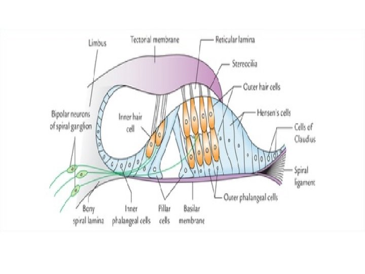

• The reticular lamina is a tough membrane supported by rods of Corti and presents tiny holes for the passage of stereocilia. • The spaces other than the tunnel of Corti in the organ of Corti are filled with the corticolymph.

SUPPORTING CELLS • The supporting Hensen's cells are elongated columnar cells and lie outside the external row of outer hair cells, succeeded more laterally by cubical cells of Claudius. • Cells of Deiter(phalangeal cells)form cap like investments around the bases of hair cells.

Steps Involved In Hearing • The sound waves are collected by the auricle and are conducted through the external auditory meatus to the tympanic membrane to make it vibrate. • The vibrating tympanic membrane causes 3 ear ossicles in the middle ear (malleus, incus and stapes) to vibrate. • The vibrations of the footplate of stapes produces vibrations in the perilymph of the scala vestibuli (here sound waves are converted into fluid waves).

• The vibrations of the perilymph cause simultaneous vibrations of the vestibular membrane and the endolymph in the cochlear duct which causes basilar membrane to vibrate. • As the basilar membrane vibrates the hair cells of the organ of Corti attached to it move. Consequently the stereocilia of hair cells embedded in the tectorial membrane become bent.

• Bending of the stereocilia causes depolarization of the hair cells. • The hair cells induce action potentials in the neurons of cochlear nerve. • The action potential generated in the cochlear nerve neurons is conducted to the cerebral cortex via auditory pathway, where it is interpreted and perceived as sound.
Ascending pathways: a)primary auditory pathway b)non-primary auditory pathway 2)Descending")
AUDITORY PATHWAY Is classified as: 1)Ascending pathways: a)primary auditory pathway b)non-primary auditory pathway 2)Descending pathways

PRIMARY AUDITORY PATHWAY It exclusively transmits the stimulus responsible for hearing from the hearing receptor in the internal ear (spiral organ or organ of Corti) to the auditory area of the cerebral cortex.
. • Dorsal and ventral cochlear nuclei.")
STRUCTURES INCLUDED • Organ of Corti (hearing receptor). • Dorsal and ventral cochlear nuclei. • Sup: olivary nucl: & Trapezoid body and its nuclei. • Lateral lemniscus. • Inferior colliculus. • Medial geniculate body. • Auditory radiation. • Auditory cortex.

PATHWAY • Cell bodies of the first order sensory neurons of the auditory pathway lie in the spiral or cochlear ganglion which is located within the cochlear modiolus. • The peripheral processes of these neurons reach the organ of Corti which is the end organ (receptor organ) for hearing, while the central processes (axons) constitute the cochlear nerve.

• Cochlear nerve ends in the dorsal cochlear nucleus and the ventral cochlear nucleus. • The second order sensory neurons arise from the cells of the cochlear nuclei and end in the nucleus of the trapezoid body and superior olivary nucleus of the same side as well as of the opposite side.

• The third order sensory neurons arise from the dorsal nucleus of the trapezoid body & superior olivary nucleus of both sides and ascend throug post part of pons & midbrain as lateral lamniscus. • The lateral lamniscus is the principal ascending auditory tract : It ascends from the region of superior olivary nucleus, through the brainstem. A few of its fibres relay in the nucleus of lateral lemnisus. on reaching midbrain, the fibers relay in nuleus of inferior colliculus (centre for auditory reflexes) for the reflex activity – which sends new set of axons to the medial geniculate body through the brachium of inferior colliculus.

However most of the fibres of the lateral lamniscus pass directly to the medial geniculate body without being relayed in the inferior colliculus. • The MGB are the final relay stations of the hearing pathway. • The fibres arising from the MGB (fourth order sensory neurons) form auditory radiations which run through the sublentiform part of the internal capsule to project into the anterior transverse temporal gyrus (Heschl’s gyrus). • The Herschl’s gyrus contains primary auditory cortex – area 41 & 42 of Broadman.

Unique feature of the primary auditory pathway • More than three neurons in its course

NON-PRIMARY AUDITORY PATHWAY • Also k/a Reticular sensory pathway carries all types of sensory messages.

DESCENDING AUDITORY PATHWAYS • Descending fibres originating in the auditory cortex & in other nuclei in the auditory pathway accompany the ascending pathway. • These fibres are bilateral & end on nerve cells at different levels of the auditory pathway & on hair cells of the organ of corti. • These fibres serve as feedback mechanism & inhibit the reception of sound.

• The two inferior colliculi are connected to each other by commissural fibres. Each inferior colliculus is connected to the superior colliculus of its own side. From the tectum of midbrain the fibres descend in the brainstem & spinal cord as tectobulbar & tectospinal tracts. These connections bring about reflex movement of the head & neck in response to sound. • The lateral lamniscus is connected by collateral branches with the medial longitudinal fasciculus & reticular formation of the brain stem. Likewise superior olivary nucleus is also connected with them. Through the medial longitudinal fasciculus they are connected with the nuclei of the ocular muscles. These connections bring about reflex conjugate movement of the eyes in response to sound. AUDITORY REFLEXES

TESTING OF 8 th Cranial nerve • RINNE’ s test • WEBER’s Test • ABC test

APPLIED • Infranuclear lesions Ø The cochlear nerve may be damaged in fractures of the petrous part of the temporal bone or affected by acoustic tumor. Ø There will be deafness on the affected side. Ø Incomplete or irritative lesions of the cochlear nerve will give rise to reduced hearing & tinnitus (ringing in the ear) on the affected side. • Supranuclear lesions Ø This will not produce any appreciable hearing defect because of the bilateral representation in the auditory cortex of each side.
- Slides: 64