
Veseptl kezelsek acut vesekrosodsban Dr Ujhelyi Lszl egyetemi

Vesepótló kezelések acut vesekárosodásban Dr. Ujhelyi László egyetemi docens Debreceni Egyetem OEC Belgyógyászati Intézet Nephrologiai Tanszék, FMC Extracorporalis Szervpótló Centrum, Debrecen
Vesepótló kezelések acut vesekárosodásban Ujhelyi László DE OEC Belgyógyászati Intézet Nephrologia Tanszék

Vázlat • • • Modalitások elnevezése Méregtelenítés fizikája Primer – szekunder akut vesekárosodás Intermittáló - folyamatos kezelések KDIGO 2012 AKI recommendations Összefoglalás
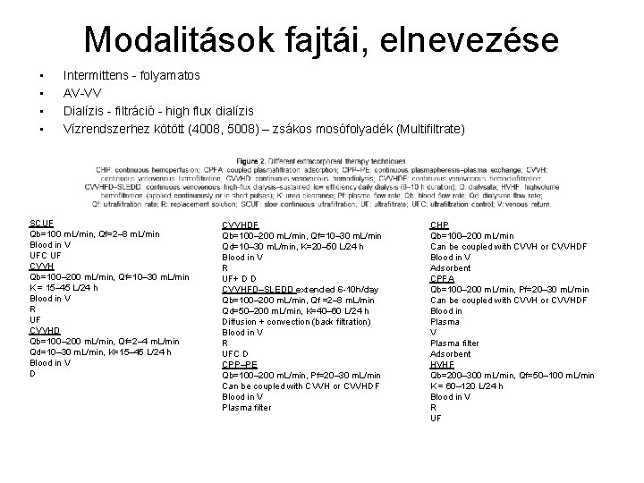
Modalitások fajtái, elnevezése • • Intermittens - folyamatos AV-VV Dialízis - filtráció - high flux dialízis Vízrendszerhez kötött (4008, 5008) – zsákos mosófolyadék (Multifiltrate) SCUF Qb=100 m. L/min, Qf=2– 8 m. L/min Blood in V UFC UF CVVH Qb=100– 200 m. L/min, Qf=10– 30 m. L/min K = 15– 45 L/24 h Blood in V R UF CVVHD Qb=100– 200 m. L/min, Qf=2– 4 m. L/min Qd=10– 30 m. L/min, K=15– 45 L/24 h Blood in V D CVVHDF Qb=100– 200 m. L/min, Qf=10– 30 m. L/min Qd=10– 30 m. L/min, K=20– 50 L/24 h Blood in V R UF+ D D CVVHFD–SLEDD extended 6 -10 h/day Qb=100– 200 m. L/min, Qf =2– 8 m. L/min Qd=50– 200 m. L/min, K=40– 60 L/24 h Diffusion + convection (back filtration) Blood in V R UFC D CPP–PE Qb=100– 200 m. L/min, Pf=20– 30 m. L/min Can be coupled with CVVH or CVVHDF Blood in V Plasma filter CHP Qb=100– 200 m. L/min Can be coupled with CVVH or CVVHDF Blood in V Adsorbent CPFA Qb=100– 200 m. L/min, Pf=20– 30 m. L/min Can be coupled with CVVH or CVVHDF Blood in Plasma V Plasma filter Adsorbent HVHF Qb=200– 300 m. L/min, Qf=50– 100 m. L/min K = 60– 120 L/24 h Blood in V R UF

Méregtelenítés fizikája Diffuzió Fick első törvénye J anyagáram sűrűség (mol/m 2 s) D diffuziós együttható (m 2/s) Ci koncentráció (mol/m 3) x távolság (m) Stokes-Einstein egyenlet k. B Boltzmann állandó T hőmérséklet (K) éta viszkozitás r részecske sugara (m) Konvektív transzport Sieving koefficiens (S) Cr recipiens oldali koncentráció Cd donor oldali koncentráció Konvektív klírensz (Kx = Qf Sx) Qf filtrációsáram Sx sieving koefficiens x anyagra Adsorptio
Urémiás toxinok Kidney International, Vol. 63 (2003), pp. 1934– 1943 Review on uremic toxins: Classification, concentration, and interindividual variability R. Vanholder, R. De Smet, G. Glorieux, et al, For the Euopean Uremoc Toxin Work Group (EUTOX) • • Szabadon vízoldékony kis molekulasúlyú anyagok, MW<500 D, N=45 – Urea 60 D – Malondialdehid 71 D – Oxalát 90 D – Dimetilglicin 103 D – Benzilalkohol 108 D – Kreatinin 113 D és egyéb guanidinek – Poliolok pl. mioinozitol – Pirimidinek – Purinok – Ribonukleozidok – Béta-lipotropin peptid 461 D Fehérjéhez kötött anyagok MW<500 D N=23 – Homocisztein 135 D – CMPF 240 D – AGE – Fenolok – Indolok – Hippurátok – poliaminok • Közepes molekulasúlyú anyagok MW>500 D N=22 – Metionin-enkephalin 555 D – Delta. sleep inducing peptide 848 D – Atrial natriuretic peptide 3080 D – Beta-endorphin 3465 D – Cholecystokinin 3866 D – Neuropeptid Y 4272 D – Endothelin 4283 D – PTH 9225 D – Beta 2 -microglobulin 11818 D – Cystatin. C 13300 D – Leptin 16000 D fehérjéhez kötött – Retinol binding protein 21200 D fehérjéhez kötött – Complement faktor D 23750 D – Interleukin-6 24500 D – Kappa Ig könnyű lánc 25000 D – Lambda Ig könnyű lánc 25000 D – Tumor necrosis factor-alpha 26000 D – Interleukin-1 beta 32000 D

Akut vesekárosodásban akkumulálódó egyéb eredetű metabolitok • • • Laktát 89 D Bilirubin diglukuronid 937 D Mioglobin 17699 D Pro- és anti-inflammatorikus mediatorok sepsisben • • • • • Pro-inflammatory TNF-a IL 1 b, IL-2, IL-6, IL-8, IL-15 Neutrophil elastase IFN-g Thromboxane Platelet-activating factor Vasoactive neuropeptides Phospholipase A 2 Plasminogen activator inhibitor-1 Prostaglandins Prostacyclin Free radical generation Soluble adhesion molecules Tyrosine kinase Protein kinase H 2 S NO HMGB 1 protein Anti-inflammatory IL-1 Ra IL-4 IL-10 IL-13 Type II IL-1 receptor Transforming growth factor-b Epinephrine Soluble TNF-a receptors Leukotriene B 4 -receptor antagonist LPS binding protein Soluble recombinant CD-14
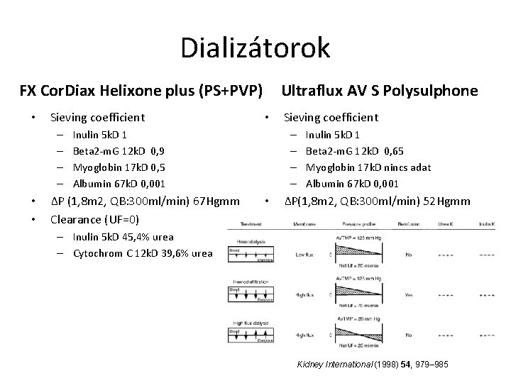
Dializátorok FX Cor. Diax Helixone plus (PS+PVP) • Sieving coefficient – – • • Ultraflux AV S Polysulphone • Inulin 5 k. D 1 Beta 2 -m. G 12 k. D 0, 9 Myoglobin 17 k. D 0, 5 Albumin 67 k. D 0, 001 ΔP (1, 8 m 2, QB: 300 ml/min) 67 Hgmm Clearance (UF=0) Sieving coefficient – – • Inulin 5 k. D 1 Beta 2 -m. G 12 k. D 0, 65 Myoglobin 17 k. D nincs adat Albumin 67 k. D 0, 001 ΔP(1, 8 m 2, QB: 300 ml/min) 52 Hgmm – Inulin 5 k. D 45, 4% urea – Cytochrom C 12 k. D 39, 6% urea Kidney International (1998) 54, 979– 985
Primer – szekunder akut vesekárosodás • Primer akut vesekárosodás: gyógyszer, vegyszer toxicitás, akut intersticialis nephritis, akut glomerulonephritis, akut vaszkuláris károsodás, hipoxiás vesekárosodás • Szekunder akut vesekárosodás, több szervi elégtelenség része – hepatorenalis szindróma, kardiorenális szindróma – posztoperativ akut vesekárosodás megelőző vesebetegséggel vagy anélkül, szívsebészeti, érsebészeti, hasi sebészeti esetek leggyakrabban infekcióval súlyosbítva (shock, SIRS, sepsis)
Intermittáló - folyamatos kezelések • Folyamatos kezelések elméleti előnyei: – – fokozott hemodinamikai stabilitás megfelelő folyadék eltávolítás veseműködés rendeződését elősegíti hatékonyabb kis- és közép mólsúlyú salakanyag eltávolítás • Intermittáló kezelések előnyei – – – – kevesebb vérzéses szövődmény rövid idő alatt hatékony kis mólsúlyú anyag eltávolítás (K, laktát) állandó géphez kötöttség nincs: diagnosztikus vizsgálatok, ápolás mobilizálhatóság flexibilitás jobb gépkihasználás költségkímélő • Sustained low-efficiency daily dialysis – CVVHFD 6 -10 h/nap – előnyöket egyesíti, ha vízrendszer mellett végzik
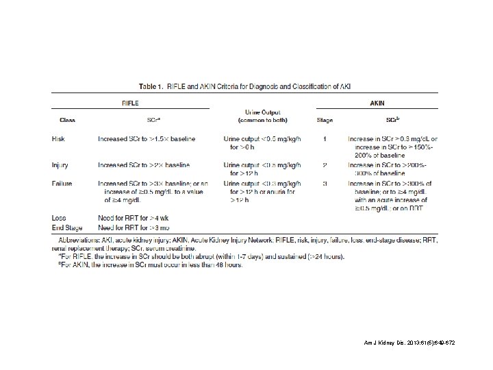
Am J Kidney Dis. 2013; 61(5): 649 -672
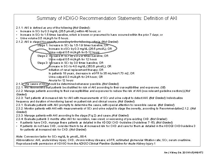
Summary of KDIGO Recommendation Statements: Definition of AKI 2. 1. 1: AKI is defined as any of the following (Not Graded): • Increase in SCr by 0. 3 mg/d. L (26. 5 µmol/L) within 48 hours; or • Increase in SCr to 1. 5 times baseline, which is known or presumed to have occurred within the prior 7 days; or • Urine volume 0. 5 m. L/kg/h for 6 hours. 2. 1. 2: AKI is staged for severity according to the following criteria. (Not Graded) Stage 1: Increase in SCr by 1. 5 -1. 9 times baseline; OR Increase in s. SCr by 0. 3 mg/d. L (26. 5 µmol/L); OR Urine output 0. 5 m. L/kg/h for 6 -12 hours Stage 2: Increase in SCr by 2. 0 -2. 9 times baseline; OR Urine output 0. 5 m. L/kg/h for 12 hours Stage 3: Increase in SCr by 3. 0 times baseline; OR Increase in SCr to 4. 0 mg/d. L (353. 6 µmol/L); OR Initiation of renal replacement therapy; OR In patients 18 years, decrease in e. GFR to 35 m. L/min/1. 73 m 2; OR Urine output 0. 3 m. L/kg/h for 24 hours; OR Anuria for 12 hours 2. 1. 3: The cause of AKI should be determined whenever possible. (Not Graded) 2. 2. 1: We recommend that patients be stratified for risk of AKI according to their susceptibilities and exposures. (1 B) 2. 2. 2: Manage patients according to their susceptibilities and exposures to reduce the risk of AKI (see relevant guideline sections). (Not Graded) 2. 2. 3: Test patients at increased risk for AKI with measurements of SCr and urine output to detect AKI. (Not Graded) Individualize frequency and duration of monitoring based on patient risk and clinical course. (Not Graded) 2. 3. 1: Evaluate patients with AKI promptly to determine the cause, with special attention to reversible causes. (Not Graded) 2. 3. 2: Monitor patients with AKI with measurements of SCr and urine output to stage the severity, according to Recommendation 2. 1. 2. (Not Graded) 2. 3. 3: Manage patients with AKI according to the stage (Fig 2) and cause. (Not Graded) 2. 3. 4: Evaluate patients 3 months after AKI for resolution, new onset, or worsening of pre-existing CKD. (Not Graded) • If patients have CKD, manage these patients as detailed in the KDOQI CKD Guideline (Guidelines 7 -15). (Not Graded) • If patients do not have CKD, consider them to be at increased risk for CKD and care for them as detailed in the KDOQI CKDGuideline 3 for patients at increased risk for CKD. (Not Graded) Note: Conversion factor for SCr mg/d. L to µmol/L, 88. 4. Abbreviations: AKI, acute kidney injury; CKD, chronic kidney disease; e. GFR, estimated glomerular filtration rate; SCr, serum creatinine. Reproduced with permission of KDIGO from the KDIGO Clinical Practice Guideline for Acute Kidney Injury. 1 Am J Kidney Dis. 2013; 61(5): 649 -672
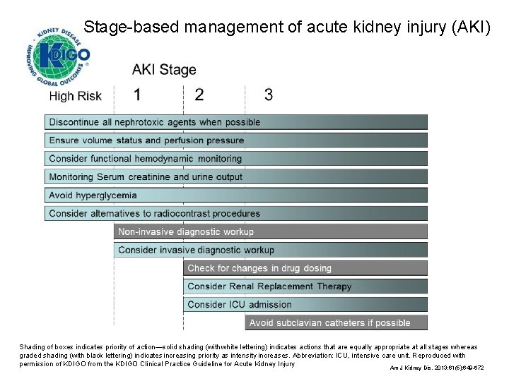
Stage-based management of acute kidney injury (AKI) Shading of boxes indicates priority of action—solid shading (with white lettering) indicates actions that are equally appropriate at all stages whereas graded shading (with black lettering) indicates increasing priority as intensity increases. Abbreviation: ICU, intensive care unit. Reproduced with permission of KDIGO from the KDIGO Clinical Practice Guideline for Acute Kidney Injury Am J Kidney Dis. 2013; 61(5): 649 -672
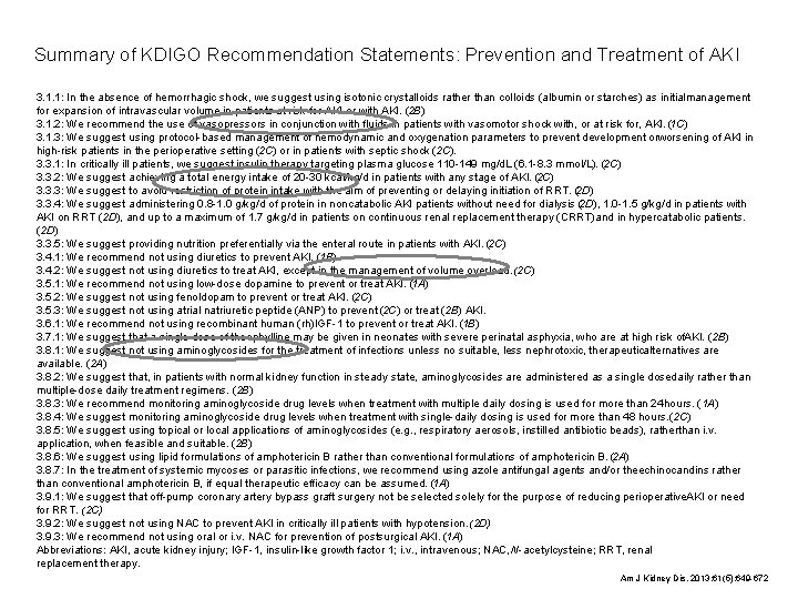
Summary of KDIGO Recommendation Statements: Prevention and Treatment of AKI 3. 1. 1: In the absence of hemorrhagic shock, we suggest using isotonic crystalloids rather than colloids (albumin or starches) as initialmanagement for expansion of intravascular volume in patients at risk for AKI or with AKI. (2 B) 3. 1. 2: We recommend the use of vasopressors in conjunction with fluids in patients with vasomotor shock with, or at risk for, AKI. (1 C) 3. 1. 3: We suggest using protocol-based management of hemodynamic and oxygenation parameters to prevent development orworsening of AKI in high-risk patients in the perioperative setting (2 C) or in patients with septic shock (2 C). 3. 3. 1: In critically ill patients, we suggest insulin therapy targeting plasma glucose 110 -149 mg/d. L (6. 1 -8. 3 mmol/L). (2 C) 3. 3. 2: We suggest achieving a total energy intake of 20 -30 kcal/kg/d in patients with any stage of AKI. (2 C) 3. 3. 3: We suggest to avoid restriction of protein intake with the aim of preventing or delaying initiation of RRT. (2 D) 3. 3. 4: We suggest administering 0. 8 -1. 0 g/kg/d of protein in noncatabolic AKI patients without need for dialysis (2 D), 1. 0 -1. 5 g/kg/d in patients with AKI on RRT (2 D), and up to a maximum of 1. 7 g/kg/d in patients on continuous renal replacement therapy (CRRT) and in hypercatabolic patients. (2 D) 3. 3. 5: We suggest providing nutrition preferentially via the enteral route in patients with AKI. (2 C) 3. 4. 1: We recommend not using diuretics to prevent AKI. (1 B) 3. 4. 2: We suggest not using diuretics to treat AKI, except in the management of volume overload. (2 C) 3. 5. 1: We recommend not using low-dose dopamine to prevent or treat AKI. (1 A) 3. 5. 2: We suggest not using fenoldopam to prevent or treat AKI. (2 C) 3. 5. 3: We suggest not using atrial natriuretic peptide (ANP) to prevent (2 C) or treat (2 B) AKI. 3. 6. 1: We recommend not using recombinant human (rh)IGF-1 to prevent or treat AKI. (1 B) 3. 7. 1: We suggest that a single dose of theophylline may be given in neonates with severe perinatal asphyxia, who are at high risk of. AKI. (2 B) 3. 8. 1: We suggest not using aminoglycosides for the treatment of infections unless no suitable, less nephrotoxic, therapeuticalternatives are available. (2 A) 3. 8. 2: We suggest that, in patients with normal kidney function in steady state, aminoglycosides are administered as a single dosedaily rather than multiple-dose daily treatment regimens. (2 B) 3. 8. 3: We recommend monitoring aminoglycoside drug levels when treatment with multiple daily dosing is used for more than 24 hours. (1 A) 3. 8. 4: We suggest monitoring aminoglycoside drug levels when treatment with single-daily dosing is used for more than 48 hours. (2 C) 3. 8. 5: We suggest using topical or local applications of aminoglycosides (e. g. , respiratory aerosols, instilled antibiotic beads), ratherthan i. v. application, when feasible and suitable. (2 B) 3. 8. 6: We suggest using lipid formulations of amphotericin B rather than conventional formulations of amphotericin B. (2 A) 3. 8. 7: In the treatment of systemic mycoses or parasitic infections, we recommend using azole antifungal agents and/or theechinocandins rather than conventional amphotericin B, if equal therapeutic efficacy can be assumed. (1 A) 3. 9. 1: We suggest that off-pump coronary artery bypass graft surgery not be selected solely for the purpose of reducing perioperative. AKI or need for RRT. (2 C) 3. 9. 2: We suggest not using NAC to prevent AKI in critically ill patients with hypotension. (2 D) 3. 9. 3: We recommend not using oral or i. v. NAC for prevention of postsurgical AKI. (1 A) Abbreviations: AKI, acute kidney injury; IGF-1, insulin-like growth factor 1; i. v. , intravenous; NAC, N-acetylcysteine; RRT, renal replacement therapy. Am J Kidney Dis. 2013; 61(5): 649 -672
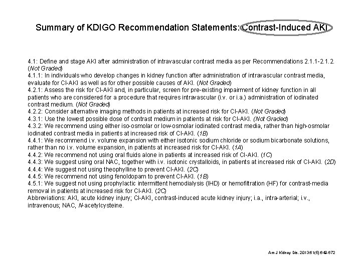
Summary of KDIGO Recommendation Statements: Contrast-Induced AKI 4. 1: Define and stage AKI after administration of intravascular contrast media as per Recommendations 2. 1. 1 -2. 1. 2. (Not Graded) 4. 1. 1: In individuals who develop changes in kidney function after administration of intravascular contrast media, evaluate for CI-AKI as well as for other possible causes of AKI. (Not Graded) 4. 2. 1: Assess the risk for CI-AKI and, in particular, screen for pre-existing impairment of kidney function in all patients who are considered for a procedure that requires intravascular (i. v. or i. a. ) administration of iodinated contrast medium. (Not Graded) 4. 2. 2: Consider alternative imaging methods in patients at increased risk for CI-AKI. (Not Graded) 4. 3. 1: Use the lowest possible dose of contrast medium in patients at risk for CI-AKI. (Not Graded) 4. 3. 2: We recommend using either iso-osmolar or low-osmolar iodinated contrast media, rather than high-osmolar iodinated contrast media in patients at increased risk of CI-AKI. (1 B) 4. 4. 1: We recommend i. v. volume expansion with either isotonic sodium chloride or sodium bicarbonate solutions, rather than no i. v. volume expansion, in patients at increased risk for CI-AKI. (1 A) 4. 4. 2: We recommend not using oral fluids alone in patients at increased risk of CI-AKI. (1 C) 4. 4. 3: We suggest using oral NAC, together with i. v. isotonic crystalloids, in patients at increased risk of CI-AKI. (2 D) 4. 4. 4: We suggest not using theophylline to prevent CI-AKI. (2 C) 4. 4. 5: We recommend not using fenoldopam to prevent CI-AKI. (1 B) 4. 5. 1: We suggest not using prophylactic intermittent hemodialysis (IHD) or hemofiltration (HF) for contrast-media removal in patients at increased risk for CI-AKI. (2 C) Abbreviations: AKI, acute kidney injury; CI-AKI, contrast-induced acute kidney injury; i. a. , intra-arterial; i. v. , intravenous; NAC, N-acetylcysteine. Am J Kidney Dis. 2013; 61(5): 649 -672
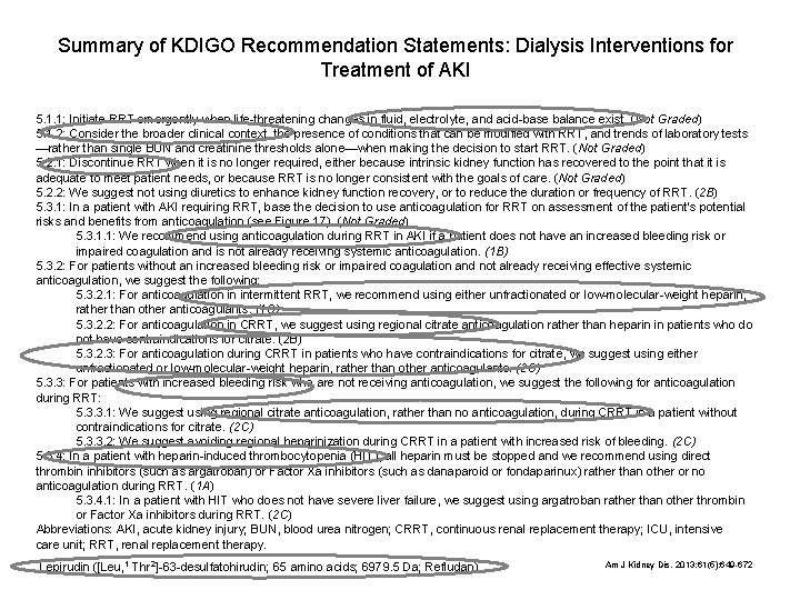
Summary of KDIGO Recommendation Statements: Dialysis Interventions for Treatment of AKI 5. 1. 1: Initiate RRT emergently when life-threatening changes in fluid, electrolyte, and acid-base balance exist. (Not Graded) 5. 1. 2: Consider the broader clinical context, the presence of conditions that can be modified with RRT, and trends of laboratory tests —rather than single BUN and creatinine thresholds alone—when making the decision to start RRT. (Not Graded) 5. 2. 1: Discontinue RRT when it is no longer required, either because intrinsic kidney function has recovered to the point that it is adequate to meet patient needs, or because RRT is no longer consistent with the goals of care. (Not Graded) 5. 2. 2: We suggest not using diuretics to enhance kidney function recovery, or to reduce the duration or frequency of RRT. (2 B) 5. 3. 1: In a patient with AKI requiring RRT, base the decision to use anticoagulation for RRT on assessment of the patient’s potential risks and benefits from anticoagulation (see Figure 17). (Not Graded) 5. 3. 1. 1: We recommend using anticoagulation during RRT in AKI if a patient does not have an increased bleeding risk or impaired coagulation and is not already receiving systemic anticoagulation. (1 B) 5. 3. 2: For patients without an increased bleeding risk or impaired coagulation and not already receiving effective systemic anticoagulation, we suggest the following: 5. 3. 2. 1: For anticoagulation in intermittent RRT, we recommend using either unfractionated or low-molecular-weight heparin, rather than other anticoagulants. (1 C) 5. 3. 2. 2: For anticoagulation in CRRT, we suggest using regional citrate anticoagulation rather than heparin in patients who do not have contraindications for citrate. (2 B) 5. 3. 2. 3: For anticoagulation during CRRT in patients who have contraindications for citrate, we suggest using either unfractionated or low-molecular-weight heparin, rather than other anticoagulants. (2 C) 5. 3. 3: For patients with increased bleeding risk who are not receiving anticoagulation, we suggest the following for anticoagulation during RRT: 5. 3. 3. 1: We suggest using regional citrate anticoagulation, rather than no anticoagulation, during CRRT in a patient without contraindications for citrate. (2 C) 5. 3. 3. 2: We suggest avoiding regional heparinization during CRRT in a patient with increased risk of bleeding. (2 C) 5. 3. 4: In a patient with heparin-induced thrombocytopenia (HIT), all heparin must be stopped and we recommend using direct thrombin inhibitors (such as argatroban) or Factor Xa inhibitors (such as danaparoid or fondaparinux) rather than other or no anticoagulation during RRT. (1 A) 5. 3. 4. 1: In a patient with HIT who does not have severe liver failure, we suggest using argatroban rather than other thrombin or Factor Xa inhibitors during RRT. (2 C) Abbreviations: AKI, acute kidney injury; BUN, blood urea nitrogen; CRRT, continuous renal replacement therapy; ICU, intensive care unit; RRT, renal replacement therapy. Lepirudin ([Leu, 1 Thr 2]-63 -desulfatohirudin; 65 amino acids; 6979. 5 Da; Refludan) Am J Kidney Dis. 2013; 61(5): 649 -672
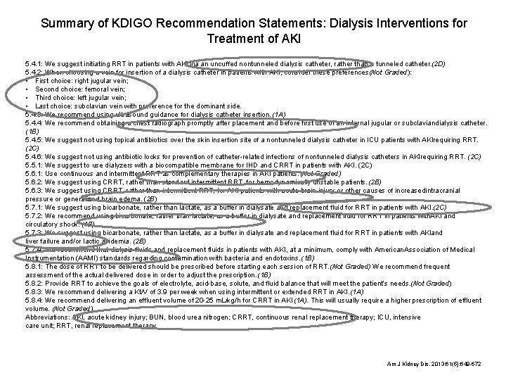
Summary of KDIGO Recommendation Statements: Dialysis Interventions for Treatment of AKI 5. 4. 1: We suggest initiating RRT in patients with AKI via an uncuffed nontunneled dialysis catheter, rather than a tunneled catheter. (2 D) 5. 4. 2: When choosing a vein for insertion of a dialysis catheter in patients with AKI, consider these preferences(Not Graded): • First choice: right jugular vein; • Second choice: femoral vein; • Third choice: left jugular vein; • Last choice: subclavian vein with preference for the dominant side. 5. 4. 3: We recommend using ultrasound guidance for dialysis catheter insertion. (1 A) 5. 4. 4: We recommend obtaining a chest radiograph promptly after placement and before first use of an internal jugular or subclaviandialysis catheter. (1 B) 5. 4. 5: We suggest not using topical antibiotics over the skin insertion site of a nontunneled dialysis catheter in ICU patients with AKIrequiring RRT. (2 C) 5. 4. 6: We suggest not using antibiotic locks for prevention of catheter-related infections of nontunneled dialysis catheters in AKIrequiring RRT. (2 C) 5. 5. 1: We suggest to use dialyzers with a biocompatible membrane for IHD and CRRT in patients with AKI. (2 C) 5. 6. 1: Use continuous and intermittent RRT as complementary therapies in AKI patients. (Not Graded) 5. 6. 2: We suggest using CRRT, rather than standard intermittent RRT, for hemodynamically unstable patients. (2 B) 5. 6. 3: We suggest using CRRT, rather than intermittent RRT, for AKI patients with acute brain injury or other causes of increasedintracranial pressure or generalized brain edema. (2 B) 5. 7. 1: We suggest using bicarbonate, rather than lactate, as a buffer in dialysate and replacement fluid for RRT in patients with AKI. (2 C) 5. 7. 2: We recommend using bicarbonate, rather than lactate, as a buffer in dialysate and replacement fluid for RRT in patients with. AKI and circulatory shock. (1 B) 5. 7. 3: We suggest using bicarbonate, rather than lactate, as a buffer in dialysate and replacement fluid for RRT in patients with AKIand liver failure and/or lactic acidemia. (2 B) 5. 7. 4: We recommend that dialysis fluids and replacement fluids in patients with AKI, at a minimum, comply with American Association of Medical Instrumentation (AAMI) standards regarding contamination with bacteria and endotoxins. (1 B) 5. 8. 1: The dose of RRT to be delivered should be prescribed before starting each session of RRT. (Not Graded) We recommend frequent assessment of the actual delivered dose in order to adjust the prescription. (1 B) 5. 8. 2: Provide RRT to achieve the goals of electrolyte, acid-base, solute, and fluid balance that will meet the patient’s needs. (Not Graded) 5. 8. 3: We recommend delivering a Kt/V of 3. 9 per week when using intermittent or extended RRT in AKI. (1 A) 5. 8. 4: We recommend delivering an effluent volume of 20 -25 m. L/kg/h for CRRT in AKI (1 A). This will usually require a higher prescription of effluent volume. (Not Graded) Abbreviations: AKI, acute kidney injury; BUN, blood urea nitrogen; CRRT, continuous renal replacement therapy; ICU, intensive care unit; RRT, renal replacement therapy. Am J Kidney Dis. 2013; 61(5): 649 -672
Összefoglalás • Tisztán akut vesekárosodásban szenvedő, jó állapotú betegek intermittáló kezelése az ESZC-ben • Több szervi elégtelenségben szenvedő betegek kezelése ITO-n – Vesepótló kezelés idejét, fajtáját, paramétereit a szakorvos (sebész, kardiológus, gasztroenterologus, neurológus), az intenzíves szakorvos – aneszteziologus és nephrologus konszenzussal határozzák meg – Több szervrendszer súlyos érintettsége esetén a vesepótló kezelés az AKI megállapításakor elkezdendő – A döntések naponta reviziót igényelnek és alkalmazkodnak a beteg változó állapotához és igényéhez
- Slides: 18