Ventilation perfusion scan Definition is a physiologic map


 • Cyclotron produced, principal photon energy is 203, 172 and")


to become")


- Slides: 9
Ventilation perfusion scan Definition: is a physiologic map that evaluates the segmental bronchio-alveolar tree ventilation and pulmonary vascular perfusion. Indications: 1. suspected lung embolism. 2. to monitor pulmonary function of lung transplants. 3. to provide preoperative estimates of lung function in ca lung prior to pneumonectomy. 4. to assess right to left shunts. 5. to conduct serial assessment of inflammatory lung diseases. CXR should be done prior to the study.
1. Xenon 133: • produced in a nuclear reactor, the principle photon energy is 81 Kev. • Is expensive, unavailable, allows only posterior imaging (significant attenuation effects) and has to performed before perfusion study.
2. Xenon 127 (XE-127) • Cyclotron produced, principal photon energy is 203, 172 and 365 Kev, can be done following the perfusion scan. • Expensive and unavailable. 3. krypton 81 m • Produced by a generator, short half life 13 s. • Strong photon energy 191 Kev. • Can be done after perfusion scan. • Unfortuntely, expensive.
4. Technitium-99 m aerosols. Produced by nebulizing radiopharmaceuticals into a fine mist that is inhaled. Tc-99 m DTPA diethylenetriaminepentaacetic acid is the most commonly used as it is available and inexpensive. It has a photopeak of 140 Kev. 210% of the aerosolized radioisotope is deposited within the lungs. . Tc-99 m DTPA is absorbed across the alveolar membrane. Its half life is 20 min.
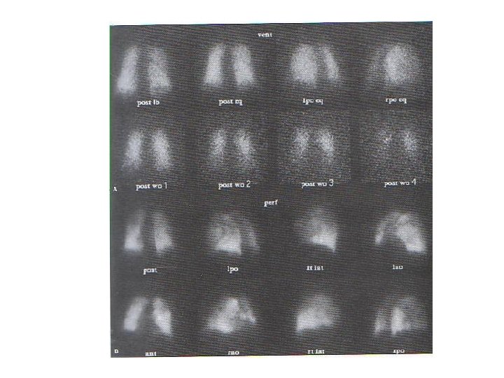
Ventilation scan technique. First, patient inhales the aerosol for 3 -5 min in supine position through a tight facemask? To avoid the normal apex-to-base gravity gradient. . 5 -10 m. Ci, 30 m. Ci if after perfusion scan. . After 5 min of re-breathing, a posterior equilibrium image is obtained that represent the aerated lung volume. . Then the ventilation system is readjusted allowing the patient to breath in fresh air and exhales Tc 99 m DTPA into the trap. . Serial posterior 30 s washout images are obtained over a 5 min interval. . Same views as perfusion study are taken.
Perfusion lung scan • Concept: particles larger than the pulmonary capillaries (>8 um)to become lodged in the precapillary arterioles, thus reflecting the relative blood flow to pulmonary segments. • Tc 99 m-MAA is mostly used, prepared by heat denaturation of human serum albumin. It is prepared by adding Tc-9 m-pertechnetate to the MAA kit. • It leaves the lungs by breaking down into smaller particles/ alveolar capillaries/RECs • Physical half life is 6 hrs. • 3 -5 m. Ci
Technique of perfusion lung scan • Shake the syringe prior to injection to resuspend the particles. • The patient is asked to breath slowly and deeply in supine position. • Examined using a large field of view and a high resolution gamma camera. • Views are obtained.
Normal V/Q scan