
VENOUS MALFORMATION Pathophysiology hemodynamic assessment and treatment strategy



- Slides: 32
VENOUS MALFORMATION Pathophysiology, hemodynamic assessment and treatment strategy C. FRANCESCHI R. DELFRATE A. BAHNINI C. LAURIAN M. BRICCHI CL. MASSONI A. BISDORFF
Les malformations vasculaires congénitales in Encyclopédie pratique d'échotomographie et de Doppler vasculaire. Bristol-Meyers Squibb 1995 C. FRANCESCHI
PATHOPHYSIOLOGAL CLASSIFICATION
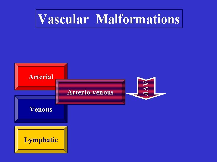
Vascular Malformations Arterial Venous Lymphatic AVF Arterio-venous
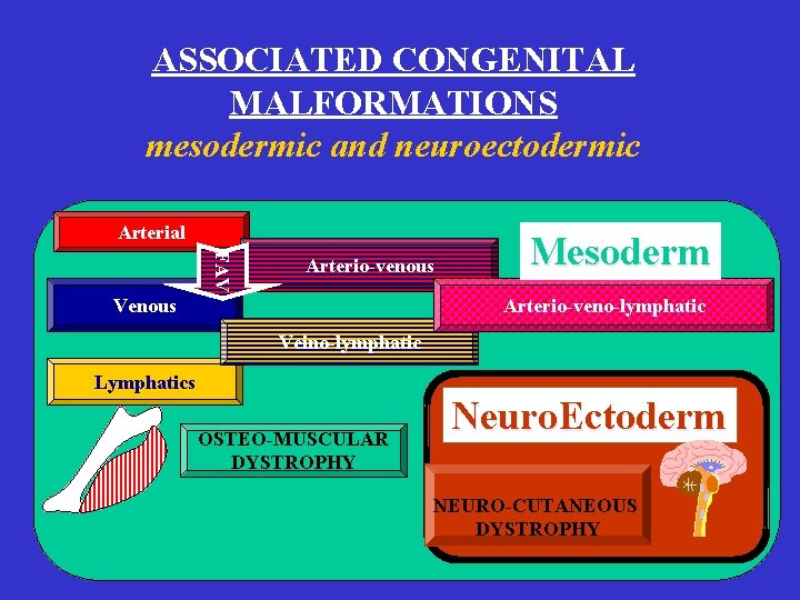
ASSOCIATED CONGENITAL MALFORMATIONS mesodermic and neuroectodermic Arterial FAV Arterio-venous Venous Mesoderm Arterio-veno-lymphatic Veino-lymphatic Lymphatics OSTEO-MUSCULAR DYSTROPHY Neuro. Ectoderm NEURO-CUTANEOUS DYSTROPHY
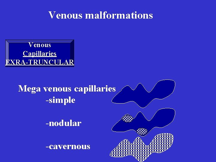
Venous malformations Venous Capillaries EXRA-TRUNCULAR Mega venous capillaries -simple -nodular -cavernous
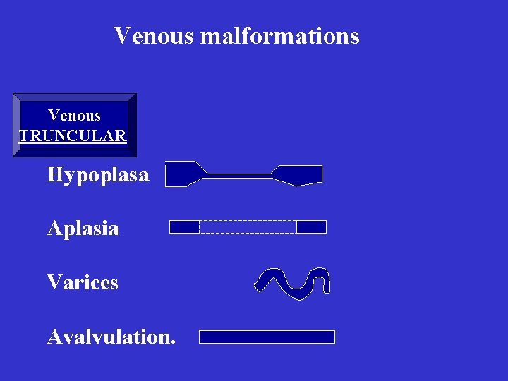
Venous malformations Venous TRUNCULAR Hypoplasa Aplasia Varices Avalvulation.
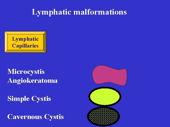
Lymphatic malformations Lymphatic Capillaries Microcystis Angiokeratoma Simple Cystis Cavernous Cystis
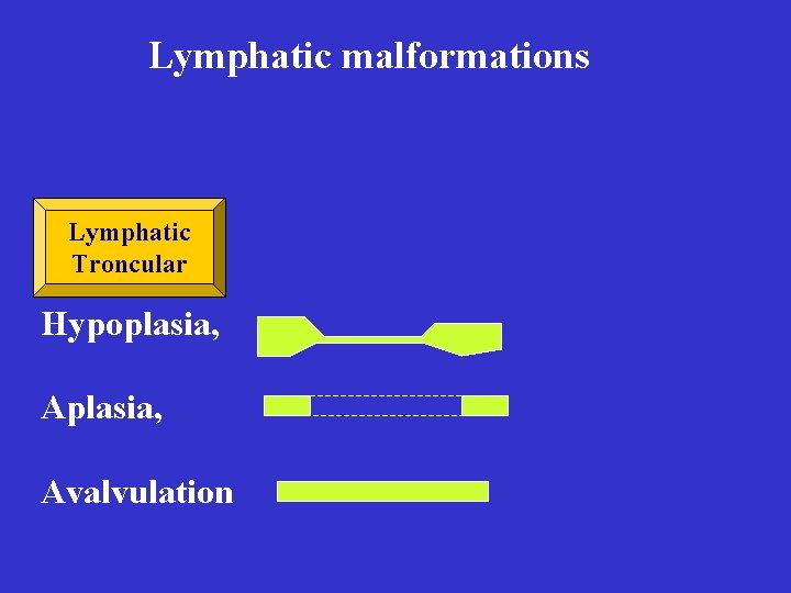
Lymphatic malformations Lymphatic Troncular Hypoplasia, Avalvulation
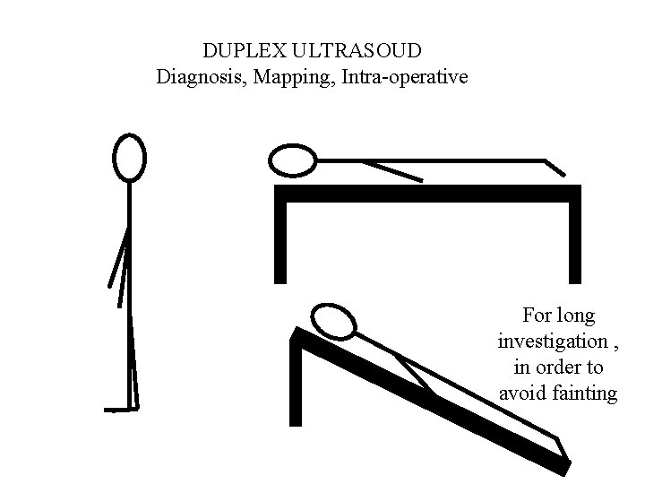
DUPLEX ULTRASOUD Diagnosis, Mapping, Intra-operative For long investigation , in order to avoid fainting
Venous malformations: Topography: Supra-aponeurotiques Sub- aponeurotiques Supra and sub-aponevrotiques.
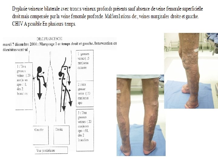
Most frequent Venous Malformations - Superficial and deep valve agenesia -True Klippel Trenaunay Weber Syndrom KTW - Pseudo K. T. W -Congénitales varices -Various extartruncular malformation -Some associations as varices + synnovial extratruncular malformation ( Knee).
Klippel-Trenaunay- Syndrome features: - No AVF - Inconstant lymphatic malformations - Varicose veins Limb hypertrophia , particularly skeletal -(angio-osteo-hypertrophic syndrom) - Capillaire superficial Angioma plan (angiomatous naevus)
Troncular VM
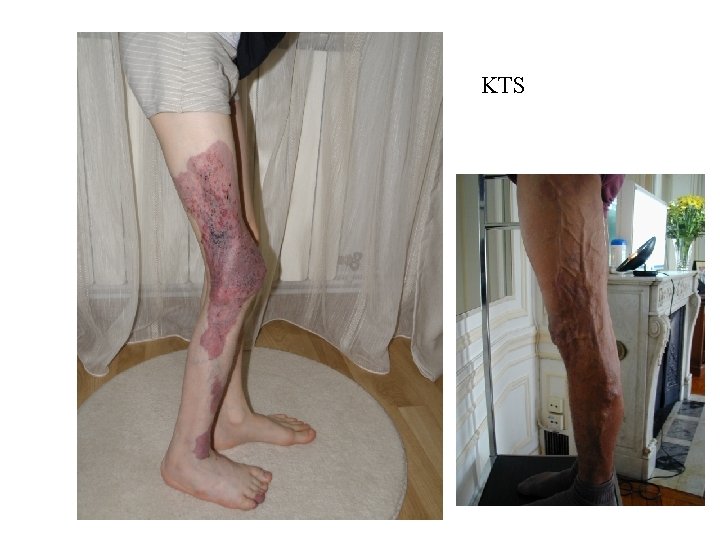
KTS
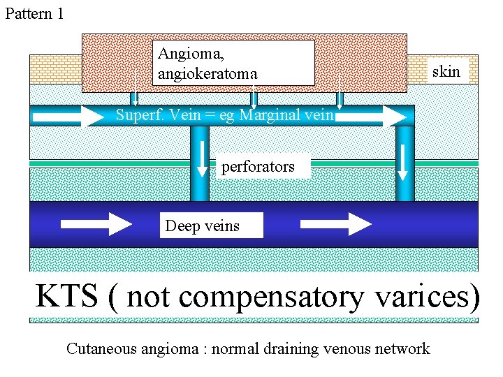
Pattern 1 Angioma, angiokeratoma skin Superf. Vein = eg Marginal vein perforators Deep veins KTS ( not compensatory varices) Cutaneous angioma : normal draining venous network
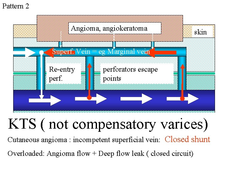
Pattern 2 Angioma, angiokeratoma skin Superf. Vein = eg Marginal vein Re-entry perforators escape points KTS ( not compensatory varices) Cutaneous angioma : incompetent superficial vein: Closed shunt Overloaded: Angioma flow + Deep flow leak ( closed circuit)
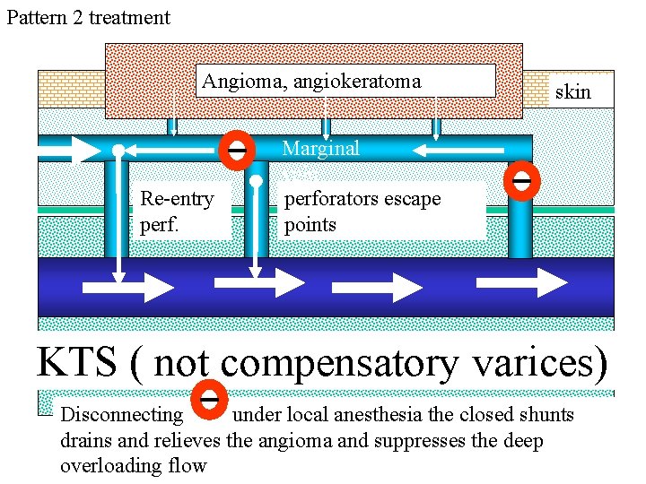
Pattern 2 treatment Angioma, angiokeratoma Re-entry perf. skin Marginal vein perforators escape points KTS ( not compensatory varices) Disconnecting under local anesthesia the closed shunts drains and relieves the angioma and suppresses the deep overloading flow
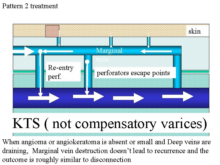
Pattern 2 treatment skin Marginal vein Re-entry perforators escape points KTS ( not compensatory varices) When angioma or angiokeratoma is absent or small and Deep veins are draining, Marginal vein destruction doesn’t lead to recurrence and the outcome is roughly similar to disconnection
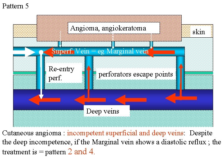
Pattern 5 Angioma, angiokeratoma skin Superf. Vein = eg Marginal vein Re-entry perforators escape points Deep veins Cutaneous angioma : incompetent superficial and deep veins: Despite the deep incompetence, if the Marginal vein shows a diastolic reflux ; the treatment is = pattern 2 and 4.
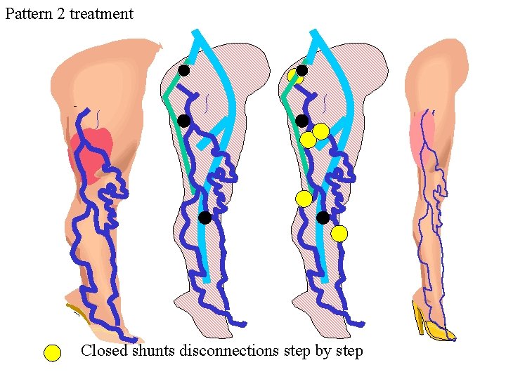
Pattern 2 treatment Closed shunts disconnections step by step
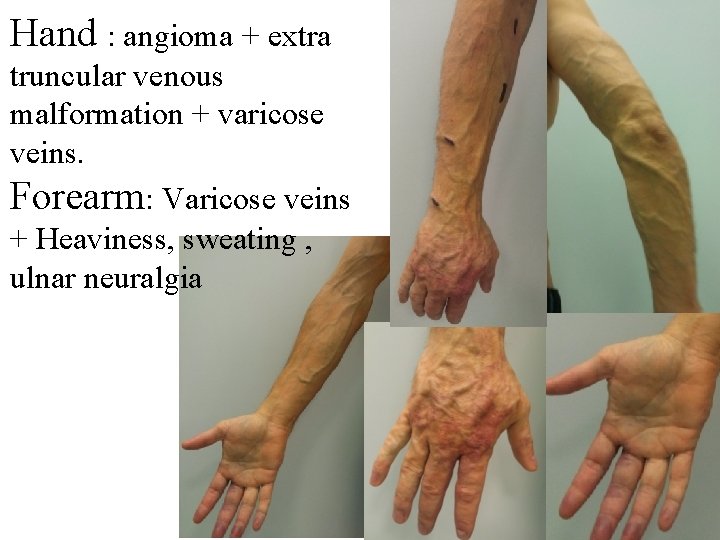
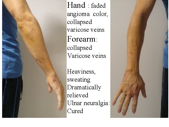
Hand : angioma + extra truncular venous malformation + varicose veins. Forearm: Varicose veins + Heaviness, sweating , ulnar neuralgia
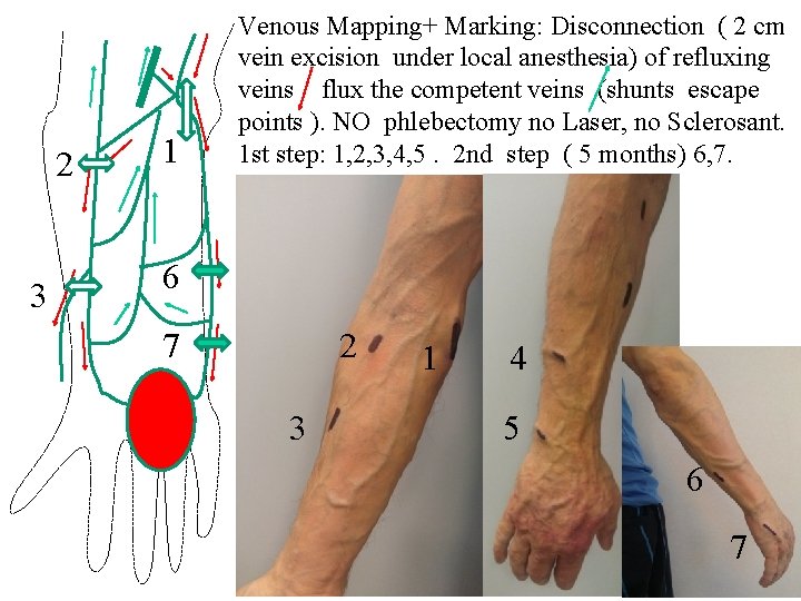
2 3 1 Venous Mapping+ Marking: Disconnection ( 2 cm vein excision under local anesthesia) of refluxing veins flux the competent veins (shunts escape points ). NO phlebectomy no Laser, no Sclerosant. 1 st step: 1, 2, 3, 4, 5. 2 nd step ( 5 months) 6, 7. 6 7 2 3 1 4 5 6 7
Hand : faded angioma color, collapsed varicose veins Forearm: collapsed Varicose veins Heaviness, sweating Dramatically relieved Ulnar neuralgia: Cured
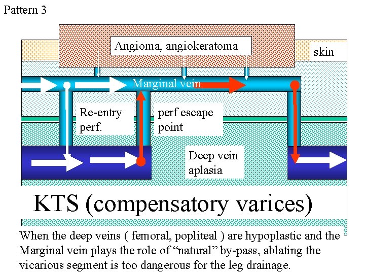
Pattern 3 Angioma, angiokeratoma skin Marginal vein Re-entry perf escape point Deep vein aplasia KTS (compensatory varices) When the deep veins ( femoral, popliteal ) are hypoplastic and the Marginal vein plays the role of “natural” by-pass, ablating the vicarious segment is too dangerous for the leg drainage.
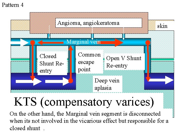
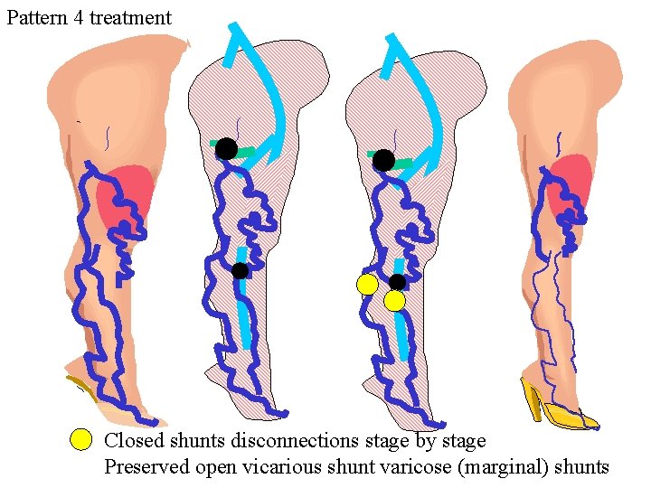
Pattern 4 Angioma, angiokeratoma skin Marginal vein Closed Shunt Reentry Common Open V Shunt escape Re-entry point Deep vein aplasia KTS (compensatory varices) On the other hand, the Marginal vein segment is disconnected when its not involved in the vicarious effect but responsible for a closed shunt.
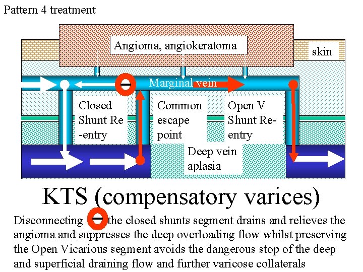
Pattern 4 treatment Angioma, angiokeratoma skin Marginal vein Closed Shunt Re -entry Common Open V escape Shunt Repoint entry Deep vein aplasia KTS (compensatory varices) Disconnecting the closed shunts segment drains and relieves the angioma and suppresses the deep overloading flow whilst preserving the Open Vicarious segment avoids the dangerous stop of the deep and superficial draining flow and further varicose collaterals
Pattern 4 treatment Closed shunts disconnections stage by stage Preserved open vicarious shunt varicose (marginal) shunts
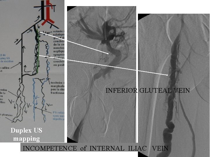
INFERIOR GLUTEAL VEIN Duplex US mapping INCOMPETENCE of INTERNAL ILIAC VEIN
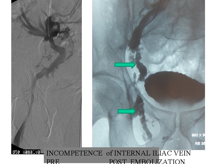
INCOMPETENCE of INTERNAL ILIAC VEIN
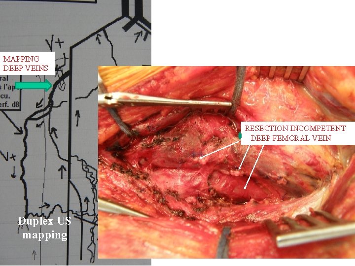
MAPPING DEEP VEINS RESECTION INCOMPETENT DEEP FEMORAL VEIN Duplex US mapping