VENOUS LYMPHATIC DRAINAGE OF LOWER LIMB BY DR

VENOUS & LYMPHATIC DRAINAGE OF LOWER LIMB BY DR SHIVARAMA BHAT P


VEINS OF LOWER LIMB • • • SUPERFICIAL VEINS DEEP VEINS PERFORATING VEINS

The great and small saphenous veins and their tributaries

SUPERFICIAL VEINS • • They lie in the superficial fascia They possess many valves along their course • They communicate with deep veins by perforating veins • They consist of: 1. Dorsal venous arch 2. Great saphenous vein & its tributaries 3. Small saphenous vein & its tributaries

SUPERFICIAL VEINS Dorsal venous arch: • It lies in the superficial fascia of dorsum of foot over the heads of metatarsal bones • Drains on the medial side into great saphenous vein • Drains on the lateral side into small saphenous vein • Receives blood from the foot via digital veins & communicating veins from the sole

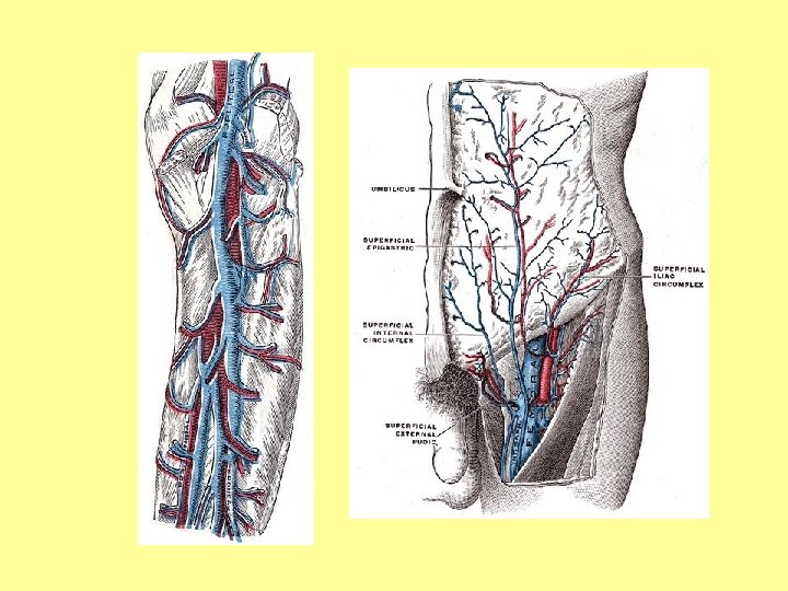
SUPERFICIAL VEINS Great saphenous vein: see front of thigh Small saphenous vein: • It arises from the lateral end of dorsal venous arch • It ascends in superficial fascia, behind lateral malleolus (with sural nerve) • It ascend in the back of thigh • It pierces deep fascia of the lower part of politeal fossa (between 2 heads of gastrocnemius • It ends in popliteal vein • It receives small veins from the back of leg • It communicates with: deep veins of foot, great saphenous vein

DEEP VEINS • They consist of: 1. Venae comitantes to anterior & posterior tibial arteries + their tributaries 2. Popliteal vein + its tributaries 3. Femoral vein + its tributaries

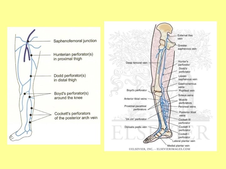
PERFORATING VEINS • They are communicating vessels between superficial & deep veins • They are found mainly in region of ankle & medal side of lower part of leg • They possess valves that prevent flow of blood from deep to superficial veins

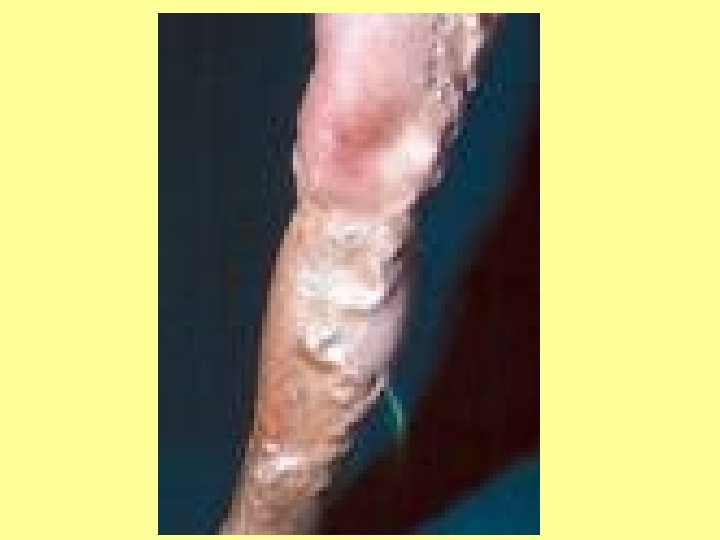
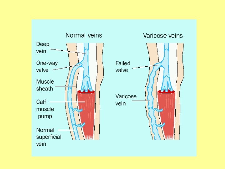
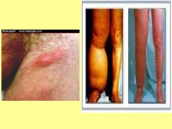
VARICOSE VEINS OF LOWER LIMB • • 1. 2. 3. 4. A condition in which superficial veins of lower limb are elongated & tortuous Causes: Weakness of the walls of veins & incompetence of their valves Incompetence of valves in perforating veins Lack of power of calf muscles Treatment: ligation & division of superficial veins

LYMPHATIC DRAINAGE

LYMPHATIC DRAINAGE OF LOWER LIMB Superficial inguinal lymph nodes: see front of thigh I Deep inguinal lymph nodes: see front of thigh II

The superficial lymph glands and lymphatic vessels of the lower extremity

LYMPHATIC DRAINAGE OF LOWER LIMB Popliteal lymph nodes: • They lie in popliteal fossa • They receive lymph from: 1. knee joint 2. Deep lymph vessels from leg along anterior & posterior tibial arteries 3. Some superficial lymph vessels from leg & foot along small saphenous vein • Their efferents drain into deep inguinal lymph nodes
 drain into")
Lymphatic drainage of the lower limb. The lymphatic channel (deep and superficial) drain into the three group of glands in the lower limb. The glands of the lower limb consist of the anterior tibial gland, the popliteal glands and inguinal glands. The anterior tibial gland is small and inconstant. It lies on the interosseous membrane in relation to the upper part of the anterior tibial vessels, and drains the anterior tibial lymphatic trunks.
and imbedded in popliteal fossa fat.")
The popliteal glands are small in size (6 -7)and imbedded in popliteal fossa fat. Tributaries are from the area drained by the small saphenous vein, knee joint, anterior and posterios tibial vessels. The efferents channels of the popliteal glands pass almost entirely alongside the femoral vessels to the deep inguinal glands, but a few may accompany the great saphenous vein, and end in the glands of the superficial subinguinal group. The inguinal glands (12 -20) are situated at the upper part of the femoral triangle. They may be divided into two groups by a horizontal line at the level of the termination of the great saphenous vein; those lying above this line are termed the superficial inguinal glands, and those below it the subinguinal glands

The sub-inguinal group consist of a superficial and a deep set. The superficial inguinal glands form a chain immediately below the inguinal ligament receiving lymphatic vessels from the integument of the penis, scrotum, perineum, buttock, and abdominal wall below the level of the umbilicus. The superficial sub-inguinal glands are placed on either side of the upper part of the great saphenous vein receiving chiefly the superficial lymphatic vessels of the lower limb and also some of the vessels which drain the integument of the penis, scrotum,
 are placed under the fascia lata,")
• The deep sub-inguinal glands (1 -3) are placed under the fascia lata, on the medial side of the femoral vein. When three are present, the lowest is situated just below the junction of the great sa the middle in the femoral canal, and the highest in the lateral part of the femoral ring. The middle one is the most inconstant of the three, but the highest, the gland of Cloquet or Rosenmüller, is also frequently absent. • They receive as deep lymphatic trunks which accompany the femoral vessels, the lymphatics from the glans penis and clitoris, and also some from the superficial subinguinal glands.

The lymphatic vessels of the lower limbs consist of two sets, superficial and deep and their distribution correspond closely with the veins. The superficial lymphatic vessels lie in the superficial fascia, and are divisible into two groups: medial and lateral. The medial group follows the course of the great saphenous vein commencing on the tibial side and dorsum of the foot and ends in the superficial sub-inguinal glands. The lateral group accompanies the small saphenous vein arising from the fibular side of the foot to join the lymphatics on the medial side of the thigh accompanying the small saphenous vein to the popliteal glands.

The deep lymphatic vessels are few in number, and accompany the deep blood vessels. In the leg, they consist of three sets, the anterior tibial, posterior tibial, and peroneal accompanying the corresponding vessels to drain into the popliteal glands

Clinical application venography. varicose veins lymphedema lymph node enlargement catherisation venepuncture

Questions • Great saphenous vein. • Lymphatic drainage of lower loimb.
- Slides: 27