Venous and lymphatic drainage of Upper Limb Dr

Venous and lymphatic drainage of Upper Limb Dr Anita Rani Professor, Department of Anatomy 20 th October 2016

Superficial Veins • Preaxial : Cephalic vein • Post axial: Basilic vein • Run away from pressure points • Accompanied by cutaneous nerves and lymphatics NOT ARTERIES • Best utilized for IV injections

Dorsal Venous Arch/ Network • Lies on Dorsum of hand Tributaries: • 3 dorsal metacarpal veins • A Dorsal digital vein from medial side of little finger • A Dorsal digital vein from lateral side of index finger • 2 dorsal digital veins from thumb • Most of the blood of palm - perforating veins - veins passing around margin of hand Efferent: • cephalic and basilic veins

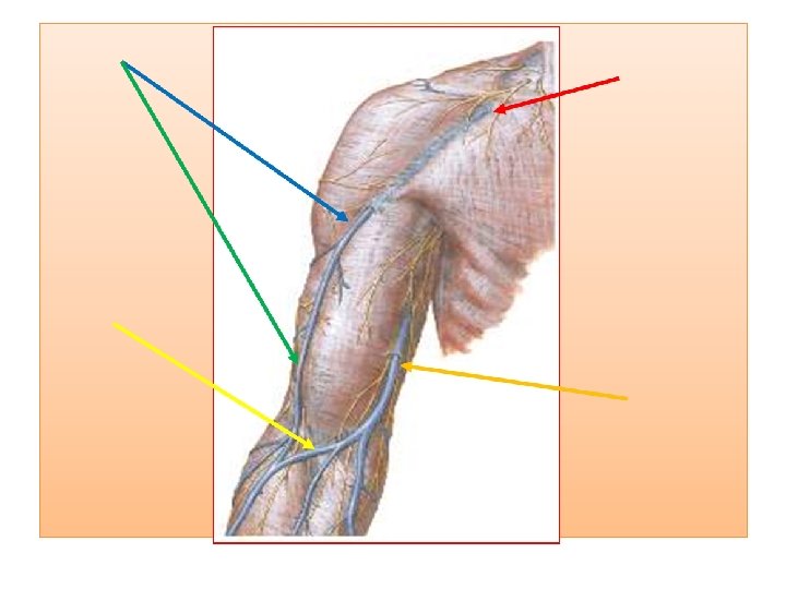
Cephalic vein: spends all of its time in subcutaneous tissues - drains the radial dorsum of hand • Preaxial vein (cf. great saphenous vein) longer vein • Begins from lateral end of dorsal venous arch • Runs upwards through roof of anatomical snuff box • Winds round the forearm • Reaches infront of elbow along lateral border of biceps brachii • Pierces deep fascia at lower border of pectoralis major • Runs in delto-pectoral groove • Pierces clavi-pectoral fascia to join axillary vein.

Cephalic vein • At elbow communicate to basilic vein through MEDIAN CUBITAL VEIN • Accompanied by lateral cutaneous nerve of forearm and terminal branch of radial nerve • An accessory cephalic vein may join it near elbow

Basilic Vein: drains the ulnar dorsum of the hand • Postaxial vein( cf. short saphenous vein of lower limb) • Smaller vein • Begins from medial end of dorsal venous arch • Runs upwards along back of medial border of forearm • Winds round at elbow and comes infront of medial epicondyle along medial margin of biceps brachii • At midarm pierces deep fascia • Runs along medial side of brachial artery upto lower border of teres major to continue as axillary vein

Basilic Vein • 2. 5 cm above medial epicondyle joined by MEDIAN CUBITAL VEIN. • Accompanied by medial cutaneous nerve of forearm and dorsal branch of ulnar nerve

Median Cubital Vein • Large communicating vein which shunts blood from cephalic to basilic vein • Begins from cephalic Vein 2. 5 cm below the bend of elbow • Runs obliquely upwards and medially • Ends in basilic vein 2. 5 cm above the medial epicondyle

Median Cubital Vein • Seperated from brachial artery by bicipital aponeurosis • May receive median vein of forearm • Connected to deep veins through perforating vein, which pierces bicipital aponeurosis. • Perforator vein helps in fixing the median cubital vein: making it ideal for IV injections

Median Vein of Forearm • Begins from palmar venous network • Ends in any one of the veins of front of elbow

Variational Anatomy

Deep Veins

Applied Anatomy • Cephalic vein communicates with External Jugular Vein by a small vein, superficial to clavicle. • In operations of removal of breast in carcinoma cases, axillary vein is also cut. • This communication enlarges and helps in maintaining the circulation. • In cases of fracture of clavicle, rupture of this channel may lead to large hematoma.

Superficial Lymphatic Vessels • The superficial lymphatic vessels of the upper limb initially arise from lymphatic plexuses in the skin of the hand (networks of lymphatic capillaries beginning in the extracellular spaces). • They then ascend up the arm, in close proximity to the major superficial veins.

Superficial Lymphatic Vessels • The vessels shadowing the basilic vein go on to enter the cubital lymph nodes. • These lie medial to the vein, and proximally to the medial epicondyle of the humerus. • Vessels carrying on from these nodes then continue up the arm, terminating in the lateral axillary lymph nodes.

Superficial Lymphatic Vessels • The vessels shadowing the cephalic vein cross the proximal part of the arm and shoulder to enter the apical axillary lymph nodes, though some exceptions instead enter the more superficial deltopectoral lymph nodes.

Deep Lymphatic Vessels • Follow the major deep veins (i. e. radial, ulnar and brachial veins) to terminating in the humeral axillary lymph nodes. • They drain lymph from joint capsules, periosteum, tendons and muscles. • Some additional lymph nodes may be found along the ascending path of the deep vessels.

Lymph Nodes • The majority of the upper lymph nodes are located in the axilla. • Divided anatomically into 5 groups: • Pectoral (anterior) • Subscapular (posterior) • Humeral (lateral) • Central • Apical
 – 3 -5 nodes, located in the medial wall")
Lymph Nodes • Pectoral (anterior) – 3 -5 nodes, located in the medial wall of the axilla. They receive lymph primarily from the anterior thoracic wall, including most of the breast.
 – 6 -7 nodes, located along the posterior axillary")
Lymph Nodes • Subscapular (posterior) – 6 -7 nodes, located along the posterior axillary fold and subscapular blood vessels. They receive lymph from the posterior thoracic wall and scapular region.
 – 46 nodes, located in the lateral wall of")
Lymph Nodes • Humeral (lateral) – 46 nodes, located in the lateral wall of the axilla, posterior to the axillary vein. They receive the majority of lymph drained from the upper limb.

Lymph Nodes • Central 3 -4 large nodes, located near the base of the axilla (deep to pectoralis minor, close to the 2 nd part of the axillary artery). They receive lymph via efferent vessels from the pectoral, subscapular and humeral axillary lymph node groups.

Lymph Nodes • Apical – Located in the apex of the axilla, close to the axillary vein and 1 st part of the axillary artery. They receive lymph from efferent vessels of the central axillary lymph nodes, therefore from all axillary lymph node groups. The apical axillary nodes also receive lymph from those lymphatic vessels accompanying the cephalic vein.

• Efferent vessels from the apical axillary nodes travel through the cervico-axillary canal, before converging to form the subclavian lymphatic trunk. • The right subclavian trunk continues to form the right lymphatic duct, and enters the right venous angle (junction of internal jugular and subclavian veins) directly. • The left subclavian trunk drains directly into the thoracic duct.

Summarizing the drainage…. .

Lymphatic drainage of breast

Applied Anatomy Lymphangitis: inflammation of lymph vessel

Applied Anatomy Lymphadenitis: inflammation of lymph nodes

Applied Anatomy Lymphoedema: obstruction to lymph vessels leading to edema


Which vein of upper limb is most commonly used for IV injections? A. Median Cubital Vein B. Cephalic vein C. Basilic vein D. Dorsal venous arch

Which of the following vein of upper limb is postaxial in location? A. Median Cubital Vein B. Cephalic vein C. Basilic vein D. Dorsal venous arch

Which of the following vein is most likely to bleed if a sharp cut occurs at anatomical snuff box? A. Median Cubital Vein B. Cephalic vein C. Basilic vein D. Dorsal venous arch

At which point basilic vein pierces deep fascia ? A. lower border of pectoralis major B. just below clavicle C. lower border of teres major D. middle of arm E. middle of forearm

Which of the following axillary lymph nodes primarily drain the upper limb? A. pectoral B. subscapular C. central D. lateral

Which of the following lymph node DOES NOT receive lymph from breast? A. pectoral B. subscapular C. humeral D. central

• Please visit university website for downloading today’s presentation. • Give your feedback. • Thanks
- Slides: 38