Vellc eredet a perifris idegrendszer az agyidegek kivtelvel

 szimpatikus /paraszimpatikus dúcok;")


 jelátviteli útvonal Wnt: 16 családtag; idegi fejlődésben Wnt 1, Wnt 3 a")
: Morgan")

 menti NC-ből származnak Chick embryo rhombomere neural")


 medullaris cord : a farokkezdemény végén. (B)")
- Slides: 18
Velőléc eredetű a perifériás idegrendszer – az agyidegek kivételével Szimpatikus dúc-lánc Háti érző dúcok myenterikus plexusok (Auerbach, Meissner)
Velőléc eredetű: Teljes perifériás idegrendszer (kivéve az agyidegek és preganglionáris rostok) szimpatikus /paraszimpatikus dúcok; postganglionáris rostok mellékvesevelő háti érző dúcok érző receptor-készülékek Bőr pigment sejtjei Thymus Hasnyálmirigy Langerhans szigetei Arc-csontok; mimikai izmok jó része Gégeporc Szív nagyerek simaizomzata Stb Anne K. Knecht & Marianne Bronner-Fraser; 2002 Velőléc eredetű fejlődési rendellenességek sokasága ismert: palatalis, facialis, szív rendellenességek (pl. velocardiofacial syndrome (VCFS)/ Di. George syndrome, Hirschsprung’s Disease) Velőléc eredetű tumorok: melanoma, neuroblastoma, Ewing’s family tumors, primitiv perifériás neuroectoderma tumorok, schwannoma-k, pheochromocytoma-k, neurofibroma-k, medullaris thyroid carcinoma. . .
Anne K. Knecht & Marianne Bronner-Fraser; 2002
Neural grove Pax 3 Neural tube Liem et al. , 1995
BMP 4, 7; Wnt → Pax 3, Foxd 3, Sox 8, 9, 10, Slug, → Rho. B → leválás az idegi csőről Rho. B HNK 1 Liu and Jessell 1998 HNK 1 + Rho. B human natural killer-1, CD 57, LEU 7 Slug (SNAI 2) Transzcripciós faktor; Represszor: E-box motivumokhoz kötődik, represszálja az E-cadherin transzcripciót is Sox 8, 9, 10 : Sox. E –csoport; CCTTGAG szekvenciához kötődő (HMGbox csoport ) DNS-kötő fehérje
http: //php. med. unsw. edu. au/embryology/index. php? title=Neural_Crest_Development Cranialis NC: melanocyta, feji ganglionok, fej-nyak kötőszövet, craniofacialis csont, porc, koponyaboltozat, fogdentin, hallócsontocskák, szem szaruhártya, sugárizom, szemizmok, pajzsmirigy parafollicularis sejtjei Nyaki (vagalis; halló placod – 7 somita) NC: enterikus/mesenterikus ganglionok, ggl. coeliaca, aorta és renális ganglionok, nagy-artériák simaizom sejtjei és pericyta-i Törzs-régió NC: pigment sejtek, háti érző dúc, szimpatikus ganglionok, mellékvesevelő Szakrális NC: enterikus ganglionok, paraszimpatikus ganglionok a tápcsatorna és érrendszer mentén Dupin et al. , 2006 E 10, 5 egér embrió; Sox 10 Feji, nyaki, törzs-szintű és szakrális NC régiók: Az A-P eredet meghatározza a sejt lehetséges in vivo „sorsát”; In vitro azonban sok más fejlődési útvonalra vihető A plasztikus NC sejtek sorsát a környezet „dönti” el.
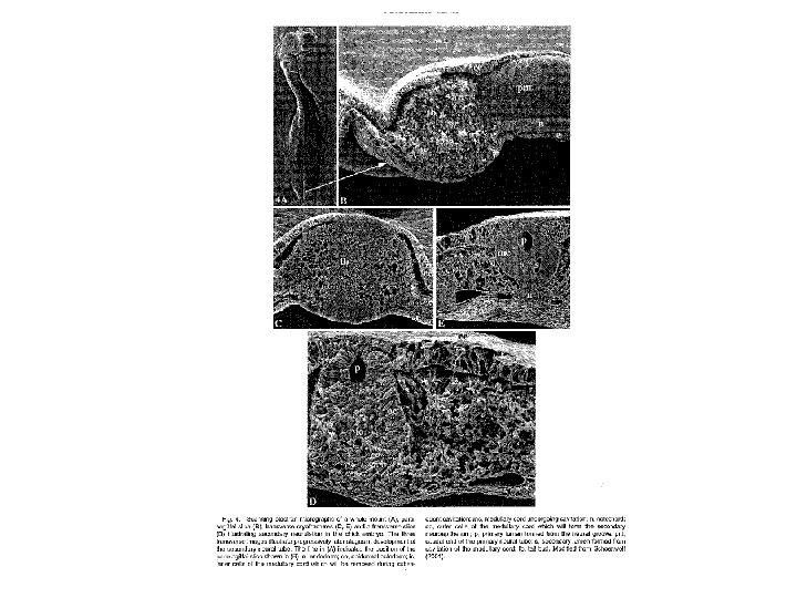
Gilbert; Developmental Biology 6 th Ed Bronner-Fraser 1986; 38 szomita csirke embrió; HNK-1 festés a 21. szomita anterior (D) és poszterior (C) régiójából. HNK-1: Galactosylgalactosylxylosylprotein 3 -beta-glucuronosyltransferase 1 (B 3 GAT 1), CD 57; Human nature killer sejt -1 epitop
Ephrin Wang and Anderson 1997; Gilbert; Developmental Biology 6 th Ed
Wnt/b-catenin (canonicus) jelátviteli útvonal Wnt: 16 családtag; idegi fejlődésben Wnt 1, Wnt 3 a Wnt 1, Wnt-3 A KO. : loss midbrain, loss cerebellum (Mc. Mahon 1990 Thomas KR, 1990, Mc. Mahon 1992 ) deficiency in neural crest derivatives, reduction in dorsolateral neural precursors in the neural tube together with a decrease in the number of thymocytes (Mulroy 2002) Heuberger J, Birchmeier W. ; 2010
Transcriptional Control of Neural Crest Development. Nelms BL, Labosky PA. San Rafael (CA): Morgan & Claypool Publishers; 2010. http: //www. ncbi. nlm. nih. gov/books/NBK 53148/#s 1. 1
Hypothetical lineage restriction in the cells of the quail cephalic neural crest. 533 clones, each derived from a single cell, were observed for the cell types derived from each cell. The results are consistent with the progressive restriction in cell fate from a pluripotent stem cell through more restricted stem cells to a "unipotential" progenitor cell. A, adrenergic neuron; C, cartilage; G, glial cells; M, melanocytes; N, cholinergic neurons. (After Le Douarin et al. , 1994. )
Az arc szilárd képletei a kopoltyúívek (rhombomérák) menti NC-ből származnak Chick embryo rhombomere neural crest cells and their musculoskeletal "packets. " (A) 2 -day chick embryo : contribution of the rhombomere neural crest cells to the pharyngeal arches. (Most of the neural crest cells from r 3 and r 5 undergo apoptosis, while the rest of these cells contribute to the larger population of r 4 neural crest cells. ) (B) 10 -day embryo showing the bones of the upper and lower jaws, tongue skeleton, and middle ear derived from the rhombomeric crest cells. The muscles derived from r 4 are attached to bones from the same rhombomere, and the part of the braincase attached to the r 4 -derived jaw-opening muscle is also derived from r 4. (Other muscles have been omitted for clarity. ) (After Ahlberg 1997. )
Feji plakódok Pán-plakód primordiumból fejlődnek; Pán-plakód primordium: az idegcső menti epithelium-duzzanatból alakul Pan-plakód markerek: Six 1 Eya 1 G. Schlosser / Developmental Biology 294 (2006) 303– 351
Placodes: Adenohypophyseal Olfactory Lens Trigeminal Profundal Otic Lateral line Epibranchial Pharyngeal derivatives (e. g. thymus) G. Schlosser / Developmental Biology 294 (2006) 303– 351
Velőléc epitheliuma: elkötelezetlen őssejtek Csak gerincesekben! In vivo is őssejtként fennmaradó NC-populációk: Bőr-prekurzorok a dermalis papillákban (Fernandes and Miller, 2009; Toma et al. , 2005), szőr-follikulusokban (Sieber-Blum and Hu, 2008; Sieber-Blum et al. , 2006) Felnőtt szájpad NCSCs (Widera et al. , 2009), Cornea limbus-őssejtek (Brandl et al. , 2009), Felnőtt fogpulpa NCSC- populációi (Coura et al. , 2008; Techawattanawisal et al. , 2007) IPS-sejtek NC-ből visszaprogrammozva: hatalmas lehetőség!
Másodlagos neuruláció 25 -somita csirke embrio. (A) medullaris cord : a farokkezdemény végén. (B) ) medullaris cord : kicsit anterior irányban. (C) Cavitáció → neural tube , ezzel egyidőben megjelenik a notochord. (D) Az üregek – idegcső és új üreg - egyesülnek. (From Catala et al. 1995