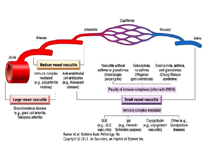
Vasculitis Inflammation of the vessel wall Signs and


= c.")

 Arteritis • is the most common form of vasculitis among the")
 Arteritis • Pathogenesis: T cell-mediated immune response to unknown vessel wall")
 Arteritis: A> granuloma; B> fragmented internal elastic lamina")


 • a systemic vasculitis of small or medium-sized muscular arteries •")


 is detected > 95% of cases -")


 • a disorder of severe vascular insufficiency and gangrene of")

- Slides: 25
Vasculitis • Inflammation of the vessel wall. • Signs and symptoms: 1 - local: according to the involved tissue 2 - systemic: (fever, myalgia, arthralgias, and malaise)
Pathogenesis 1 - immune-mediated inflammation 2 - infectious pathogens. Ø It is critical to distinguish between infectious and immunologic mechanisms due to the huge difference in management. 3 - Physical injury (radiation, mechanical trauma) 4 - chemical injury (toxins)
• The main immunologic mechanisms underlying vasculitis are: 1 - Immune complex deposition 2 -Antineutrophil cytoplasmic antibodies (ANCA) 3 -Anti-endothelial cell antibodies 4 -Auto-reactive T cells
Immune complex deposition • Example: Drug hypersensitivity vasculitis. - e. g. , penicillin - vary from mild and self-limiting, to severe and even fatal - skin lesions are most common. - Treatment: discontinuation of the offending drug.
Anti-Neutrophil Cytoplasmic Antibodies • ANCAs = circulating antibodies that react with neutrophil cytoplasmic antigens (mainly enzymes) • ANCAs blood levels are very useful markers for diagnosis, clinical severity, and as predictive of disease recurrence.
• two types are most important: 1 - Antiproteinase-3 (PR 3 -ANCA)= c. ANCA. - proteinase-3 is a neutrophil azurophilic granule constituent; - e. g. Wegener granulomatosis. 2 -Anti-myeloperoxidase (MPO-ANCA)= p -ANCA. - MPO is a lysosomal granule constituent; - e. g. Churg-Strauss syndrome
Anti-Endothelial Cell Antibodies • Antibodies against endothelial cells • Associated with certain vasculitides, such as Kawasaki disease (discussed later).
Giant Cell (Temporal) Arteritis • is the most common form of vasculitis among the elderly in developed countries. • chronic, granulomatous, inflammation of large arteries • mainly the temporal arteries, vertebral and ophthalmic arteries, as well as the aorta also can be involved. • ophthalmic artery involvement sudden and permanent blindness (rapid diagnosis and treatment are mandatory)
Giant Cell (Temporal) Arteritis • Pathogenesis: T cell-mediated immune response to unknown vessel wall antigen. • Morphology: • granulomatous inflammation (75%) within the inner media centered on the internal elastic membrane (( lymphocytes and macrophages, with multinucleate giant cells )) • fragmentation of the internal elastic lamina. • lesions at different stages of development are seen within the same artery
Giant Cell (Temporal) Arteritis: A> granuloma; B> fragmented internal elastic lamina
Giant Cell Arteritis- clinical picture • • • rare before the age of 50. Signs and symptoms: fever, fatigue, weight loss facial pain or headache (superficial temporal artery). Ocular symptoms (ophthalmic artery) in 50% of patients; range from diplopia complete vision loss. • Diagnosis: - Vessel biopsy and histology • Treatment: - Corticosteroid or anti-TNF therapies
Takayasu Arteritis • vasculitis of medium-sized and large arteries • scarring and thickening of the aorta- especially the aortic arch with severe luminal narrowing of the major branch vessels. • marked weakening of the pulses in the upper extremities (= the pulseless disease).
Takayasu arteritis • Pathogenesis: An autoimmune etiology is likely • affects the aortic arch and arch vessels (2/3) • the distinction from giant cell aortitis is made largely on the basis of a patient's age: >50 years giant cell aortitis <50 years Takayasu aortitis. • Treatment: immunosuppressives
Takayasu arteritis -MORPHOLOGY
Polyarteritis nodosa (PAN) • a systemic vasculitis of small or medium-sized muscular arteries • typically involves the renal and visceral vessels and spares the pulmonary circulation. • There is no association with ANCAs - (1/3) chronic hepatitis B infection immune complexes containing hepatitis B antigens deposit in affected vessels. - (2/3) The cause is unknown
PAN- The clinical course • • - episodic, with long symptom-free intervals. malaise, fever, and weight loss the vascular involvement is widely scattered. Usually present as a combination of: malignant hypertension a major cause of death abdominal pain and bloody stools (GIT lesions) muscular aches and pains peripheral neuritis. • Treatment: if untreated fatal - immunosuppression remission or cure in 90% of the cases
Kawasaki disease • acute, febrile illness of infancy and childhood (80% of cases < 4 years) • arteritis of mainly large to medium-sized vessels. • Its clinical significance: involvement of coronary arteries aneurysms rupture or thrombosis myocardial infarction. • Originally in Japan, the disease is now recognized worldwide
• Also called mucocutaneous lymph node syndrome: - conjunctival and oral erythema and blistering - erythema of the palms and soles - a desquamative rash - cervical lymph node enlargement • Pathogenesis: anti-endothelial cell antibodies • Treatment: - intravenous immunoglobulin therapy and aspirin
Wegener granulomatosis • a necrotizing vasculitis with a specific triad of: 1 - Granulomas of the lung and/or the upper respiratory tract (ear, nose, sinuses, throat) 2 - Vasculitis of small to medium-sized vessels (capillaries, venules, arterioles, and arteries) mostly in lungs and upper respiratory tract 3 - renal vasculitis (Glomerulonephritis):
Wegener granulomatosispathogenesis • PR 3 -ANCAs (c-ANCA) is detected > 95% of cases - useful markers of disease activity (After immunosuppressive therapy, ANCA levels fall dramatically, while rising titers are predictive of relapse) • The typical patient is >40 year old and male, although women and all ages can be affected. • If untreated, the mortality rate at 1 year is 80%.
• • Wegener granulomatosis- clinical picture Rash, myalgias, articular involvement, neuritis, and fever bilateral pneumonitis, nodules and cavitary lesions (95%) chronic sinusitis (90%) mucosal ulcerations of nasopharynx (75%) renal disease (80%) rapidly progressive renal failure. Treatment: steroids, cyclophosphamide, TNF inhibitors. . . Most patients with Wegener granulomatosis now survive, but remain at high risk for relapses that can ultimately lead to renal failure.
Churg-Strauss syndrome • is a small vessel necrotizing vasculitis • classically associated with asthma, allergic rhinitis, lung infiltrates, peripheral eosinophilia, necrotizing granulomas, infiltration by eosinophils. • extremely rare disorder. • purpura, GIT bleeding, and renal disease are the major associations. • Cardiomyopathy (60% of patients) a major cause of morbidity and death. • Pathogenesis: p-ANCA associated
Thromboangiitis obliterans (Buerger disease) • a disorder of severe vascular insufficiency and gangrene of the extremities. • focal acute and chronic inflammation of medium-sized and small arteries, especially the tibial and radial arteries, associated with thrombosis • secondary extension into adjacent veins and nerves may be seen. • Pathogenesis: almost exclusively in heavy tobacco smokers and usually < age 35. • The etiology is unknown: - components of tobacco- ? Direct endothelial cell toxicity ? -an immune response. -? A genetic predilection
Thromboangiitis obliterans/clinical manifestations • Early : Raynaud phenomenon, foot pain induced by exercise, superficial nodular phlebitis (venous inflammation). • severe pain-even at rest neural involvement. • Chronic ulcerations • Gangrene of fingers and toes • Treatment: Smoking abstinence in the early stages of the disease