Vascular mechanism of hypertension Mohammad Saifur Rohman MD


")


![Stretch Receptor (↑ = opens. ↓ = closes) [3] On vascular smooth muscle cellsif](https://slidetodoc.com/presentation_image/15f57861afa102646211f1eabaad80e5/image-11.jpg "Stretch Receptor (↑ = opens. ↓ = closes) [3] On vascular smooth muscle cellsif")


- Slides: 17
Vascular mechanism of hypertension Mohammad Saifur Rohman MD. Ph. D. FICA Department of Cardiology and Vascular Medicine Faculty of medicine Brawijaya University
Vasoconstriction • Alterations in the structure and function of both small and large arteries plays a pivotal role in the origin and progression of hypertension
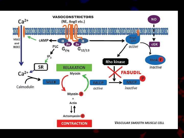
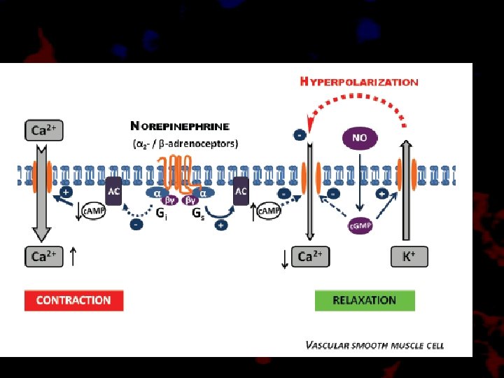
Cellular Mechanism of Vasoconstriction
Vascular vs. Renal • Increased vascular resistance in genetically altered mice blood vessel constriction alone-without renal involvement, can cause hypertension
Endothelial dysfunction • Impaired release of endothelial derived relaxing factors (NO, endothelial-derived relaxing factor) • Super oxide anion production reducing NO bioavailability Enzymatic source of superoxide : • NADPH osidases • Uncoupled e. NOS • Xantine oxidase • Mitochondria electron trasnport • Inflamed in hypertension : genesis and complication of high BP
Normal Endothelial Function
Endothelial Dysfunction
Ang II induced Vasoconstriction
Stretch Receptor (↑ = opens. ↓ = closes) [3] On vascular smooth muscle cellsif not otherwise specified ↑Stretch-activated ion channels ATP (intracellular) ↓ATP-sensitive K+ channel ATP (extracellular) muscarinic agonists e. g. acetylcholine ↑P 2 X receptor NPY receptor adrenergic agonists e. g. epinephrine, norepinephrine anddopamine ↑α 1 adrenergic receptor thromboxane endothelin ↑thromboxane receptor ↑endothelin receptor ETA Vasoconstrictor [3] angiotensin II ↑muscarinic receptor M 2 ↑Angiotensin receptor 1 Asymmetric dimethylarginine Antidiuretic hormone (ADH or Vasopressin) • Products of platelet activation [4] • Endotoxin[4] • Thrombin[4] • insulin[4] • Hypoxia [4] Arginine vasopressin receptor 1(V 1) on smooth muscle cells Transduction (↑ = increases. ↓ = decreases) [3] • depolarization -->open VDCCs (primarily) --> ↑intracellular Ca 2+ • ↑Voltage-gated Na+ channels --> • more depolarization --> open VDCCs -> ↑intracellular Ca 2+ • ↓Na+-Ca 2+ exchanger activity --> ↑intracellular Ca 2+ ↑Ca 2+ Activation of Gi --> ↓c. AMP --> ↓PKA activity --> ↓phosphorylation of MLCK --> ↑MLCK activity --> ↑phosphorylation of MLC (calcium-independent) Activation of Gq --> ↑PLC activity --> ↑IP 3 and DAG --> activation of IP 3 receptor in SR --> ↑intracellular Ca 2+ • On smooth muscle cells: Activation of Gq --> ↑PLC activity --> ↑IP 3 and. DAG --> activation of IP 3 receptor in SR --> ↑intracellular Ca 2+ • On endothelium: endothelin synthesis[4] open VDCCs --> ↑intracellular Ca 2+[5] Reduced production of nitric oxide Activation of Gq --> ↑PLC activity --> ↑IP 3 and DAG --> activation of IP 3 receptor in SR --> ↑intracellular Ca 2+ Arginine vasopressin receptoron endothelium Endothelin production[4] Various receptors onendothelium[4] Endothelin production[4]
Structure: Vascular Remodeling
Cytoplasmic vs. Nuclear events
Ang II induced Remodeling
Super oxide and remodeling
Vascular remodeling in Hypertension
Thank You