Vascular Compromise in Chronic Solar Retinopathy Mehreen Adhi


- Slides: 28
Vascular Compromise in Chronic Solar Retinopathy Mehreen Adhi, MD November 17, 2017 Department of Ophthalmology and Visual Sciences
Patient Presentation Chief Complaint: • “I have blurry vision when I read and when I am working on my computer” HPI: • 31 year old otherwise healthy female originally from Georgia initially presented in 2011 with the above complaint. No visual distortion/blurring at distance. No pain, floaters and flashes of light
Patient Presentation Review of Systems: • Unremarkable
Patient Presentation Past Ocular History: • Unremarkable Past Medical/Surgical History: • Unremarkable Family History: • Unremarkable Medications: • Artificial tears PRN Allergies: • No known drug allergies
External Exam OD VA Refraction OS 20/20 -3 20/25 -3 +1. 00+0. 50 x 65 – 20/20 +1. 25 – 20/20 Pupils 4→ 3 mm IOP 13 mm. Hg EOM Full CVF Full No r. APD 4→ 3 mm Full
Anterior Segment Exam SLE OD OS WNL White and quiet Clear Deep and quiet Iris WNL Lens Clear Vitreous Clear External Exam/ Eyelids Conjunctiva/ Sclera Cornea Ant Chamber
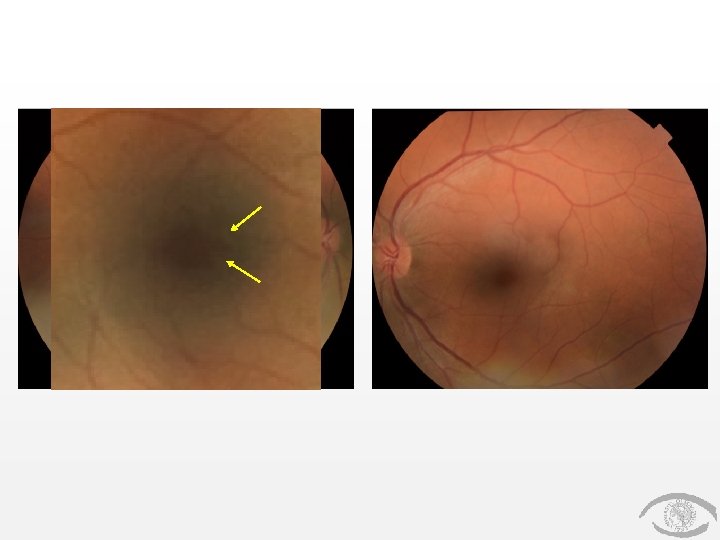
Posterior Segment Exam Dilated Fundus Exam Optic Nerve OD OS Pink and sharp Macula Retinal pigment epithelial changes Good foveal reflex Vessels WNL Choroidal nevus at ~12: 30 -1: 00 o’c (flat, +drusen, no orange pigment) WNL Periphery
OD OS
Assessment and Plan • 31 year old healthy female with blurry vision. Exam significant for latent hyperopia. Incidental finding of retinal pigment epithelial changes in the right eye on clinical examination with a history of gazing directly at a solar eclipse in her home country of Georgia a few years ago • Solar retinopathy • Yearly evaluation
2017
2011 2013 2015 2017
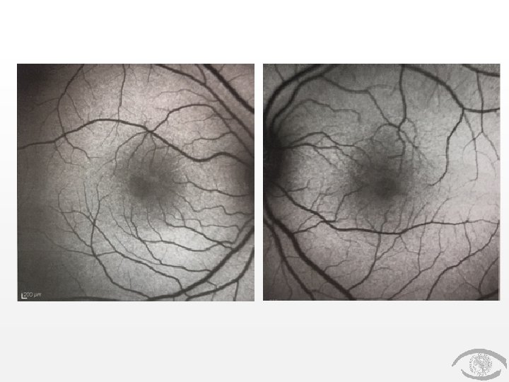
OD OS
OD OS
OD OS
OD OS
Solar Retinopathy • Also known as foveomacular retinitis; is a rare but well-recognized clinical entity of vision loss and macular damage due to exposure to solar radiation • Exposure to welding arc, gazing at a solar eclipse or direct sun-gazing in religious ritual participants, military personnel patients with mental disturbances • Symptoms typically are bilateral, but may be asymmetric and include blurred vision, glare, central or paracentral scotoma, metamorphopsia, photophobia and headache
Solar Retinopathy • Histopathological studies have demonstrated that both retinal pigment epithelium and outer segments of the photoreceptor layer are most susceptible to solar damage • A thermally enhanced phototoxic reaction at the retinal pigment epithelium has been proposed to be the principal pathogenic mechanism
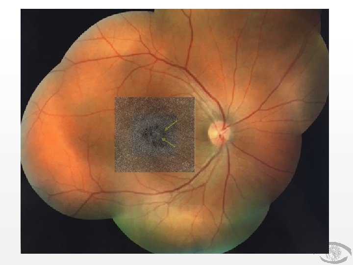
Solar Retinopathy • Fundus photography: small yellowishwhite spot with surrounding gray area or pigmentary changes in the central fovea • Fundus autofluorescence: may show a well-circumscribed hypoautofluorescent fovea surrounded by an irregular ring of hyperautofluorescence • Fluorescein angiography: may reveal a window/transmission defect
• 38 year-old man was referred 2 weeks after unprotected viewing of a solar eclipse • Visual acuity was 20/30 in both eyes
• Case of bilaterally symmetric foveomacular retinitis followed over 5 years • Symptoms persisted for 4 weeks (visual acuity at presentation 20/25) and fundus changes persisted for 3 months after which visual acuity was 20/20 • Optical coherence tomography changes persisted over 5 years of follow up
Solar Retinopathy • No treatment • Visual prognosis is generally good • Depends on the extent of photoreceptor damage
Conclusion • Solar retinopathy is a rare clinical entity, usually bilateral but may be unilateral; exposure to solar radiation • Clinical and imaging findings may be subtle; structural optical coherence imaging is the most sensitive modality • Optical coherence tomography angiography may be useful to determine the extent of vascular damage
References • • • Badhani, Anurag, et al. “Correlation of Macular Structure and Function in a Boy with Primary Foveomacular Retinitis and Sequence of Changes over 5 Years. ” Documenta Ophthalmologica, vol. 135, no. 1, 2017, pp. 43– 52. Ovid, doi: 10. 1007/s 10633 -017 -9590 -1 Bechmann, M. “Optical Coherence Tomography Findings in Early Solar Retinopathy. ” British Journal of Ophthalmology, vol. 84, no. 5, Jan. 2000, doi: 10. 1136/bjo. 84. 5. 546 b Bonyadi, Mohammad Hossein Jabbarpoor, et al. “Spectral-Domain Optical Coherence Tomography Features of Mild and Severe Acute Solar Retinopathy. ” Ophthalmic Surgery, Lasers, and Imaging, Aug. 2011, doi: 10. 3928/15428877 -20110901 -05 Chen, Kevin C. “High Definition Spectral Domain Optical Coherence Tomography Findings in Three Patients with Solar Retinopathy and Review of the Literature. ” The Open Ophthalmology Journal, vol. 6, no. 1, 2012, pp. 29– 35. , doi: 10. 2174/1874364101206010029 Chen, Royce W. S. , et al. “High-Speed Ultrahigh-Resolution Optical Coherence Tomography Findings In Chronic Solar Retinopathy. ” Retinal Cases & Brief Reports, vol. 2, no. 2, 2008, pp. 103– 105. , doi: 10. 1097/icb. 0 b 013 e 3181506993
Acknowledgement • • • Henry Kaplan MD Wei Wang MD Janelle Adeniran MD Efrat Fleissig MD Ashwini Kini MD
Thank you