Vascular Access Dr Mueen Ullah Khan Associate Professor


")


 monitoring Arterial blood sampling")

 Full-thickness")


. Advancement of catheter over guide wire.")
 Hematoma/bleeding (14.")


- Slides: 79
Vascular Access Dr. Mueen Ullah Khan Associate Professor and Consultant Department of Anesthesia King Saud University Riyadh
Lecture Objectives. . At the end of the lecture you will be able to: 1. 2. 3. 4. 5. 6. 7. 8. 9. Examine the construction of the commonly used venous catheters. Anatomical considerations regarding peripheral and central venous access. Choice of catheter size. Prepare and set-up an IV infusion set. The choice of sites for placement of IV catheters. What are the different sites suitable for central venous catheter and arterial catheter placement? Universal precautions. Indications and complications of central venous access Indications and complications of arterial access
Medical Asepsis Removal or destruction of disease-causing organisms or infected material Sterile technique (surgical asepsis) Clean technique Copyright © 2007, 2006, 2001, 1994 by Mosby, Inc. , an affiliate of Elsevier Inc.
Antiseptics and Disinfectants Chemical agents used to kill specific microorganisms Disinfectants • Used on nonliving objects • Toxic to living tissue Antiseptics • Applied to living tissue • More dilute to prevent cell damage Some chemical agents have antiseptic and disinfectant properties Copyright © 2007, 2006, 2001, 1994 by Mosby, Inc. , an affiliate of Elsevier Inc.
Universal Precautions Universal standard precautions on every patient • Observe hand washing and gloving procedures • Face shields indicated during clean procedures • Sterile gowns plus above all for sterile procedures. Copyright © 2007, 2006, 2001, 1994 by Mosby, Inc. , an affiliate of Elsevier Inc.
Types of IV Catheters Hollow needles • Butterfly type Indwelling plastic catheter over hollow needle Indwelling plastic catheter inserted through a hollow needle • Intracath Copyright © 2007, 2006, 2001, 1994 by Mosby, Inc. , an affiliate of Elsevier Inc.
Needles Vary in length and gauge • Larger gauge means a smaller needle Copyright © 2007, 2006, 2001, 1994 by Mosby, Inc. , an affiliate of Elsevier Inc.
Peripheral IV Insertion Common sites: • Hands and arms • Antecubital fossa (AC space) Copyright © 2007, 2006, 2001, 1994 by Mosby, Inc. , an affiliate of Elsevier Inc.
Peripheral IV Insertion Alternate sites: • Long saphenous veins • External jugular veins Embolism and infection rates higher Copyright © 2007, 2006, 2001, 1994 by Mosby, Inc. , an affiliate of Elsevier Inc.
Peripheral IV Insertion Avoid sites that have injury or disease: • Trauma • Dialysis fistula • History of mastectomy Copyright © 2007, 2006, 2001, 1994 by Mosby, Inc. , an affiliate of Elsevier Inc.
Peripheral IV Procedure Explain procedure Assemble equipment Inspect fluid for contamination, appearance, and expiration date Prepare infusion set • Attach infusion set to bag of solution Copyright © 2007, 2006, 2001, 1994 by Mosby, Inc. , an affiliate of Elsevier Inc.
Peripheral IV Procedure Clamp tubing and squeeze reservoir on infusion set until it fills half way Open clamp and flush air from tubing Close clamp Maintain aseptic technique Copyright © 2007, 2006, 2001, 1994 by Mosby, Inc. , an affiliate of Elsevier Inc.
Peripheral IV Procedure Select catheter: • Large-bore catheter used for fluid replacement 14 to 16 gauge • Smaller bore catheter used for “keep open” lines 18 to 20 gauge Prepare other equipment Copyright © 2007, 2006, 2001, 1994 by Mosby, Inc. , an affiliate of Elsevier Inc.
Peripheral IV Procedure � Put on gloves � Select site � Apply tourniquet above antecubital space � Prepare site � Cleanse area with alcohol or iodine wipes (per protocol) q Check for iodine allergy Copyright © 2007, 2006, 2001, 1994 by Mosby, Inc. , an affiliate of Elsevier Inc.
Peripheral IV Procedure Stabilize vein Apply pressure and tension to point of entry Copyright © 2007, 2006, 2001, 1994 by Mosby, Inc. , an affiliate of Elsevier Inc.
Peripheral IV Insertion Bevel of the needle up in adults • May be down in infants and children Pass needle through skin into vein from side or directly on top Copyright © 2007, 2006, 2001, 1994 by Mosby, Inc. , an affiliate of Elsevier Inc.
Peripheral IV Procedure �Advance needle and catheter about 2 mm past point where blood return is seen in hub of needle �Slide catheter over needle and into vein Copyright © 2007, 2006, 2001, 1994 by Mosby, Inc. , an affiliate of Elsevier Inc.
Peripheral IV Procedure Withdraw needle while stabilizing catheter Lock in protective sheath if present Apply pressure on proximal end of catheter to stop escaping blood Obtain blood samples if needed Copyright © 2007, 2006, 2001, 1994 by Mosby, Inc. , an affiliate of Elsevier Inc.
Peripheral IV Procedure Release tourniquet Attach IV tubing Copyright © 2007, 2006, 2001, 1994 by Mosby, Inc. , an affiliate of Elsevier Inc.
Peripheral IV Procedure Open tubing clamp and allow fluid infusion to begin at prescribed flow rate Copyright © 2007, 2006, 2001, 1994 by Mosby, Inc. , an affiliate of Elsevier Inc.
Peripheral IV Procedure � Cover puncture site dressing • Antibiotic ointment if indicated by protocol � Anchor tubing � Secure catheter � Document procedure � Monitor flow Copyright © 2007, 2006, 2001, 1994 by Mosby, Inc. , an affiliate of Elsevier Inc.
Local Complications Pain and irritation Infiltration and extravasation Phlebitis Thrombosis and thrombophlebitis Hematoma formation Venous spasm Vessel collapse Cellulitis Nerve, tendon, ligament, and limb damage Copyright © 2007, 2006, 2001, 1994 by Mosby, Inc. , an affiliate of Elsevier Inc.
Infiltration—Causes Dislodgement of catheter or needle cannula during venipuncture Puncture of vein wall during venipuncture Leakage of solution into surrounding tissue from insertion site Poor vein or site selection Irritating solution inflames vein’s intima Improper cannula size High delivery rate or pressure Poorly secured IV Copyright © 2007, 2006, 2001, 1994 by Mosby, Inc. , an affiliate of Elsevier Inc.
Infiltration—Signs & Symptoms Cool skin around IV site Swelling at IV site • With or without pain Sluggish or absent flow Infusion flows when pressure is applied to vein above tip of cannula No backflow of blood into IV tubing when clamp is fully opened and solution container is lowered below IV site Copyright © 2007, 2006, 2001, 1994 by Mosby, Inc. , an affiliate of Elsevier Inc.
Infiltration—Management Lower fluid reservoir to check for presence of backflow of blood into the tubing • Absence of backflow suggests infiltration Discontinue IV infusion Remove needle or catheter Apply a pressure dressing to the site Choose new site Initiate IV therapy with new equipment Document Copyright © 2007, 2006, 2001, 1994 by Mosby, Inc. , an affiliate of Elsevier Inc.
Central Venous Access �Requires special training �Authorization from medical direction �Not for rapid fluid replacement in prehospital setting �Within scope of paramedic practice in some EMS systems Copyright © 2007, 2006, 2001, 1994 by Mosby, Inc. , an affiliate of Elsevier Inc.
Central Venous Access Common Sites include: • Femoral vein • Internal jugular vein • Subclavian vein Copyright © 2007, 2006, 2001, 1994 by Mosby, Inc. , an affiliate of Elsevier Inc.
Central Venous Access Prepare as for peripheral veins Sterile procedure Success depends on: • Patient's body position • Knowledge of anatomy • Familiarity with the procedure Copyright © 2007, 2006, 2001, 1994 by Mosby, Inc. , an affiliate of Elsevier Inc.
Femoral Vein Anatomy Copyright © 2007, 2006, 2001, 1994 by Mosby, Inc. , an affiliate of Elsevier Inc.
Femoral Vein Cannulation Copyright © 2007, 2006, 2001, 1994 by Mosby, Inc. , an affiliate of Elsevier Inc.
Internal Jugular Vein Anatomy Copyright © 2007, 2006, 2001, 1994 by Mosby, Inc. , an affiliate of Elsevier Inc.
Internal Jugular Vein Cannulation Posterior approach Copyright © 2007, 2006, 2001, 1994 by Mosby, Inc. , an affiliate of Elsevier Inc.
Internal Jugular Vein Cannulation Central approach Copyright © 2007, 2006, 2001, 1994 by Mosby, Inc. , an affiliate of Elsevier Inc.
Internal Jugular Vein Cannulation Anterior approach Copyright © 2007, 2006, 2001, 1994 by Mosby, Inc. , an affiliate of Elsevier Inc.
Subclavian Vein Anatomy Copyright © 2007, 2006, 2001, 1994 by Mosby, Inc. , an affiliate of Elsevier Inc.
Subclavian Vein Cannulation Copyright © 2007, 2006, 2001, 1994 by Mosby, Inc. , an affiliate of Elsevier Inc.
Central Venous Access
Central Venous Access Indications • Available when peripheral vessels collapse • Access to central pressure measurements In-hospital procedure • Safer vasopressor administration Copyright © 2007, 2006, 2001, 1994 by Mosby, Inc. , an affiliate of Elsevier Inc.
Central Venous Access Disadvantages • Excessive time for • • placement Sterile technique Special equipment Skill deterioration High complication rate Pneumothorax, arterial injury, abnormal placement • Chest x-ray should be obtained immediately Copyright © 2007, 2006, 2001, 1994 by Mosby, Inc. , an affiliate of Elsevier Inc.
Central Venous Access Disadvantages • Can’t initiate during other patient care activities • Not generally considered to be a useful prehospital technique • Lower flow rates than peripheral IV Copyright © 2007, 2006, 2001, 1994 by Mosby, Inc. , an affiliate of Elsevier Inc.
Systemic Complications � Contamination and infection � Hypersensitivity reactions � Sepsis � Speed shock � Emboli (blood clot, air, and catheter) Copyright © 2007, 2006, 2001, 1994 by Mosby, Inc. , an affiliate of Elsevier Inc.
Air Embolism Uncommon but can be fatal Air enters bloodstream through catheter tubing Risk greatest with catheter in central circulation • Negative pressure may pull air in Copyright © 2007, 2006, 2001, 1994 by Mosby, Inc. , an affiliate of Elsevier Inc.
Air Embolism Air can enter circulation • During catheter insertion • If tubing is disconnected If enough air enters the heart chamber: • Blood flow is impeded • Shock develops Copyright © 2007, 2006, 2001, 1994 by Mosby, Inc. , an affiliate of Elsevier Inc.
Air Embolism Signs and symptoms • Hypotension • Cyanosis • Weak and rapid pulse • Loss of consciousness Copyright © 2007, 2006, 2001, 1994 by Mosby, Inc. , an affiliate of Elsevier Inc.
Air Embolism Management • Close the tubing • Turn patient on left side with head down • Check tubing for leaks • Administer 100% Oxygen • Notify medical direction Copyright © 2007, 2006, 2001, 1994 by Mosby, Inc. , an affiliate of Elsevier Inc.
Complications—Central Veins Femoral vein • Local complications • Systemic complications Internal jugular and subclavian veins • Local complications • Systemic complications Copyright © 2007, 2006, 2001, 1994 by Mosby, Inc. , an affiliate of Elsevier Inc.
IV Medications � IV injection may be given in: • An established IV line • Heparin or saline lock • Implantable port �Port-A-Cath, Hickman catheter • Directly into the vein � IV injections generally less than 5 m. L • IV push or IV bolus medications Copyright © 2007, 2006, 2001, 1994 by Mosby, Inc. , an affiliate of Elsevier Inc.
Intravenous Therapy IV solution Infusion set Macrodrip or microdrip Tubing clamp Injection port Copyright © 2007, 2006, 2001, 1994 by Mosby, Inc. , an affiliate of Elsevier Inc.
Intermittent Infusion IV piggybacks Setup is secondary to primary IV infusion Piggyback medication hung in tandem and connected to primary setup Copyright © 2007, 2006, 2001, 1994 by Mosby, Inc. , an affiliate of Elsevier Inc.
Intermittent Infusion Calculate rate of secondary infusion • Drops per minute Lower primary infusion reservoir • Center of gravity lower than secondary infusion reservoir Open piggyback line flow clamp Adjust flow rate Copyright © 2007, 2006, 2001, 1994 by Mosby, Inc. , an affiliate of Elsevier Inc.
Volume-Control IV Devices Permit accurate delivery of IV medications Electronic flow-rate regulators • Regulate precise doses of drugs that can cause toxicity Vasopressors Antidysrhythmics Follow manufacturer’s instructions Copyright © 2007, 2006, 2001, 1994 by Mosby, Inc. , an affiliate of Elsevier Inc.
Drug Pump � Slow injection of medication � Syringe with battery attachment that regulates injection of medication � To administer medication SC � Can attach to indwelling vascular devices • Port-A-Cath • Hickman catheter Copyright © 2007, 2006, 2001, 1994 by Mosby, Inc. , an affiliate of Elsevier Inc.
Indwelling Vascular Devices Heparin or saline lock Copyright © 2007, 2006, 2001, 1994 by Mosby, Inc. , an affiliate of Elsevier Inc.
Indwelling Vascular Devices Single-, dual-, and triple-lumen catheters Copyright © 2007, 2006, 2001, 1994 by Mosby, Inc. , an affiliate of Elsevier Inc.
Ultrasound guided IV insertion
Ultrasound guided CVC insertion
Arterial Line Placement Provide continuous blood pressure (BP) monitoring Arterial blood sampling
Arterial line placement Indications Continuous arterial BP monitoring - more accurate than sphygmomanometric BP Inability to use indirect BP monitoring (eg, in patients with severe burns or morbid obesity) Frequent blood sampling Frequent arterial blood gas sampling
Contraindications for arterial line placement ABSOLUTE RELATIVE Absent pulse Thromboangiitis obliterans (Buerger disease) Full-thickness burns over the cannulation site Inadequate circulation to the extremity Raynaud syndrome Anticoagulation Atherosclerosis Coagulopathy Inadequate collateral flow Infection at the cannulation site Partial-thickness burn at the cannulation site Previous surgery in the area Synthetic vascular graft
Technical Considerations Not entirely without risks, Requires appropriate knowledge of the anatomy and procedural skills. Arterial line placement is considered a safemajor complications that is below 1%. Common site of cannulation radial, ulnar, brachial, axillary, posterior tibial, femoral, and dorsalis pedis arteries.
Allen test The Allen test is a worldwide used test to determine whether the patency of the radial or ulnar artery is normal. It is performed prior to radial cannulation or catheterisation. The test is used to reduce the risk of ischemia to the hand.
Allen test Instruct the patient to clench his or her fist OR hand tightly. Using your fingers, apply occlusive pressure to both the ulnar and radial arteries, to obstruct blood flow to the hand. While applying occlusive pressure to both arteries, have the patient relax his or her hand, and check whether the palm and fingers have blanched. If this is not the case, you have not completely occluded the arteries with your fingers.
Modified Allen test NEGATIVE TEST POSITIVE TEST
Allen test- Release the occlusive pressure on the ulnar artery Positive modified Allen test – hand flushes within 5 -15 seconds it indicates that the ulnar artery has good blood flow; this normal flushing of the hand is considered to be a positive test. Negative modified Allen test – If the hand does not flush within 5 -15 seconds, it indicates that ulnar circulation is inadequate or nonexistent; in this situation, the radial artery supplying arterial blood to that hand should not be punctured.
Radial artery Aatomic consideration Originates in the cubital fossa from the brachial artery At the wrist, the radial artery sits proximal and medial to the radial styloid process and just lateral to the flexor carpi radialis tendon.
Femoral artery Anatomic consideration Originates at the inguinal ligament from the external iliac artery Medial to the femoral nerve and lateral to the femoral vein and lymphatics.
Arterial Line Placement Equipment Sterile gloves Sterile gauze Sterile towels Chlorhexidine or povidone-iodine skin preparation solution 1% Lidocaine needle 5 -m. L syringe Appropriate-sized cannula for artery Scalpel (No. 11 blade) Nonabsorbable suture (3 -0 to 4 -0) Adhesive tape or strips Sterile nonabsorbable dressing Three-way stopcock Pressure transducer kit Pressure tubing Arm board of appropriate size for the patient (eg, neonate, pediatric, adult) Needle holder Intravenous (IV) tubing T-connector
Arterial Line Placement Patient Preparation UNCOSCIOS PATIENT Anesthesia/ Sedation is not required. CONSCIOUS PATIENT provided LA -lidocaine 1% UNCOPERATIVE PATIENT sedation or general anesthesia may be required.
Arterial Line Placement Positioning The patient is placed in the supine position. The arm is placed up on a flat surface in neutral position, with the palm up and the wrist adequately exposed. The wrist is dorsiflexed to 30 -45° and supported in this position with a towel or gauze under its dorsal aspect
Arterial Line Placement The most commonly used methods Catheter over needle Catheter over wire (including direct Seldinger and modified Seldinger techniques)
Catheter over needle technique
Catheter over needle technique
Radial artery cannulation (Seldinger). Advancement of catheter over guide wire.
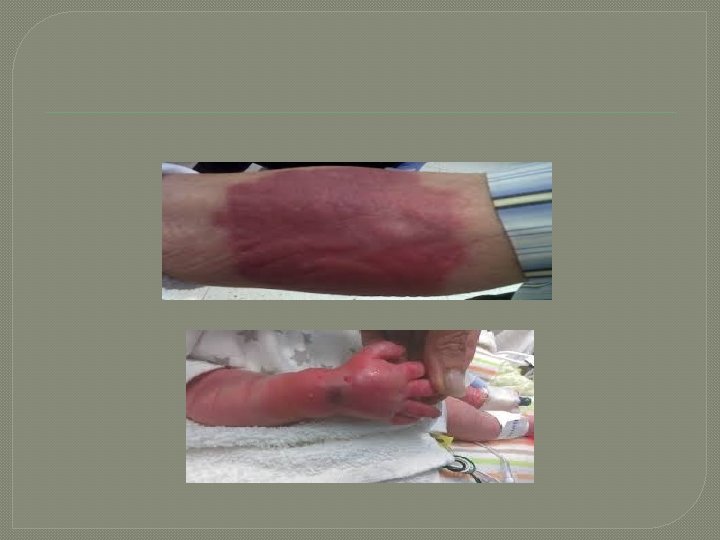
Complications of arterial line placement COMMON Temporary radial artery occlusion (19. 7%) Hematoma/bleeding (14. 4%) LESS COMMON Localized catheter site infection (0. 72%) - The risk increases with the length of time the catheter is in place Hemorrhage (0. 53%) Sepsis (0. 13%) Permanent ischemic damage (0. 09%) Pseudoaneurysm formation (0. 09%)
Reference book and the relevant page numbers. .
Questions? Copyright © 2007, 2006, 2001, 1994 by Mosby, Inc. , an affiliate of Elsevier Inc.
Thank You Dr. Date: