
Vascular Access and Basic Hardware Dr K SURESH
Vascular Access and Basic Hardware Dr. K. SURESH SK Hospital and KIMS Hospital, Trivandrum
Vascular access • First important step in diagnostic / interventional catheterization • Percutaneous approach has replaced the cutdown approach in the modern era • Transradial has emerged as the frontline vascular option in most centers –both for diagnostic catheterisation and in interventional practice
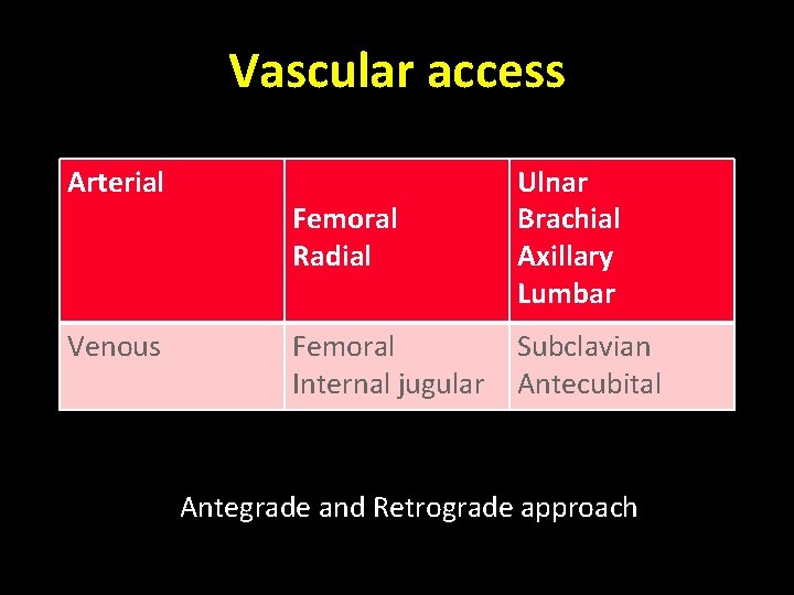
Vascular access Arterial Venous Femoral Radial Ulnar Brachial Axillary Lumbar Femoral Internal jugular Subclavian Antecubital Antegrade and Retrograde approach
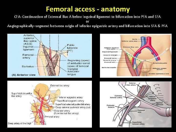
Femoral access - anatomy CFA- Continuation of External Iliac A below Inguinal ligament to bifurcation into PFA and SFA or Angiographically–segment between origin of Inferior epigastric artery and bifurcation into SFA & PFA
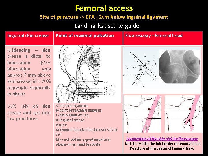
Femoral access Site of puncture -> CFA : 2 cm below inguinal ligament Landmarks used to guide Inguinal skin crease Point of maximal pulsation Fluoroscopy –femoral head Misleading – skin crease is distal to bifurcation (CFA bifurcation was approx 6 mm above skin crease) in > 70% of people, especially in obese 50% rely on skin crease and get into low punctures A-inguinal ligament B-point of maximal impulse C-bifurcation of CFA D-inguinal crease Issues: Maximum impulse maybe over SFA in 5% May not obtain a good impulse in obese –may need to rotate Localization of the skin nick by fluoroscopy Nick to overlie the inf: border of femoral head Puncture at the center of femoral head

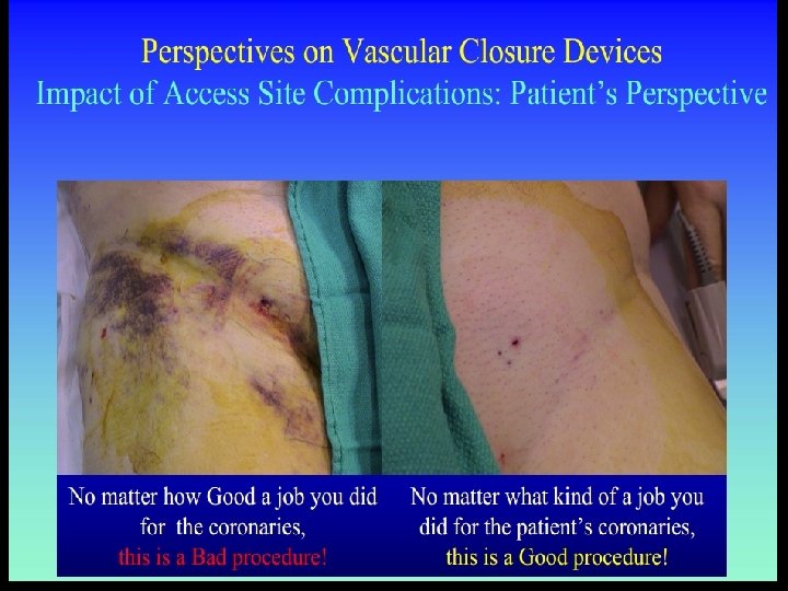
Femoral - complications • Bleeding and hematoma (5 -10%) • RPH • Local complications of femoral access (2 -10%) – Pseudoaneurysm (1 -6%) – AV fistula (1%) – Dissection acute closure (<1%) – Thrombosis distal embolisation (1%) – Infection – Nerve damage • Puncture site relation to complications – Low puncture: Pseudoaneurysm, AV fistula, Nerve damage, Hematoma – High punctures / posterior punctures: RPH , Hematoma
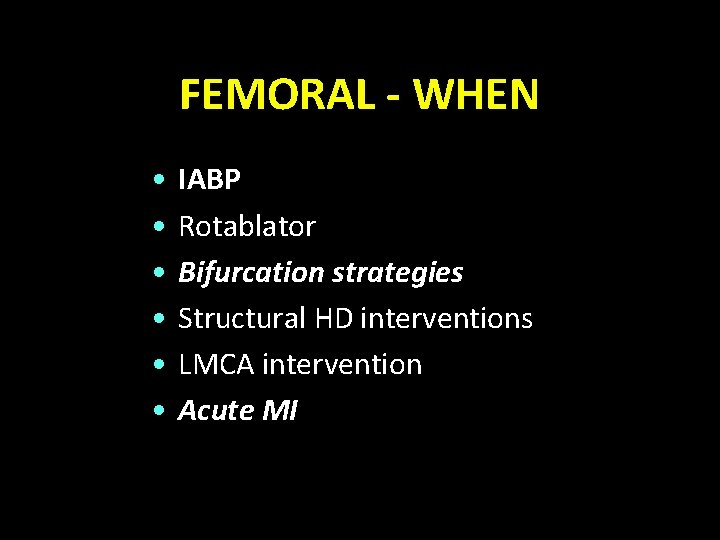
FEMORAL - WHEN • • • IABP Rotablator Bifurcation strategies Structural HD interventions LMCA intervention Acute MI
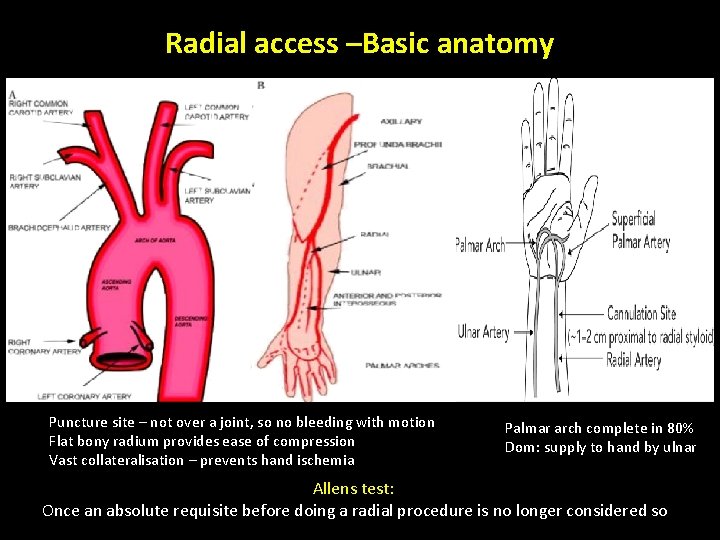
Radial access –Basic anatomy Puncture site – not over a joint, so no bleeding with motion Flat bony radium provides ease of compression Vast collateralisation – prevents hand ischemia Palmar arch complete in 80% Dom: supply to hand by ulnar Allens test: Once an absolute requisite before doing a radial procedure is no longer considered so
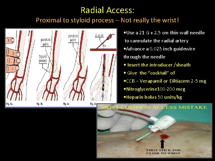
Radial Access: Proximal to styloid process – Not really the wrist! • Use a 21 G x 2. 5 cm thin wall needle to cannulate the radial artery • Advance a 0. 025 inch guidewire through the needle • Insert the introducer /sheath • Give the “cocktail” of • CCB – Verapamil or Diltiazem 2 -5 mg • Nitroglycerine 100 -200 mcg • Heparin bolus 50 units/kg
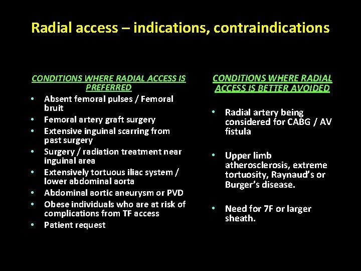
Radial access – indications, contraindications CONDITIONS WHERE RADIAL ACCESS IS PREFERRED • Absent femoral pulses / Femoral bruit • Femoral artery graft surgery • Extensive inguinal scarring from past surgery • Surgery / radiation treatment near inguinal area • Extensively tortuous iliac system / lower abdominal aorta • Abdominal aortic aneurysm or PVD • Obese individuals who are at risk of complications from TF access • Patient request CONDITIONS WHERE RADIAL ACCESS IS BETTER AVOIDED • Radial artery being considered for CABG / AV fistula • Upper limb atherosclerosis, extreme tortuosity, Raynaud’s or Burger’s disease. • Need for 7 F or larger sheath.

Trans-radial - Access Site Complications • Radial artery occlusion (≈5%, higher rates when routine doppler is used, mostly asymptomatic) • Forearm hematoma and/or pain • Radial artery pseudoaneurysm • Radial or brachial artery perforation • Uncontrolled bleeding with resultant compartment syndrome • Pain / severe spasm – precluding advancement / removal of catheters • Need for femoral conversion (5 -10%)

Radial access Advantages Decrease the incidence of major vascular complications Disadvantages Associated with a significant operator learning curve Has limited compatibility with very large Decrease the incidence of bleeding equipment complications Appears to decrease MACE in patients with Elderly patients may have tortuousity of the radial and subclavian arteries which ACS makes the procedure more challenging Better control over vascular access and hemostasis for obese and overall patients May have limited guiding catheter support in most challenging PCI scenarios (heavy Decreased time to ambulation calcifications, tortousity, complex Improved patient movement and comfort bifurcations) Access to LIMA Allows early discharge policy Associated with upper limb arterial May decrease cost complications (rare) Higher radiation exposure to the operator
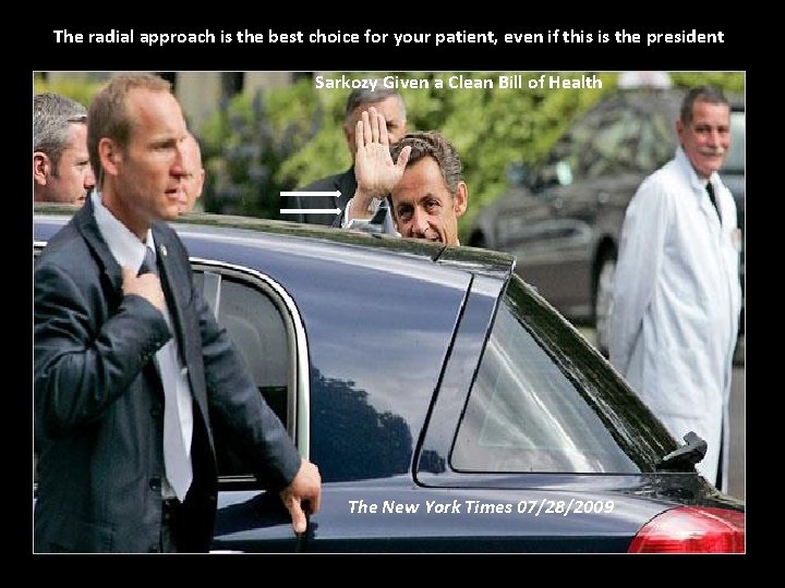
The radial approach is the best choice for your patient, even if this is the president Sarkozy Given a Clean Bill of Health The New York Times 07/28/2009
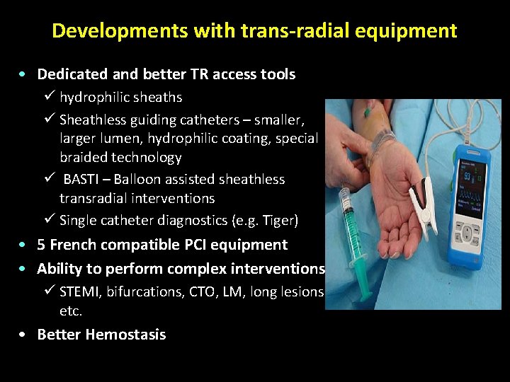
Developments with trans-radial equipment • Dedicated and better TR access tools ü hydrophilic sheaths ü Sheathless guiding catheters – smaller, larger lumen, hydrophilic coating, special braided technology ü BASTI – Balloon assisted sheathless transradial interventions ü Single catheter diagnostics (e. g. Tiger) • 5 French compatible PCI equipment • Ability to perform complex interventions ü STEMI, bifurcations, CTO, LM, long lesions etc. • Better Hemostasis
Ulnar access • SITE • 2 -3 cm above the crease of wrist • ADVANTAGES • Preservation of radial artery for CABG • PREREQUISITE • Reverse Allen’s test • Not to be used after failed ipsilateral radial attempt • COMPLICATIONS • Same as with radial artery access; nerve damage more likely • EVIDENCE – PCVI-CUBA trial radial vs ulnar • Success rate - access 96% vs 93%, • PCI – 96% vs 95%, • Complication rate 1% vs 1. 2 %.
Brachial access – seldom done Cutdown / puncture SITE OF PUNCTURE Medial aspect of cubital fossa, 2 -3 cm above the elbow crease INDICATIONS Need for upper limb or venous access, but CI for radial access Severe PVD / Renal or lower limb artery angioplasty Selective LIMA access from left arm COMPLICATIONS Hand ischemia - Due to thrombosis Compartment syndrome - Hematoma extends into forearm Median nerve injury - 0. 2 and 1. 4% Orator’s hand posture ACCESS trial – radial vs brachial access More complications with brachial approach ( 0. 2% vs 2. 6% p 0. 03 )
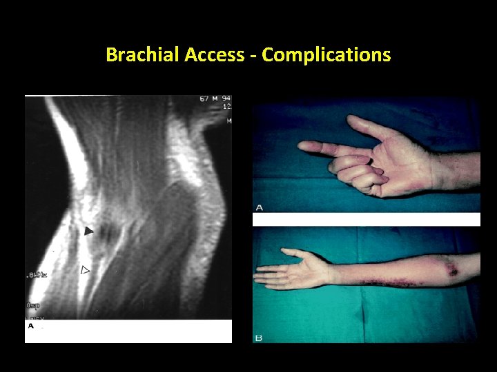
Brachial Access - Complications
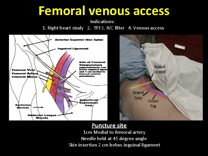
Femoral venous access Indications: 1. Right heart study 2. TPI 3. IVC filter 4. Venous access Puncture site 1 cm Medial to femoral artery Needle held at 45 degree angle Skin insertion 2 cm below inguinal ligament
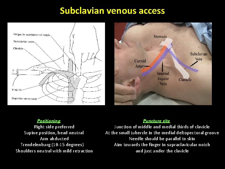
Subclavian venous access Positioning Right side preferred Supine position, head neutral Arm abducted Trendelenburg (10 -15 degrees) Shoulders neutral with mild retraction Puncture site Junction of middle and medial thirds of clavicle At the small tubercle in the medial deltopectoral groove Needle should be parallel to skin Aim towards the finger in supraclavicular notch and just under the clavicle
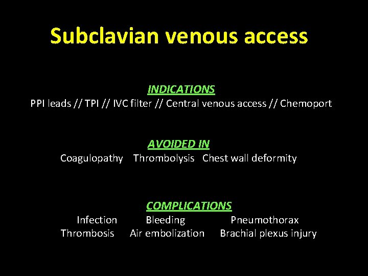
Subclavian venous access INDICATIONS PPI leads // TPI // IVC filter // Central venous access // Chemoport AVOIDED IN Coagulopathy Thrombolysis Chest wall deformity Infection Thrombosis COMPLICATIONS Bleeding Air embolization Pneumothorax Brachial plexus injury
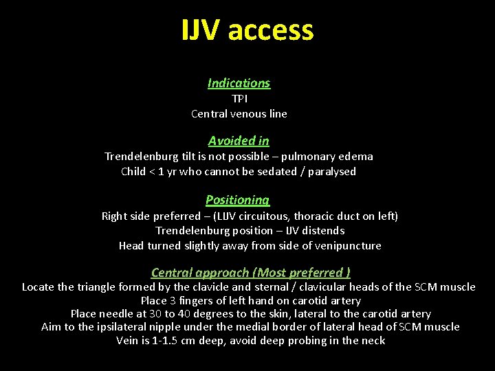
IJV access Indications TPI Central venous line Avoided in Trendelenburg tilt is not possible – pulmonary edema Child < 1 yr who cannot be sedated / paralysed Positioning Right side preferred – (LIJV circuitous, thoracic duct on left) Trendelenburg position – IJV distends Head turned slightly away from side of venipuncture Central approach (Most preferred ) Locate the triangle formed by the clavicle and sternal / clavicular heads of the SCM muscle Place 3 fingers of left hand on carotid artery Place needle at 30 to 40 degrees to the skin, lateral to the carotid artery Aim to the ipsilateral nipple under the medial border of lateral head of SCM muscle Vein is 1 -1. 5 cm deep, avoid deep probing in the neck
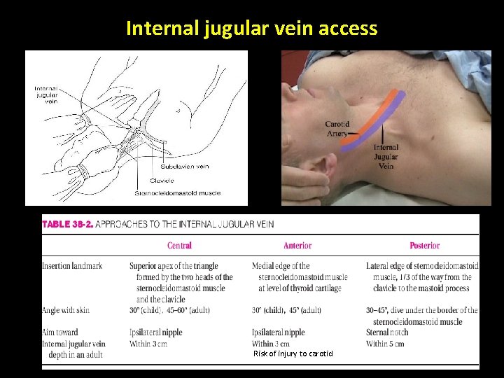
Internal jugular vein access Risk of injury to carotid
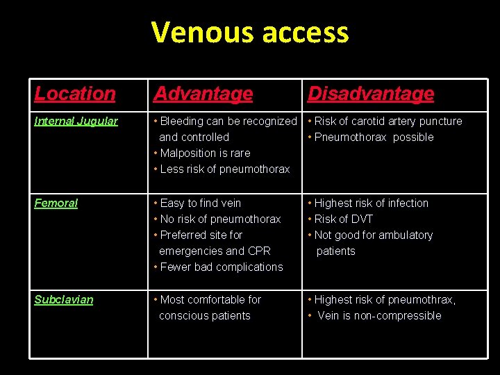
Venous access Location Advantage Disadvantage Internal Jugular • Bleeding can be recognized • Risk of carotid artery puncture and controlled • Pneumothorax possible • Malposition is rare • Less risk of pneumothorax Femoral • Easy to find vein • No risk of pneumothorax • Preferred site for emergencies and CPR • Fewer bad complications • Highest risk of infection • Risk of DVT • Not good for ambulatory patients Subclavian • Most comfortable for conscious patients • Highest risk of pneumothrax, • Vein is non-compressible
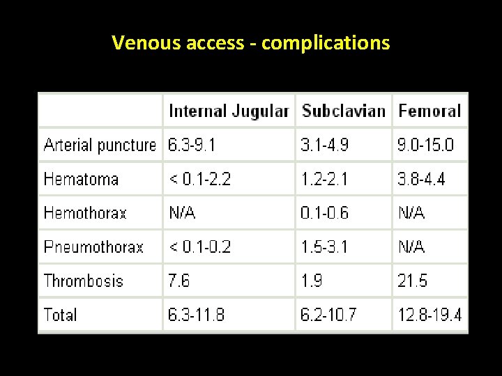
Venous access - complications
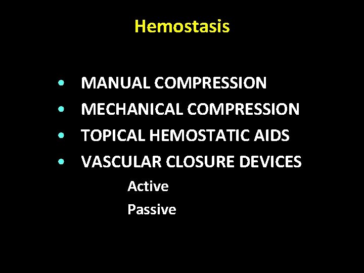
Hemostasis • • MANUAL COMPRESSION MECHANICAL COMPRESSION TOPICAL HEMOSTATIC AIDS VASCULAR CLOSURE DEVICES 1. Active 2. Passive
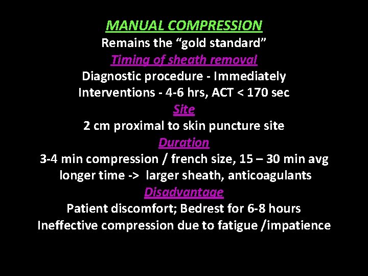
MANUAL COMPRESSION Remains the “gold standard” Timing of sheath removal Diagnostic procedure - Immediately Interventions - 4 -6 hrs, ACT < 170 sec Site 2 cm proximal to skin puncture site Duration 3 -4 min compression / french size, 15 – 30 min avg longer time -> larger sheath, anticoagulants Disadvantage Patient discomfort; Bedrest for 6 -8 hours Ineffective compression due to fatigue /impatience
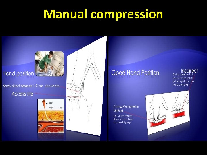
Manual compression
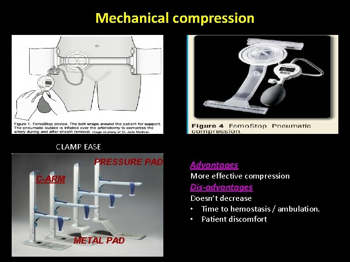
Mechanical compression CLAMP EASE PRESSURE PAD Advantages More effective compression C-ARM Dis-advantages Doesn’t decrease • Time to hemostasis / ambulation. • Patient discomfort METAL PAD
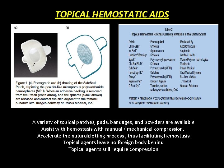
TOPICAL HEMOSTATIC AIDS A variety of topical patches, pads, bandages, and powders are available Assist with hemostasis with manual / mechanical compression. Accelerate the naturalclotting process , thus facilitating hemostasis Topical agents leave no foreign body behind Topical agents still require compression
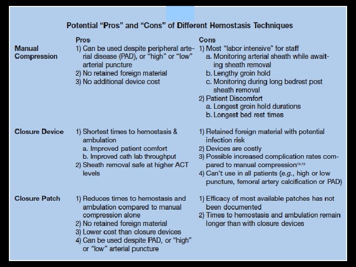
VASCULAR CLOSURE DEVICES Introduced in 1995 To decrease vascular complications and To reduce the time to hemostasis and ambulation CLASSIFICATION PASSIVE Enhance hemostasis with prothrombotic material or mechanical compression, But do not achieve prompt hemostasis or shorten the time to ambulation ACTIVE Suture (Perclose), Collagen Plugs (Angioseal), Clips (Starclose) Achieve prompt hemostasis or shorten the time to ambulation
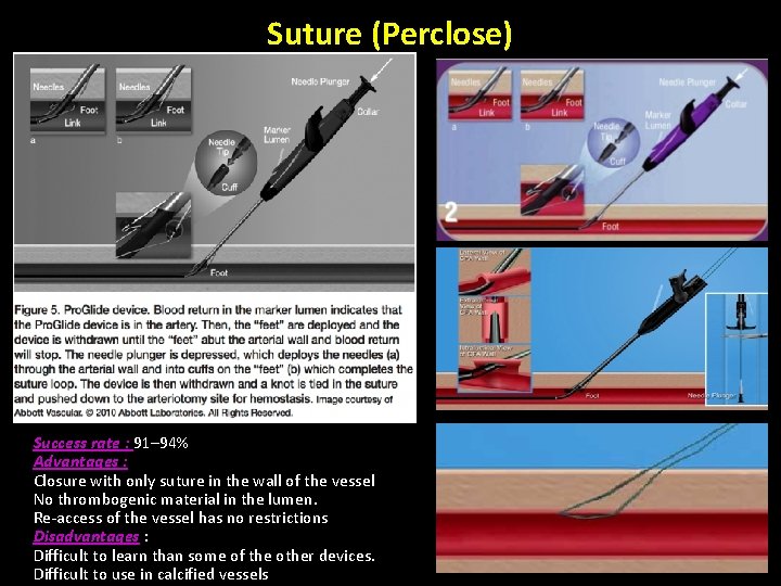
Suture (Perclose) Success rate : 91– 94% Advantages : Closure with only suture in the wall of the vessel No thrombogenic material in the lumen. Re-access of the vessel has no restrictions Disadvantages : Difficult to learn than some of the other devices. Difficult to use in calcified vessels
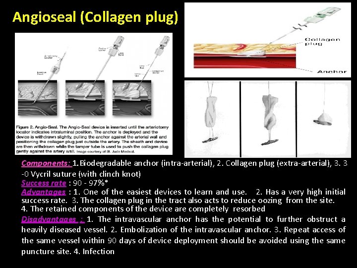
Angioseal (Collagen plug) Components: 1. Biodegradable anchor (intra-arterial), 2. Collagen plug (extra-arterial), 3. 3 -0 Vycril suture (with clinch knot) Success rate : 90 - 97%* Advantages : 1. One of the easiest devices to learn and use. 2. Has a very high initial success rate. 3. The collagen plug in the tract also acts to reduce oozing from the site. 4. The retained components of the device are completely resorbed Disadvantages : 1. The intravascular anchor has the potential to further obstruct a heavily diseased vessel. 2. Embolization of the intravascular anchor. 3. Repeat access of the same vessel within 90 days of device deployment should be avoided using the same puncture site. 4. Infection
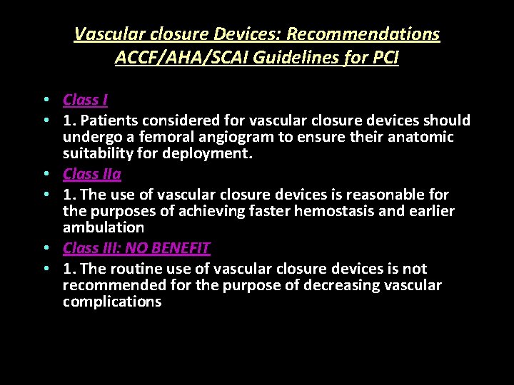
Vascular closure Devices: Recommendations ACCF/AHA/SCAI Guidelines for PCI • Class I • 1. Patients considered for vascular closure devices should undergo a femoral angiogram to ensure their anatomic suitability for deployment. • Class IIa • 1. The use of vascular closure devices is reasonable for the purposes of achieving faster hemostasis and earlier ambulation • Class III: NO BENEFIT • 1. The routine use of vascular closure devices is not recommended for the purpose of decreasing vascular complications

Thank You
- Slides: 36