
VARIATIONS IN MINERAL CRYSTALLITES OF BONE AT DISTINCT
VARIATIONS IN MINERAL CRYSTALLITES OF BONE AT DISTINCT SKELETAL SITES: POSSIBLE RELATIONSHIP WITH DISEASES LIKE OSTEOPOROSIS? DR HASAN BABER DOW UNIVERSIRY OF HEALTH SCIENCES Email: Relicpassion@yahoo. com Web: www. scienctifica. com Supervisor Maisoon Al Jawad
BACKROUND Bones are made of hard tissues that form the human skeleton Consist of both organic and inorganic mineral Bones at different sites appear to respond differently to external stimuli. Low levels mechanical load in limb – ‘disuse’ osteoporosis Low levels mechanical load in skull – no osteoporosis Weight bearing versus protective Ulna Calvaria
EVALUATING OSTEOPOROSIS AND ITS RELATIONSHIP TO BONE MINERAL ORIENTATION Osteoporosis is defined as a reduction in skeletal mass. Factors facilitating bone loss and osteoporosis 1. Hormonal imbalance 2. Calcium haemostasis imbalance 3. Loss of gonadal function 4. Ageing Mechanical stress – ‘disuse osteoporosis’ Disuse osteoporosis corresponds to the loss of bone, as a result of mechanical unloading of the skeleton. It could occur due to prolonged bed rest and space travel. The skull not affected
What causes these differences in bone metabolism at these two functionally distinct sites?
Variations in bone at different skeletal sites Major determinant of bone strength is due to 1) Amount of cancellous and cortical bone 2) Amount of organic matrix and collagen present 3) Amount and Composition of Mineral content present Skull bone has a higher ‘safety factor’ compared with limb bone ORIENTATION OF MINERAL CONTENT AND DIRECTION?
Research Question: Are there structural differences in the mineral content in bones from functionally different sites? Objectives: To identify bone mineral crystal orientation and morphology of Rat ulna, calvaria and whale rostrum, by X-ray diffraction and scanning electron microscope.
Materials & Methods Materials: The animals were male (Sprague - Dawley) rats, 110 -120 Gms, which equates to approximately 5 weeks old. Methods: Samples for Scanning Electron Microscope were prepared by fixing and dehydration in graded alcohols and storing them in 100% ethanol. X-ray diffraction carried at on X-Mas (BM 28) Beam line, at the European synchrotron radiation facility using 100 micro thick sections of fixed tissue. We first did calibrant of the samples, then did powder diffraction of each sample to produce diffraction patterns. Data Analysis: The diffraction patterns were then fitted in form of curves by software “ ORIGINS". Fitted curves were then plotted two theta, as a function of distance by software “SIGMAPLOT”.

Diagrammatic representation of the parts of bones that were scanned Ulna Calvaria Rostrum
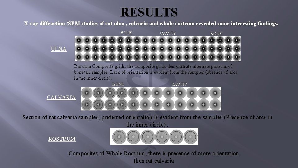
RESULTS X-ray diffraction /SEM studies of rat ulna , calvaria and whale rostrum revealed some interesting findings. BONE CAVITY BONE ULNA Rat ulna Composite grids, the composite grids demonstrate alternate patterns of bone/air samples. Lack of orientation is evident from the samples (absence of arcs in the inner circle). BONE CAVITY CALVARIA Section of rat calvaria samples, preferred orientation is evident from the samples (Presence of arcs in the inner circle). ROSTRUM Composites of Whale Rostrum, there is presence of more orientation then rat calvaria
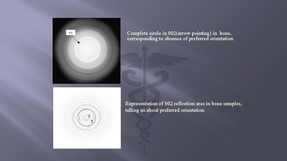
Complete circle in 002(arrow pointing) in bone, corresponding to absence of preferred orientation Representation of 002 reflection arcs in bone samples, telling us about preferred orientation
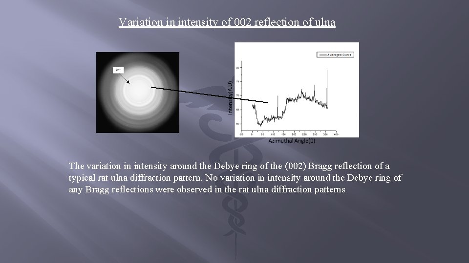
Variation in intensity of 002 reflection of ulna The variation in intensity around the Debye ring of the (002) Bragg reflection of a typical rat ulna diffraction pattern. No variation in intensity around the Debye ring of any Bragg reflections were observed in the rat ulna diffraction patterns
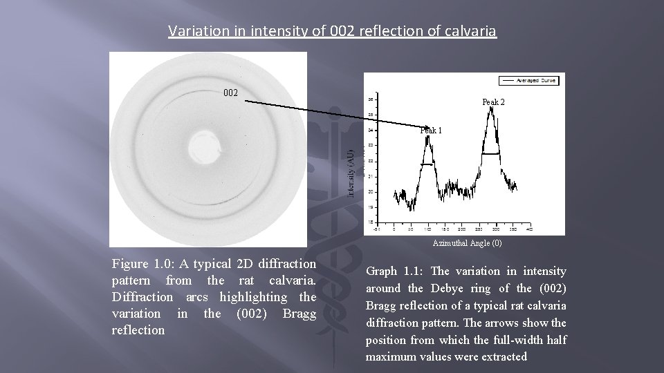
Variation in intensity of 002 reflection of calvaria 002 Peak 1 Azimuthal Angle (0) Figure 1. 0: A typical 2 D diffraction pattern from the rat calvaria. Diffraction arcs highlighting the variation in the (002) Bragg reflection Graph 1. 1: The variation in intensity around the Debye ring of the (002) Bragg reflection of a typical rat calvaria diffraction pattern. The arrows show the position from which the full-width half maximum values were extracted
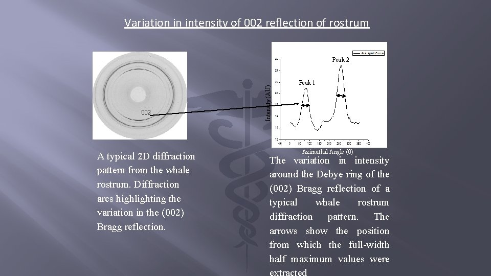
Variation in intensity of 002 reflection of rostrum 002 A typical 2 D diffraction pattern from the whale rostrum. Diffraction arcs highlighting the variation in the (002) Bragg reflection. Intensity (AU) Peak 2 Peak 1 Azimuthal Angle (0) The variation in intensity around the Debye ring of the (002) Bragg reflection of a typical whale rostrum diffraction pattern. The arrows show the position from which the full-width half maximum values were
When integrating the specimen Azimuthally, with plotted graphs of FWHM(Y-axis) against Distance (X-axis), an increase in value of FWHM corresponds to reduction in preferred orientation.
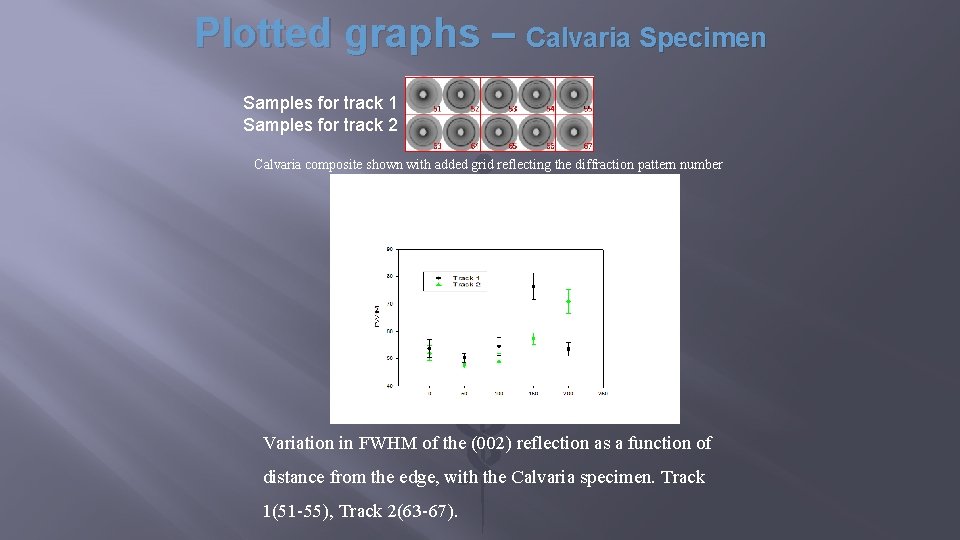
Plotted graphs – Calvaria Specimen Samples for track 1 Samples for track 2 Calvaria composite shown with added grid reflecting the diffraction pattern number Distance in Microns μm Variation in FWHM of the (002) reflection as a function of distance from the edge, with the Calvaria specimen. Track 1(51 -55), Track 2(63 -67).
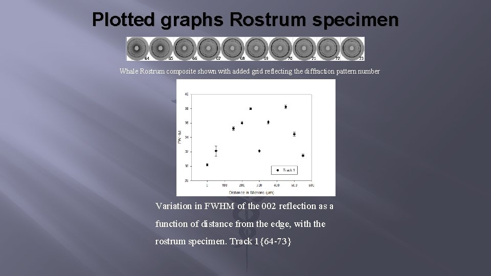
Plotted graphs Rostrum specimen Whale Rostrum composite shown with added grid reflecting the diffraction pattern number Variation in FWHM of the 002 reflection as a function of distance from the edge, with the rostrum specimen. Track 1{64 -73}
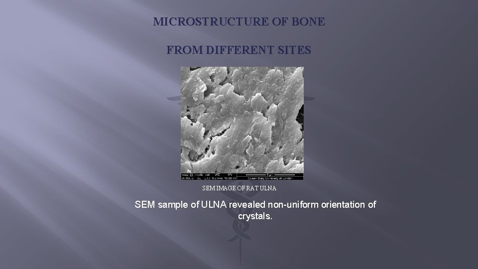
MICROSTRUCTURE OF BONE FROM DIFFERENT SITES SEM IMAGE OF RAT ULNA SEM sample of ULNA revealed non-uniform orientation of crystals.
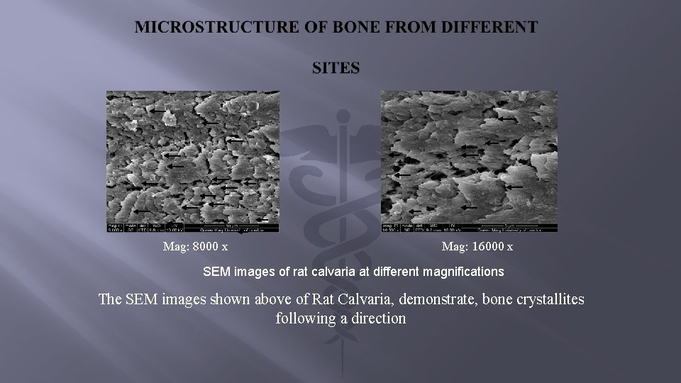
Mag: 8000 x Mag: 16000 x SEM images of rat calvaria at different magnifications The SEM images shown above of Rat Calvaria, demonstrate, bone crystallites following a direction
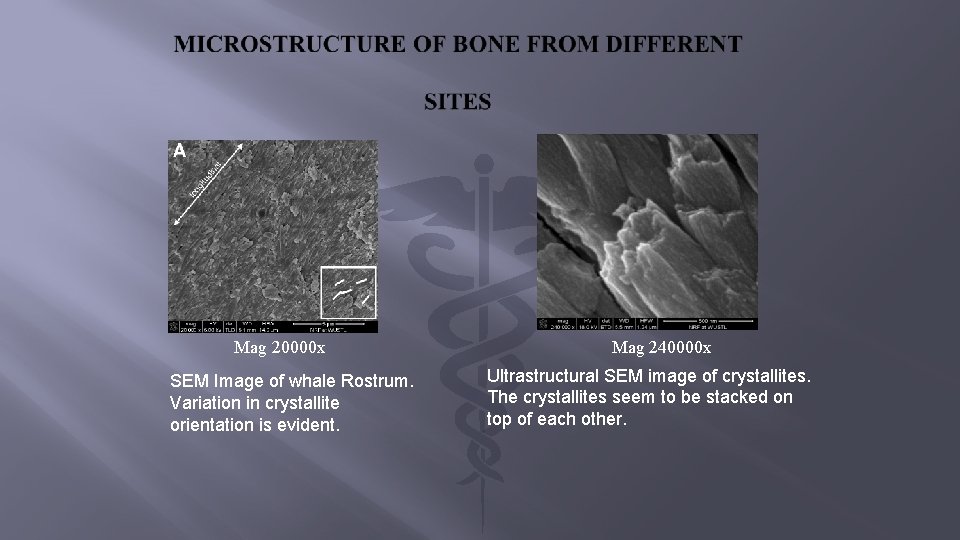
Mag 20000 x SEM Image of whale Rostrum. Variation in crystallite orientation is evident. Mag 240000 x Ultrastructural SEM image of crystallites. The crystallites seem to be stacked on top of each other.
DISCUSSION This study demonstrates differences in skull bones when compared with ulna by both SEM and X-ray Diffraction. Our studies revealed: 1. Presence of high crystallite orientation and texture in whale rostrum. 2. Rat calvaria crystal have less texture and orientation, then those in whale rostrum. 3. Little or no orientation in rat ulna samples.
Conclusion SEM AND S-XRD revealed differences in crystallite orientation that varies from bone sites. The order of crystallite ordering decreases by the order of Whale Rostrum > Rat Calvaria > Rat Ulna The difference in crystal orientation in calvaria, whale rostrum and ulna is consistent with - but does not alone explain – differences in strength and susceptibility to osteoporosis in these anatomically/functional and embryologically diverse bones.
LIMITATIONS Does these differences exist in a pathological bone samples like in osteoporosis that we have not explored in our current study We have compared normal bone via X-ray diffraction and SEM. We can further investigate their differential properties in a pathologic bone.

- Slides: 23