V ESICOURETERALREFLUX Fahimeh Kazemi Rashed kazemirashedfyahoo com Tabriz

V ESICOURETERALREFLUX Fahimeh Kazemi Rashed kazemirashedf@yahoo. com Tabriz University of Medical Sciences
? Who gets VUR? What are the types")
V ESICOURETERALREFLUX What is vesicoureteral reflux (VUR)? Who gets VUR? What are the types of VUR? What are the symptoms of VUR? What are the complications of VUR? How is VUR diagnosed? What other tests do children with VUR need? How is primary VUR treated?

L EARNING OBJECTIVES Upon completion of this activity, participants should be able to: Describe the prevalence of vesicoureteral reflux (VUR) in the general pediatric population and in children with urinary tract infections. Describe the grading system of the International Reflux Study for classification of VUR. Describe the inheritance pattern of VUR and guidelines for its management. Identify the most reliable tests for the diagnosis of VUR. Describe the techniques and efficacy of surgery for the management of VUR.

B ACKGROUND, INCIDENCE AND ETIOLOGY VUR is the retrograde flow of urine from the bladder into the ureter. It occurs in approximately 1– 3% of children and is associated with 7– 17% of children diagnosed with end -stage renal disease worldwide. Treatment of VUR is aimed at preventing the sequelae of pyelonephritis, renal parenchymal injury, hypertension, and chronic renal insufficiency. A period of 30– 40 years can pass between the first renal-scarring pyelonephritis and the development of hypertension or end-stage renal disease

B ACKGROUND, INCIDENCE AND ETIOLOGY An estimated 30– 40% of children under the age of 5 years who develop a urinary tract infection (UTI) have VUR can be further categorized as either primary or secondary. Primary VUR in children is frequently attributed to an abnormally short intravesical tunnel at the ureterovesical junction Secondary VUR occurs when reflux is induced by abnormally increased bladder pressures, such as those seen with urethral obstruction or neurogenic bladder dysfunction.

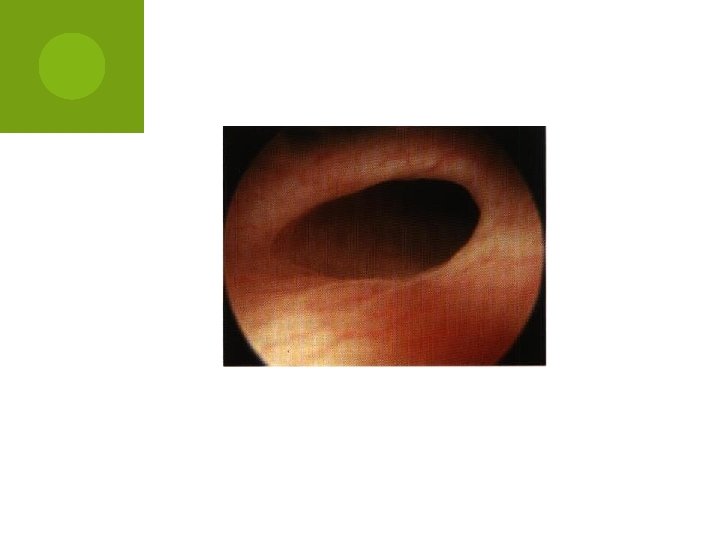
R ISK FACTORS Factors associated with VUR include the following: A short submucosal tunnel Lateral placement of the ureteral orifice Abnormal configuration of ureteral orifice (e. g. , stadium, horseshoe , and golf-hole orifices) Infection Severe bladder-outlet obstruction Young age Duplicated collecting systems (particularly from the more laterally placed orifice draining the lower pole)

BLADDER SUBMUCOSAL TUNNEL cement of the ureteral orifice
 Diagnosis")
Diagram of the bladder submucosal tunnel t submucosal tunnel Cooper, C. S. (2009) Diagnosis and management of vesicoureteral reflux in children Nat. Rev. Urol. doi: 10. 1038/nrurol. 2009. 150

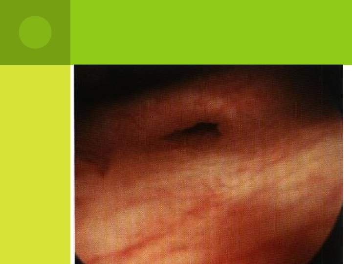
N ORMAL ORIFICE

configuration of ureteral orifice H ORSE SHOE ORIFICE

G OLF HOLE ORIFICE Abnormal configuration of ureteral orifice

PREDICTIVEOF REFLUX RESOLUTIONAND/OR THE RISK OF RENAL INJURY In general, the severity or grade of VUR has been used as the main factor to determine the likelihood of spontaneous reflux resolution and risk of renal injury. Other factors include age, sex, laterality, bladder volume and pressure at the onset of reflux, presence of renal scars, presence of voiding dysfunction, and a history of UTI.

Figure 7 User interface of a neural network for predicting the chance and timing of spontaneous resolution of vesicoureteral reflux based on a 2‑year resolution model Cooper, C. S. (2009) Diagnosis and management of vesicoureteral reflux in children Nat. Rev. Urol. doi: 10. 1038/nrurol. 2009. 150

B ACKGROUND, INCIDENCE AND ETIOLOGY strong inheritance pattern exists for primary VUR The chance of a sibling of a child with VUR also having reflux is about 25%, and the offspring of affected individuals have a 27– 51% increased risk of having reflux. appropriate clinical assessment and management of children with VUR requires the treating physician to develop an individualized approach that considers multiple factors.

C LINICAL MANAGEMENT Antibiotics versus operative intervention no significant differences exist in renal function or growth, the progression or development of new scars, or UTIs in patients treated with one intervention compared to the other. Pyelonephritic symptoms, including febrile UTIs, tended to be more common in the medically treated groups compared with the surgical groups

C LINICAL MANAGEMENT new scars occurred earlier in children treated surgically compared with those in the medical treatment groups, but, as noted, no significant difference occurred overall with longer follow-up in terms of new renal scars in those treated with antibiotics compared to those undergoing surgery. nine re-implantation procedures would be required in order to prevent one incidence of febrile UTI, with no reduction in the number of children developing any UTI or renal damage.

C LINICAL MANAGEMENT Antibiotics versus observation Antibiotic prophylaxis for reducing the likelihood of developing a UTI has been associated with a 24 -fold increased risk of Escherichia coli developing resistance to trimethoprim– sulfamethoxazole. antibiotic prophylaxis has not been proven to reduce the incidence of pyelonephritis in children with VUR.

C LINICAL EVALUATION Assessment of reflux The only tests that routinely and reliably detect reflux are voiding cystourethrography (VCUG) and nuclear cystography. An initial VCUG provides better anatomic details regarding reflux, including the presence or absence of periureteral diverticuli, ureteral duplication, and abnormalities of the bladder, such as trabeculations or urethral obstruction. Typically, follow-up studies are performed with nuclear cystography, as there is a decreased exposure to radioactivity with this study.

C LINICAL EVALUATION Assessment of renal scars

D IAGNOSTICWORK-UP basic diagnostic work-up comprises : detailed medical history (including family history, screening for LUTD), physical examination including blood pressure measurement, urinalysis (including proteinuria), and urine culture.

D IAGNOSTICWORK-UP The choice of imaging modalities varies depending on the mode of presentation.

D IAGNOSTICWORK-UP The use of VCUG is recommended : In patients with US findings of bilateral high-grade hydronephrosis, duplex kidneys, ureterocele, ureteric dilatation and abnormal bladders, as the likelihood of VUR is much higher. In all other conditions, the use of VCUG to detect reflux is optional

G RADING OF REFLUX

G RADING OF REFLUX

G RADE 1

G RADE 2

G RADE 3

G RADE 4

G RADE 5
 Diagnosis and management of vesicoureteral reflux")
International Reflux Grading System Cooper, C. S. (2009) Diagnosis and management of vesicoureteral reflux in children Nat. Rev. Urol. doi: 10. 1038/nrurol. 2009. 150

G RADE I- URETER ONLY

G RADE II-U RETER, PELVIS, CALYCES, NO DILATION, NORMAL CALYCEALFORNICES

G RADE III-M ILD OR MODERATEDILATION AND/OR TORTUOSITYOF THE URETER, AND MILD OR MODERATEDILATIONOF THE PELVIS, BUT NO OR SLIGHTBLUNTINGOF THE FORNIC

G R A D E I V - M O D E R A T ED I L A T I O NA N D/O R T U O S I T Y O F T H E U R E T E RA N D M I L D D I L A T I O NO F R E N A LP E L V I S A N D C A L Y C E; S C O M P L E T EO B L I T E R A T I OONF S H A R P A N G L EO F F O R N I C E SB U T M A I N T E N A N COE F P A P I L L A R Y I M P R E S S I O NISN M A J O R I T YO F C A L Y C E, S B U T N O O R S L I G H TB L U N T I N G

G R ADE V - G R OS S D I LA T IO NA N D T O R T U O SIT YOF U RET E R; GR O S SD IL A TI O NO F R E NA LP EL V ISAN D C ALY CE; S P A P I LLA R YIM P R ES S I ONAS R E NO L O NG E R V I SI BL EIN M A J O R I T YOF C A LY CE S

T REATMENT There are mainly two treatment approaches Conservative Interventional

S PONTANEOUS R ESOLUTION Low Grade I………………. . 82% at 5 years Grade II………………. . 80% at 5 years Intermediate Grade – III…… 50% at 5 yrs Grade IV……………… 25% at 5 yrs Grade V………………. 12% resolution

S PONTANEOUS R ESOLUTION Grade III & IV management presents the most controversy

S PONTANEOUS R ESOLUTION Age at diagnosis Younger children are more likely to have VUR is more likely to resolve in younger children Intervals of significant growth and beneficial urodynamic change are most likely to effect change Resolution usually occurs within the first few years after diagnosis Rarely resolves if continued reflux after 5 y

M ANAGEMENT D ECISION M AKING Spontaneous resolution of VUR occurs in many infants and children - rarely at puberty More severe grades are less likely to resolve Sterile reflux does not appear to cause significant nephropathy Extended courses of prophylactic antibiotics are well tolerated by children Anti-Reflux surgery is highly successful in capable hands 95 -99% success rate

M ANAGEMENT D ECISION M AKING Medical management initially recommended for prepubertal children with I, III This also may be true for Grade IV - esp. in younger children with unilateral disease If no trend in improvement is seen in 2 - 3 years, surgery is recommended Grade V is unlikely to resolve and surgery is recommended after infancy Observation may be reasonable if diagnosed perinatally

M ANAGEMENT D ECISION M AKING Surgery recommended in most girls with persistent VUR Implications for future pregnancies Especially if recurrent infections or scars present Some discontinue antibiotics at puberty in girls Surgery if UTI occurs Prophylaxis can be stopped at puberty in boys Less likely to develop UTIs

C ONSERVATIVETHERAPY The objective of conservative therapy is prevention of febrile UTI It is based on the understanding that: VUR resolves spontaneously, mostly in young patients with low-grade reflux; 81% and 48% in VUR grades I-II and III-V, respectively VUR does not damage the kidney when free of infection

C ONSERVATIVETHERAPY The conservative approach includes: watchful waiting, intermittent or continuous antibiotic prophylaxis bladder rehabilitation in those with LUTD Circumcision during early infancy may be considered as part of conservative approach

C ONSERVATIVETHERAPY The most used antibacterial agents for prophylactic treatment are : nitrofurantoin (in children > 3 months: 1 -2 mg/kg/day, orally) trimethoprim (in children > 3 months: 2 mg/kg/day, orally) In the first 3 months of life, when these agents are poorly absorbed from the gut, amoxycillin (15 mg/kg/day, orally

S URGICAL M ANAGEMENT: I NDICATIONSFOR S URGERY Breakthrough UTIs on prophylactic antibiotics Noncompliance with medical management Severe VUR (Grade IV & V), especially with pyelonephrotic changes (evidence of scarring) Failure of renal growth, new renal scars, or deterioration of renal function on serial studies Persistent VUR in girls at puberty Reflux associated with congenital abnormalities at the UVJ (e. g. bladder diverticulum)

S URGICAL P RINCIPLESOF R EFLUX C ORRECTION Exclusion of causes of ……………. . Secondary VUR Mobilization of the ureter ……………Without tension or damage to blood supply Creation of a submucosal tunnel……. . 5: 1 ratio Entry point of the ureter (hitus), the direction of the submucosal tunnel ureteromucosal anastomosis…………Stenosis, angulation, or twisting of the ureter Muscular backing of the ureter ………Effective antireflux mechanism Gentle Handling of the bladder ………………. Hematuria and bladder spasms

G IL-V ERNET T ECHNIQUE

P OST- O PERATIVE C ARE & E VALUATION Renal U/S VCUG 6 weeks 3 -6 months Periodic F/U 18 months, 3 years, 5 years check U/A, BP, U/S

E ARLY C OMPLICATIONS Reflux Contralateral or ipsilateral ureter Trigonal edema, bladder dysfunction Majority are low grade Obstruction Edema, spasms, blood clots Most are mild Occur 1 -2 weeks post-op - pain, N/V, rarely fever Renal scan shows delay in excretion Nephrostomy tubes or ureteral stents if symptoms persist

L ATE C OMPLICATIONS Reflux Short length to diameter ratio Weak muscular backing Failure to treat secondary causes of VUR CIC and anticholinergics Treatment of dysfunctional voiding Obstruction Complete obstruction Ischemia or hiatal angulation Intermittent obstruction Lateral placement of orifice obstructs with filling

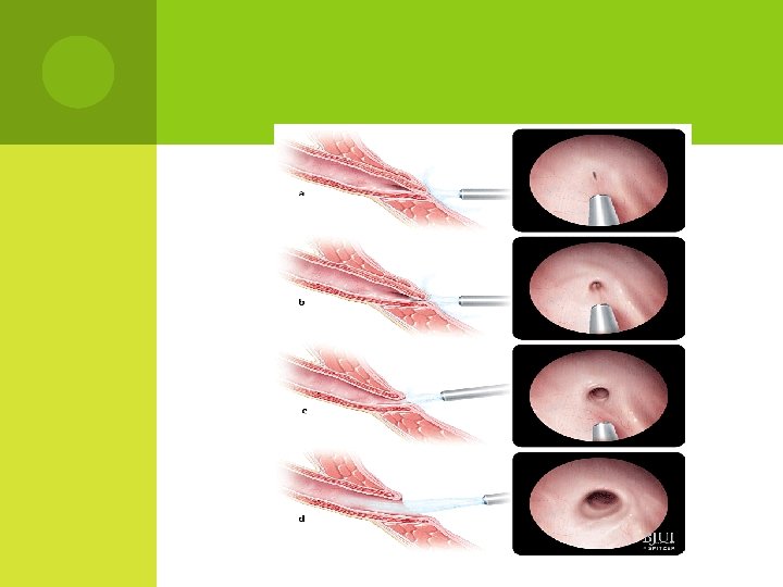
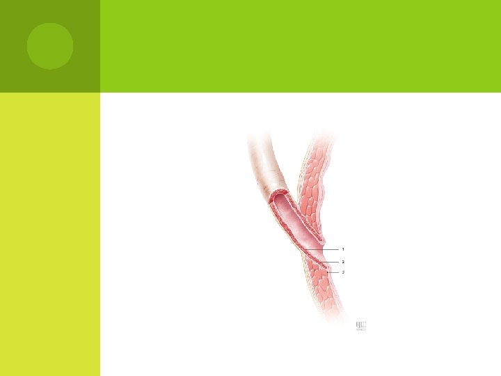
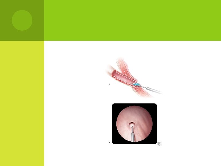
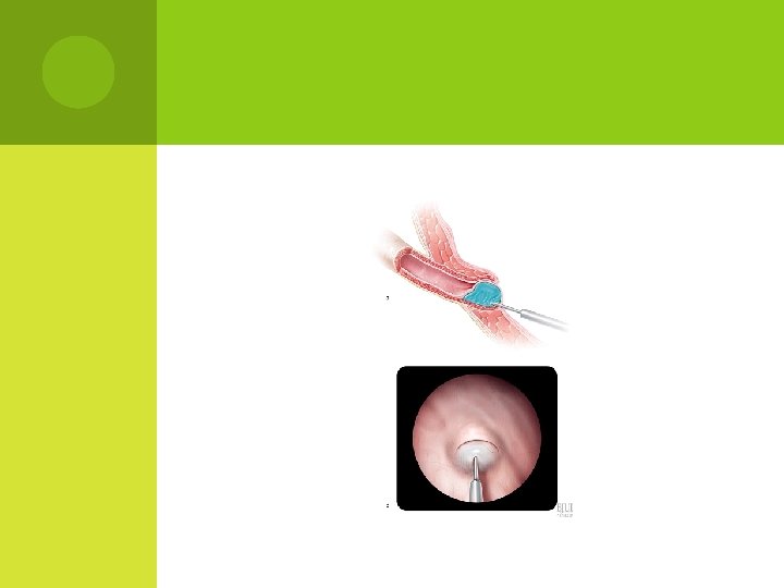
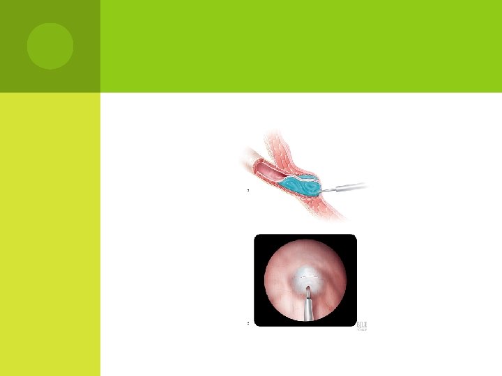
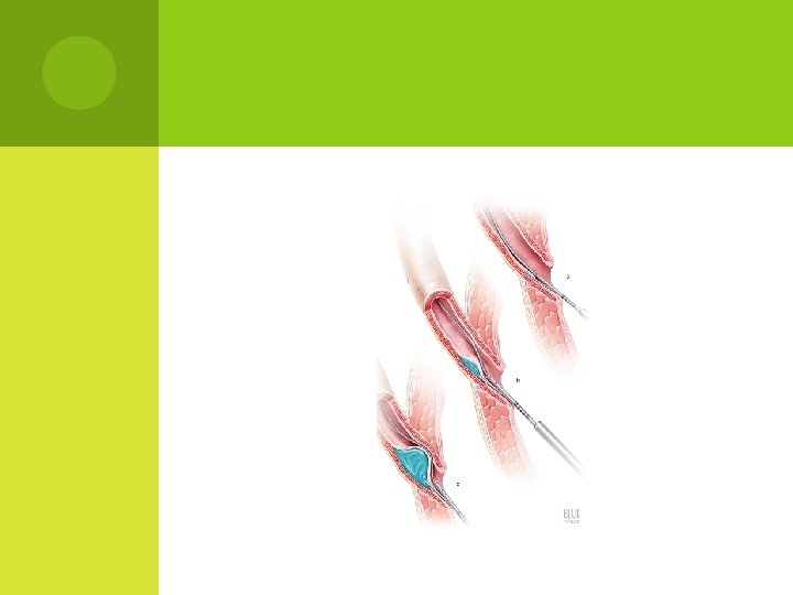
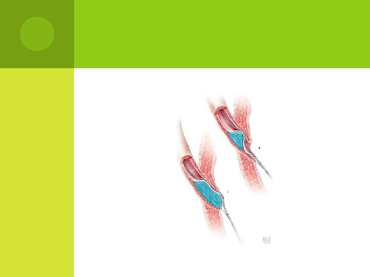
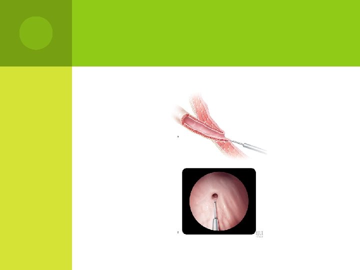
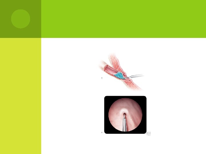
I NTERVENTIONALTREATMENT endoscopic injection of bulking agents

ENDOSCOPICINJECTIONOF BULKING AGENTS With the availability of biodegradable substances, endoscopic subureteric injection of bulking agents has become an alternative to long -term antibiotic prophylaxis and surgical intervention in the treatment of VUR in children.
, collagen, autologous fat, polydimethylsiloxane, silicone, chondrocytes and,")
BULKING AGENTS : polytetrafluoroethylene (PTFE or Teflon), collagen, autologous fat, polydimethylsiloxane, silicone, chondrocytes and, more recently, a solution of dextranomer/hyaluronic acid (Deflux).
 was FDAapproved for the treatment of")
BULKING AGENTS : bulking agents dextranomer/hyaluronic acid (Deflux) was FDAapproved for the treatment of VUR in children.
 following one")
R ESULTS OF E NDOSCOPIC ANTI-REFLUX PROCEDURES Reflux resolution rate (by ureter) following one treatment: grades I and II……………… 78. 5%, grade III……………. 72%, grade IV…………… 63% grade V……………. 51%. If the first injection was unsuccessful, the second treatment had a success rate of 68%, and the third treatment 34%

R ESULTS OF E NDOSCOPIC ANTI-REFLUX PROCEDURES The aggregate success rate with one or more injections was 85%. The success rate was significantly lower for duplicated (50%) versus single systems (73%), neuropathic (62%) versus normal bladders (74%).
")
Endoscopic injection for vesicoureteral reflux. reproduced from Deflux® patient education brochure (Q‑Med, Uppsala, Sweden) Cooper, C. S. (2009) Diagnosis and management of vesicoureteral reflux in children Nat. Rev. Urol. doi: 10. 1038/nrurol. 2009. 150

Figure 4 Dimercaptosuccinic acid scintigraphy with single photon emission computed tomography imaging demonstrating a cortical defect in the upper pole of the left kidney Cooper, C. S. (2009) Diagnosis and management of vesicoureteral reflux in children Nat. Rev. Urol. doi: 10. 1038/nrurol. 2009. 150
 Cooper, C. S. (2009)")
Voiding cystourethrogram, demonstrating right‑sided reflux with a periureteral diverticulum (arrows) Cooper, C. S. (2009) Diagnosis and management of vesicoureteral reflux in children Nat. Rev. Urol. doi: 10. 1038/nrurol. 2009. 150
. Paltiel H J et")
Sonograms of bladder demonstrate right-sided bilobed subureteric chondrocyte mound (arrow). Paltiel H J et al. Radiology 2004; 232: 390 -397 © 2004 by Radiological Society of North America
. Paltiel H J")
Transverse sonogram of bladder depicts bilateral unilobed subureteric chondrocyte mounds (arrows). Paltiel H J et al. Radiology 2004; 232: 390 -397 © 2004 by Radiological Society of North America
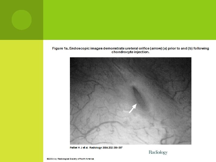
 (a) prior to and (b)")
Figure 1 b. Endoscopic images demonstrate ureteral orifice (arrow) (a) prior to and (b) following chondrocyte injection. Paltiel H J et al. Radiology 2004; 232: 390 -397 © 2004 by Radiological Society of North America

Thank you
- Slides: 77