
V 26 Regular vs alternative splicing Regular splicing
- Slides: 48
V 26 Regular vs. alternative splicing • Regular splicing mechanistic steps recognition of splice sites • Alternative splicing different mechanisms how frequent is alternative splicing? • Effect of alternative splicing on protein – protein interactions • Interplay of alternative splicing and epigenetic modifications 26. lecture SS 2018 Bioinformatics III 1
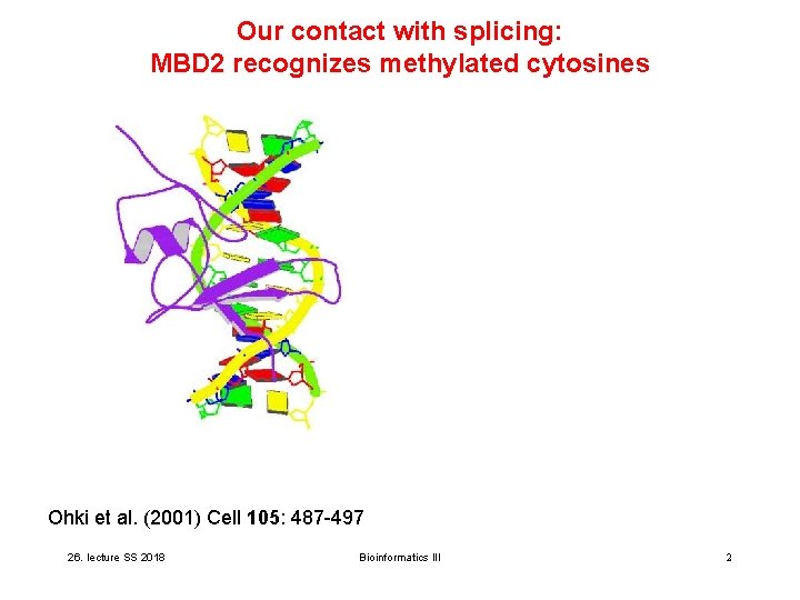
Our contact with splicing: MBD 2 recognizes methylated cytosines Ohki et al. (2001) Cell 105: 487 -497 26. lecture SS 2018 Bioinformatics III 2
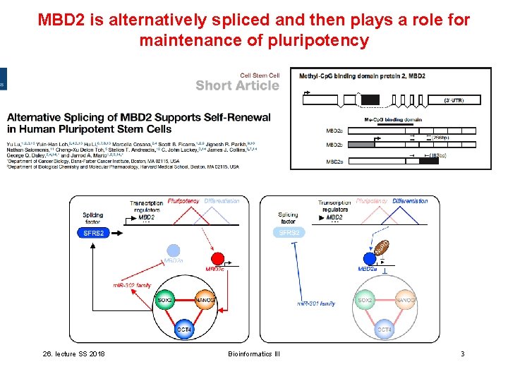
MBD 2 is alternatively spliced and then plays a role for maintenance of pluripotency 26. lecture SS 2018 Bioinformatics III 3
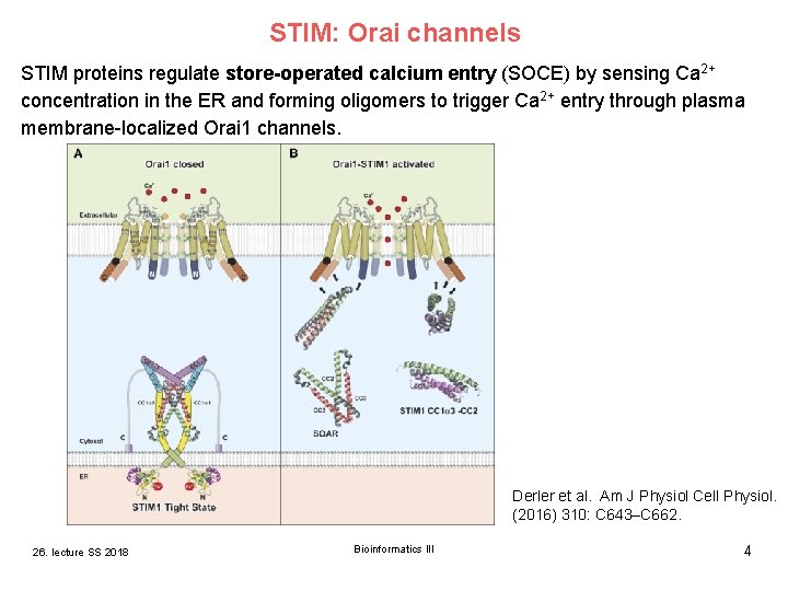
STIM: Orai channels STIM proteins regulate store-operated calcium entry (SOCE) by sensing Ca 2+ concentration in the ER and forming oligomers to trigger Ca 2+ entry through plasma membrane-localized Orai 1 channels. Derler et al. Am J Physiol Cell Physiol. (2016) 310: C 643–C 662. 26. lecture SS 2018 Bioinformatics III 4
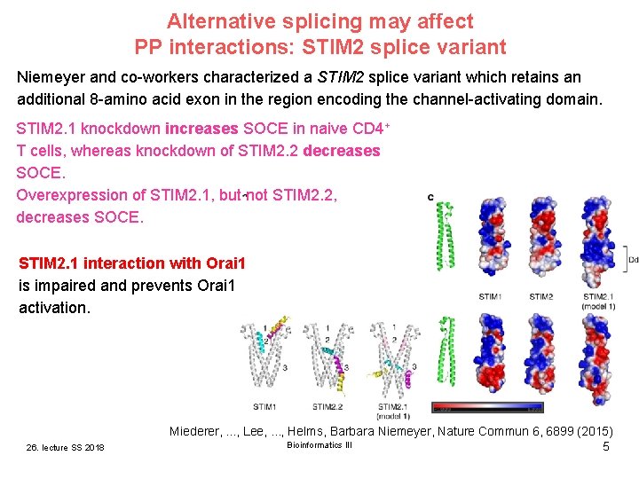
Alternative splicing may affect PP interactions: STIM 2 splice variant Niemeyer and co-workers characterized a STIM 2 splice variant which retains an additional 8 -amino acid exon in the region encoding the channel-activating domain. STIM 2. 1 knockdown increases SOCE in naive CD 4+ T cells, whereas knockdown of STIM 2. 2 decreases SOCE. Overexpression of STIM 2. 1, but not STIM 2. 2, decreases SOCE. STIM 2. 1 interaction with Orai 1 is impaired and prevents Orai 1 activation. Miederer, . . . , Lee, . . . , Helms, Barbara Niemeyer, Nature Commun 6, 6899 (2015) 26. lecture SS 2018 Bioinformatics III 5
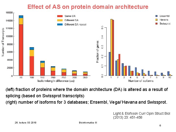
Effect of AS on protein domain architecture (left) fraction of proteins where the domain architecture (DA) is altered as a result of splicing (based on Swissprot transcripts) (right) number of isoforms for 3 databases; Ensembl, Vega/ Havana and Swissprot. Light & Elofsson Curr Opin Struct Biol (2013) 23: 451 -458 26. lecture SS 2018 Bioinformatics III 6
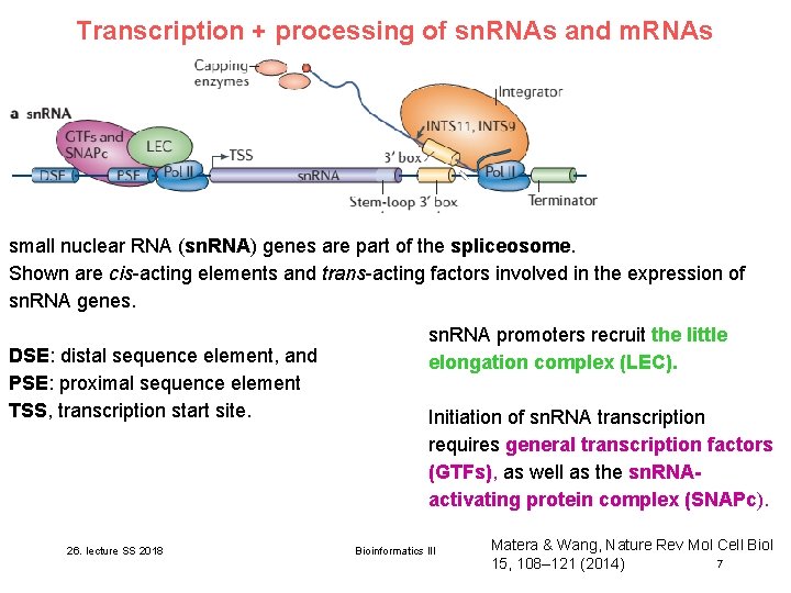
Transcription + processing of sn. RNAs and m. RNAs small nuclear RNA (sn. RNA) genes are part of the spliceosome. Shown are cis-acting elements and trans-acting factors involved in the expression of sn. RNA genes. DSE: distal sequence element, and PSE: proximal sequence element TSS, transcription start site. 26. lecture SS 2018 sn. RNA promoters recruit the little elongation complex (LEC). Initiation of sn. RNA transcription requires general transcription factors (GTFs), as well as the sn. RNAactivating protein complex (SNAPc). Bioinformatics III Matera & Wang, Nature Rev Mol Cell Biol 7 15, 108– 121 (2014)
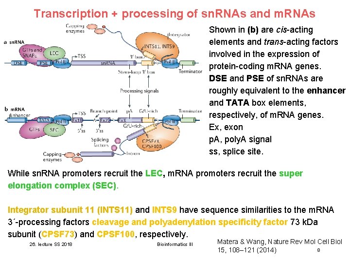
Transcription + processing of sn. RNAs and m. RNAs Shown in (b) are cis-acting elements and trans-acting factors involved in the expression of protein-coding m. RNA genes. DSE and PSE of sn. RNAs are roughly equivalent to the enhancer and TATA box elements, respectively, of m. RNA genes. Ex, exon p. A, poly. A signal ss, splice site. While sn. RNA promoters recruit the LEC, m. RNA promoters recruit the super elongation complex (SEC). Integrator subunit 11 (INTS 11) and INTS 9 have sequence similarities to the m. RNA 3ʹ-processing factors cleavage and polyadenylation specificity factor 73 k. Da subunit (CPSF 73) and CPSF 100, respectively. 26. lecture SS 2018 Bioinformatics III Matera & Wang, Nature Rev Mol Cell Biol 8 15, 108– 121 (2014)
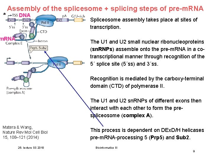
Assembly of the splicesome + splicing steps of pre-m. RNA DNA m. RNA Spliceosome assembly takes place at sites of transcription. The U 1 and U 2 small nuclear ribonucleoproteins (sn. RNPs) assemble onto the pre-m. RNA in a cotranscriptional manner through recognition of the 5ʹ splice site (5ʹss) and 3ʹss. Recognition is mediated by the carboxy-terminal domain (CTD) of polymerase II. The U 1 and U 2 sn. RNPs of different exons then interact with each other to form the prespliceosome (complex A). Matera & Wang, Nature Rev Mol Cell Biol 15, 108– 121 (2014) 26. lecture SS 2018 This process is dependent on DEx. D/H helicases pre-m. RNA-processing 5 (Prp 5) and Sub 2. Bioinformatics III 9
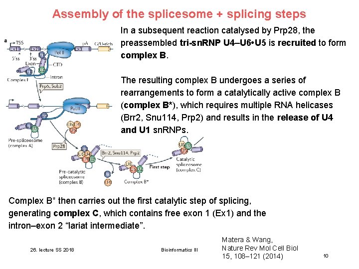
Assembly of the splicesome + splicing steps In a subsequent reaction catalysed by Prp 28, the preassembled tri-sn. RNP U 4–U 6 • U 5 is recruited to form complex B. The resulting complex B undergoes a series of rearrangements to form a catalytically active complex B (complex B*), which requires multiple RNA helicases (Brr 2, Snu 114, Prp 2) and results in the release of U 4 and U 1 sn. RNPs. Complex B* then carries out the first catalytic step of splicing, generating complex C, which contains free exon 1 (Ex 1) and the intron–exon 2 “lariat intermediate”. 26. lecture SS 2018 Bioinformatics III Matera & Wang, Nature Rev Mol Cell Biol 15, 108– 121 (2014) 10
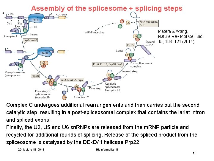
Assembly of the splicesome + splicing steps Matera & Wang, Nature Rev Mol Cell Biol 15, 108– 121 (2014) Complex C undergoes additional rearrangements and then carries out the second catalytic step, resulting in a post-spliceosomal complex that contains the lariat intron and spliced exons. Finally, the U 2, U 5 and U 6 sn. RNPs are released from the m. RNP particle and recycled for additional rounds of splicing. Release of the spliced product from the spliceosome is catalysed by the DEx. D/H helicase Prp 22. 26. lecture SS 2018 Bioinformatics III 11
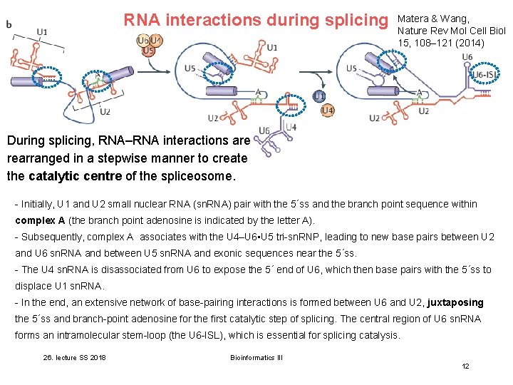
RNA interactions during splicing Matera & Wang, Nature Rev Mol Cell Biol 15, 108– 121 (2014) During splicing, RNA–RNA interactions are rearranged in a stepwise manner to create the catalytic centre of the spliceosome. - Initially, U 1 and U 2 small nuclear RNA (sn. RNA) pair with the 5ʹss and the branch point sequence within complex A (the branch point adenosine is indicated by the letter A). - Subsequently, complex A associates with the U 4–U 6 • U 5 tri-sn. RNP, leading to new base pairs between U 2 and U 6 sn. RNA and between U 5 sn. RNA and exonic sequences near the 5ʹss. - The U 4 sn. RNA is disassociated from U 6 to expose the 5ʹ end of U 6, which then base pairs with the 5ʹss to displace U 1 sn. RNA. - In the end, an extensive network of base-pairing interactions is formed between U 6 and U 2, juxtaposing the 5ʹss and branch-point adenosine for the first catalytic step of splicing. The central region of U 6 sn. RNA forms an intramolecular stem-loop (the U 6 -ISL), which is essential for splicing catalysis. 26. lecture SS 2018 Bioinformatics III 12
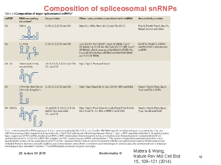
Composition of spliceosomal sn. RNPs 26. lecture SS 2018 Bioinformatics III Matera & Wang, Nature Rev Mol Cell Biol 15, 108– 121 (2014) 13
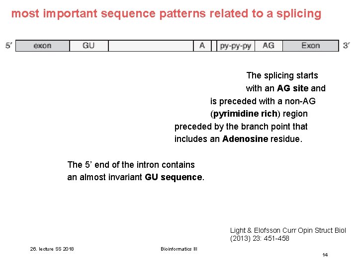
most important sequence patterns related to a splicing The splicing starts with an AG site and is preceded with a non-AG (pyrimidine rich) region preceded by the branch point that includes an Adenosine residue. The 5’ end of the intron contains an almost invariant GU sequence. Light & Elofsson Curr Opin Struct Biol (2013) 23: 451 -458 26. lecture SS 2018 Bioinformatics III 14
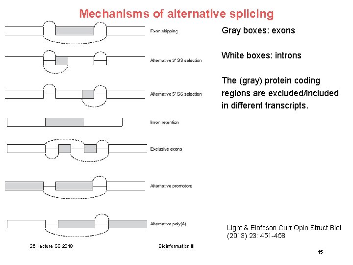
Mechanisms of alternative splicing Gray boxes: exons White boxes: introns The (gray) protein coding regions are excluded/included in different transcripts. Light & Elofsson Curr Opin Struct Biol (2013) 23: 451 -458 26. lecture SS 2018 Bioinformatics III 15
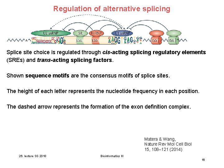
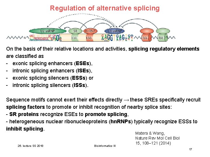
Regulation of alternative splicing Splice site choice is regulated through cis-acting splicing regulatory elements (SREs) and trans-acting splicing factors. Shown sequence motifs are the consensus motifs of splice sites. The height of each letter represents the nucleotide frequency in each position. The dashed arrow represents the formation of the exon definition complex. Matera & Wang, Nature Rev Mol Cell Biol 15, 108– 121 (2014) 26. lecture SS 2018 Bioinformatics III 16
Regulation of alternative splicing On the basis of their relative locations and activities, splicing regulatory elements are classified as - exonic splicing enhancers (ESEs), - intronic splicing enhancers (ISEs), - exonic splicing silencers (ESSs) or - intronic splicing silencers (ISSs). Sequence motifs cannot exert their effects directly → these SREs specifically recruit splicing factors to promote or inhibit recognition of nearby splice sites: - SR proteins recognize ESEs to promote splicing, - heterogeneous nuclear ribonucleoproteins (hn. RNPs) typically recognize ESSs to inhibit splicing. 26. lecture SS 2018 Bioinformatics III Matera & Wang, Nature Rev Mol Cell Biol 15, 108– 121 (2014) 17
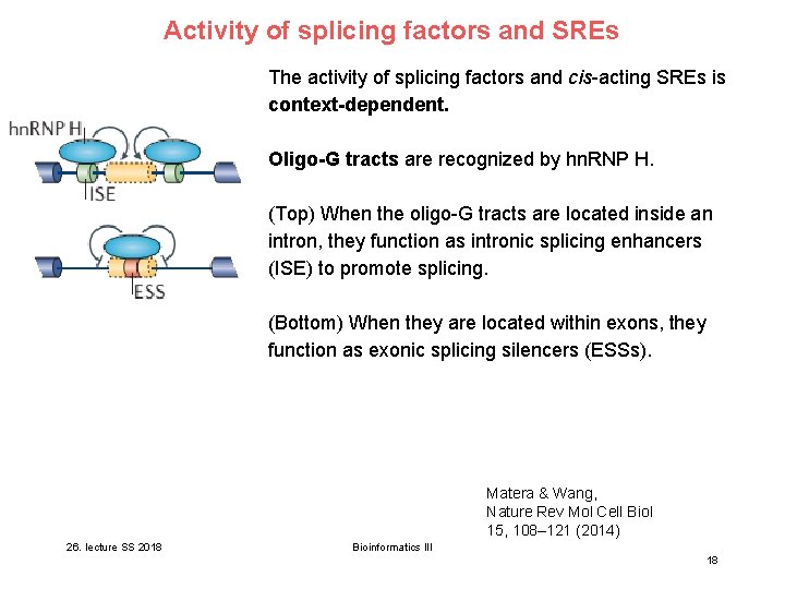
Activity of splicing factors and SREs The activity of splicing factors and cis-acting SREs is context-dependent. Oligo-G tracts are recognized by hn. RNP H. (Top) When the oligo-G tracts are located inside an intron, they function as intronic splicing enhancers (ISE) to promote splicing. (Bottom) When they are located within exons, they function as exonic splicing silencers (ESSs). Matera & Wang, Nature Rev Mol Cell Biol 15, 108– 121 (2014) 26. lecture SS 2018 Bioinformatics III 18
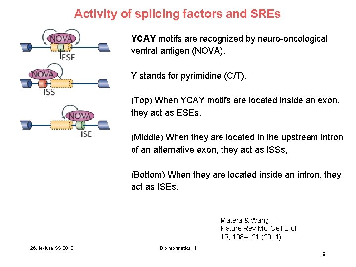
Activity of splicing factors and SREs YCAY motifs are recognized by neuro-oncological ventral antigen (NOVA). Y stands for pyrimidine (C/T). (Top) When YCAY motifs are located inside an exon, they act as ESEs, (Middle) When they are located in the upstream intron of an alternative exon, they act as ISSs, (Bottom) When they are located inside an intron, they act as ISEs. Matera & Wang, Nature Rev Mol Cell Biol 15, 108– 121 (2014) 26. lecture SS 2018 Bioinformatics III 19
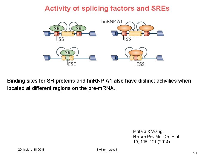
Activity of splicing factors and SREs Binding sites for SR proteins and hn. RNP A 1 also have distinct activities when located at different regions on the pre-m. RNA. Matera & Wang, Nature Rev Mol Cell Biol 15, 108– 121 (2014) 26. lecture SS 2018 Bioinformatics III 20
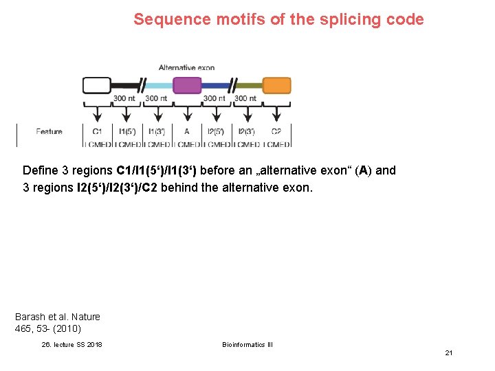
Sequence motifs of the splicing code Define 3 regions C 1/I 1(5‘)/I 1(3‘) before an „alternative exon“ (A) and 3 regions I 2(5‘)/I 2(3‘)/C 2 behind the alternative exon. Barash et al. Nature 465, 53 - (2010) 26. lecture SS 2018 Bioinformatics III 21
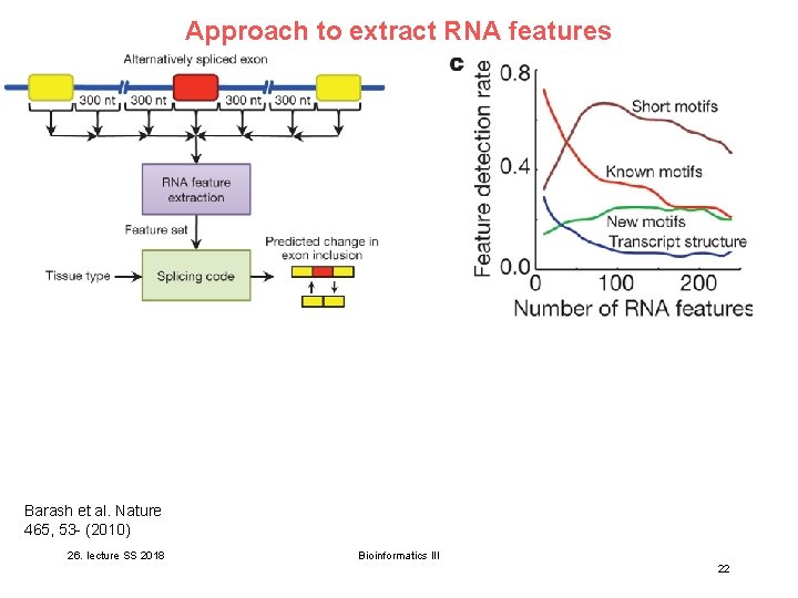
Approach to extract RNA features Barash et al. Nature 465, 53 - (2010) 26. lecture SS 2018 Bioinformatics III 22
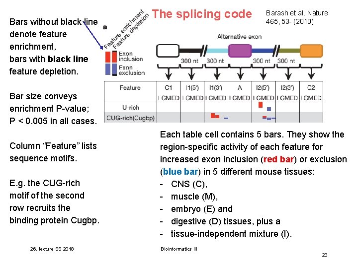
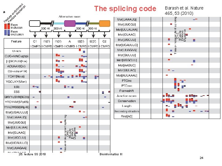
Bars without black line denote feature enrichment, bars with black line feature depletion. The splicing code Barash et al. Nature 465, 53 - (2010) Bar size conveys enrichment P-value; P < 0. 005 in all cases. Column “Feature” lists sequence motifs. E. g. the CUG-rich motif of the second row recruits the binding protein Cugbp. 26. lecture SS 2018 Each table cell contains 5 bars. They show the region-specific activity of each feature for increased exon inclusion (red bar) or exclusion (blue bar) in 5 different mouse tissues: - CNS (C), - muscle (M), - embryo (E) and - digestive (D) tissues, plus a - tissue-independent mixture (I). Bioinformatics III 23
The splicing code 26. lecture SS 2018 Barash et al. Nature 465, 53 (2010) Bioinformatics III 24
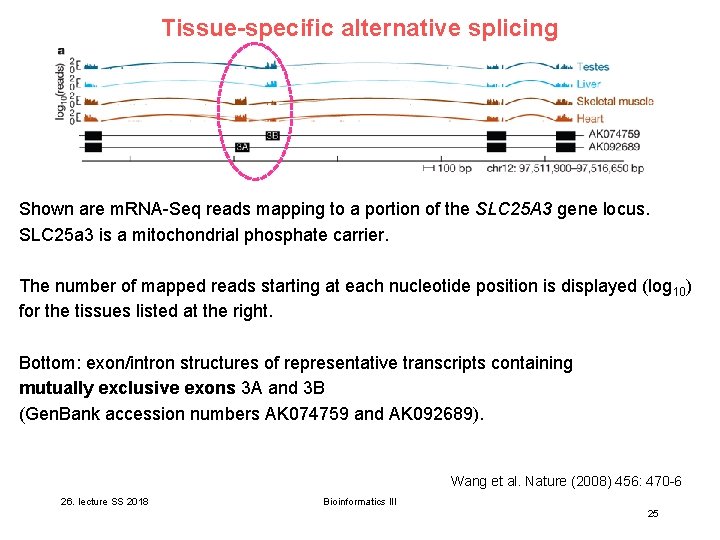
Tissue-specific alternative splicing are micro. RNAs? Shown. What are m. RNA-Seq reads mapping to a portion of the SLC 25 A 3 gene locus. SLC 25 a 3 is a mitochondrial phosphate carrier. How can one identify micro. RNAs? The number of mapped reads starting at each nucleotide position is displayed (log 10) is the function of micro. RNAs? for the. What tissues listed at the right. Bottom: exon/intron structures of representative transcripts containing mutually exclusive exons 3 A and 3 B (Gen. Bank accession numbers AK 074759 and AK 092689). Wang et al. Nature (2008) 456: 470 -6 26. lecture SS 2018 Bioinformatics III 25
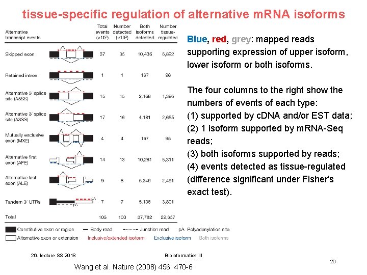
tissue-specific regulation of alternative m. RNA isoforms Blue, red, grey: mapped reads supporting expression of upper isoform, lower isoform or both isoforms. The four columns to the right show the numbers of events of each type: What are micro. RNAs? (1) supported by c. DNA and/or EST data; How can one identify micro. RNAs? (2) 1 isoform supported by m. RNA-Seq reads; What is the function of micro. RNAs? (3) both isoforms supported by reads; (4) events detected as tissue-regulated (difference significant under Fisher's exact test). 26. lecture SS 2018 Bioinformatics III Wang et al. Nature (2008) 456: 470 -6 26
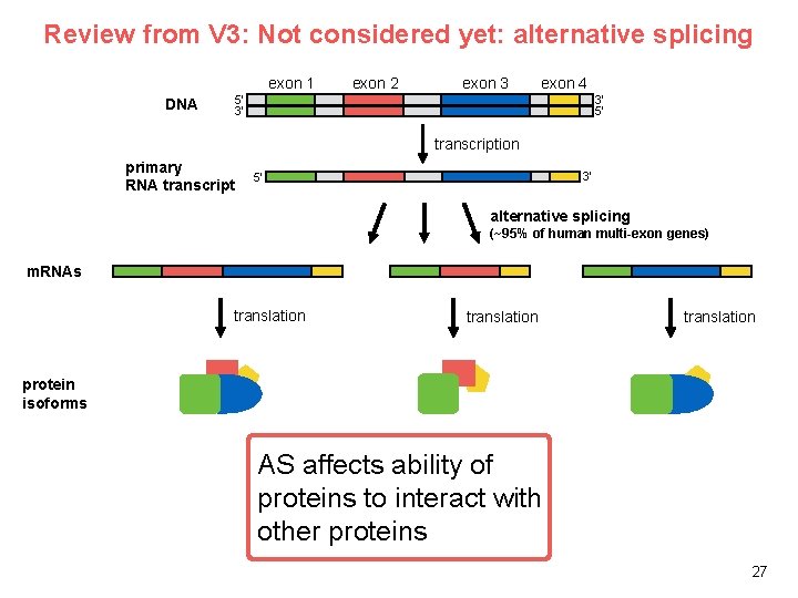
Review from V 3: Not considered yet: alternative splicing exon 1 DNA exon 2 exon 3 exon 4 3’ 5’ 5’ 3’ transcription primary RNA transcript 3’ 5’ alternative splicing (~95% of human multi-exon genes) m. RNAs translation protein isoforms AS affects ability of proteins to interact with other proteins 27
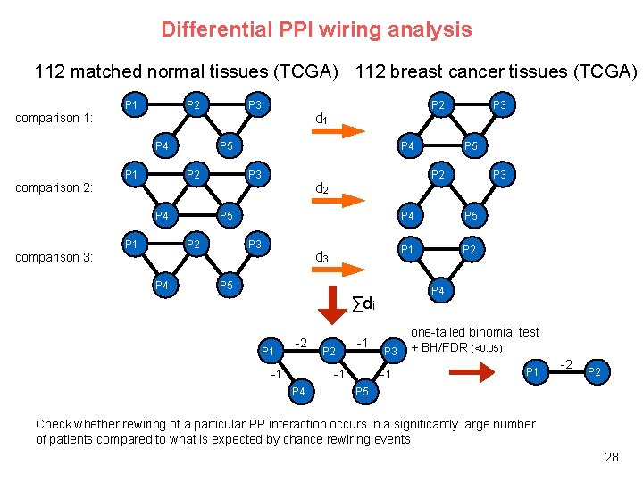
Differential PPI wiring analysis 112 matched normal tissues (TCGA) 112 breast cancer tissues (TCGA) P 1 P 2 P 3 d 1 comparison 1: P 4 comparison 2: P 1 P 5 P 2 P 4 comparison 3: P 1 P 4 P 3 d 2 P 3 d 3 P 5 P 1 -1 P 2 -1 -1 P 4 P 5 P 3 P 4 P 5 P 1 P 2 P 4 ∑di -2 P 3 P 2 P 5 P 2 P 4 P 2 P 3 -1 one-tailed binomial test + BH/FDR (<0. 05) P 1 -2 P 5 Check whether rewiring of a particular PP interaction occurs in a significantly large number of patients compared to what is expected by chance rewiring events. 28
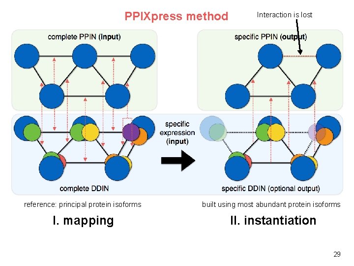
PPIXpress method Interaction is lost reference: principal protein isoforms built using most abundant protein isoforms I. mapping II. instantiation 29
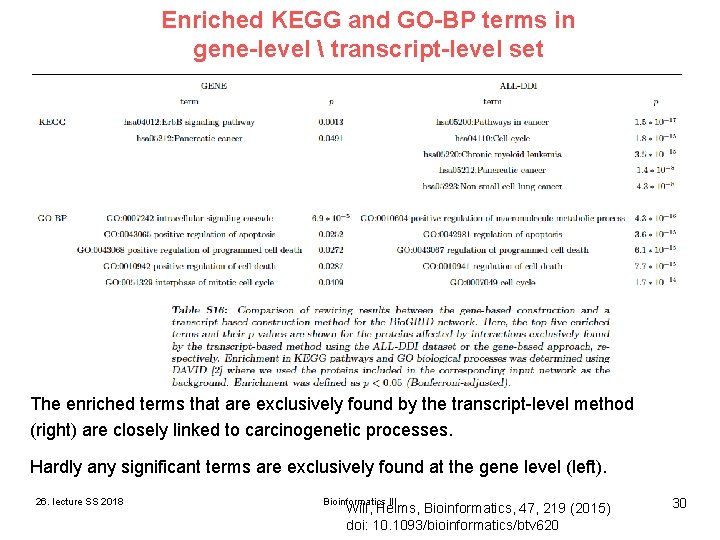
Enriched KEGG and GO-BP terms in gene-level transcript-level set The enriched terms that are exclusively found by the transcript-level method (right) are closely linked to carcinogenetic processes. Hardly any significant terms are exclusively found at the gene level (left). 26. lecture SS 2018 Bioinformatics III Will, Helms, Bioinformatics, 47, 219 (2015) doi: 10. 1093/bioinformatics/btv 620 30
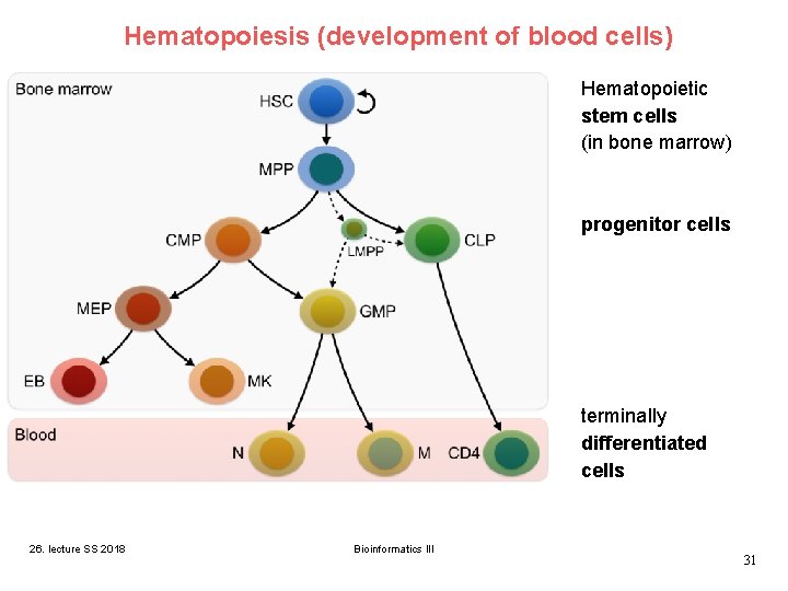
Hematopoiesis (development of blood cells) Hematopoietic stem cells (in bone marrow) progenitor cells terminally differentiated cells 26. lecture SS 2018 Bioinformatics III 31
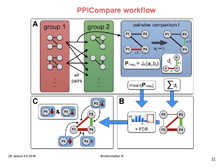
PPICompare workflow 26. lecture SS 2018 Bioinformatics III 32
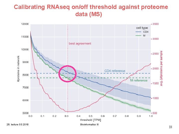
Calibrating RNAseq on/off threshold against proteome data (MS) 26. lecture SS 2018 Bioinformatics III 33
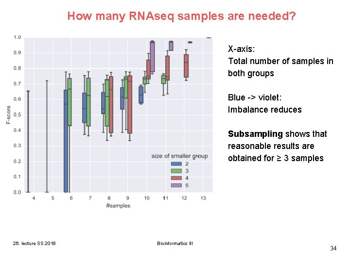
How many RNAseq samples are needed? X-axis: Total number of samples in both groups Blue -> violet: Imbalance reduces Subsampling shows that reasonable results are obtained for ≥ 3 samples 26. lecture SS 2018 Bioinformatics III 34
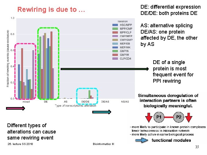
DE: differential expression DE/DE: both proteins DE Rewiring is due to … AS: alternative splicing DE/AS: one protein affected by DE, the other by AS DE of a single protein is most frequent event for PPI rewiring Different types of alterations can cause same rewiring event 26. lecture SS 2018 Bioinformatics III 35
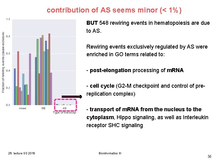
contribution of AS seems minor (< 1%) BUT 548 rewiring events in hematopoiesis are due to AS. Rewiring events exclusively regulated by AS were enriched in GO terms related to: - post-elongation processing of m. RNA - cell cycle (G 2 -M checkpoint and control of prereplication complex) - transport of m. RNA from the nucleus to the cytoplasm, Hippo signaling, as well as Interleukin receptor SHC signaling 26. lecture SS 2018 Bioinformatics III 36
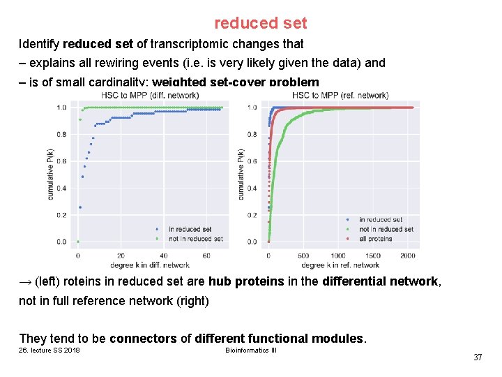
reduced set Identify reduced set of transcriptomic changes that – explains all rewiring events (i. e. is very likely given the data) and – is of small cardinality: weighted set-cover problem → (left) roteins in reduced set are hub proteins in the differential network, not in full reference network (right) They tend to be connectors of different functional modules. 26. lecture SS 2018 Bioinformatics III 37
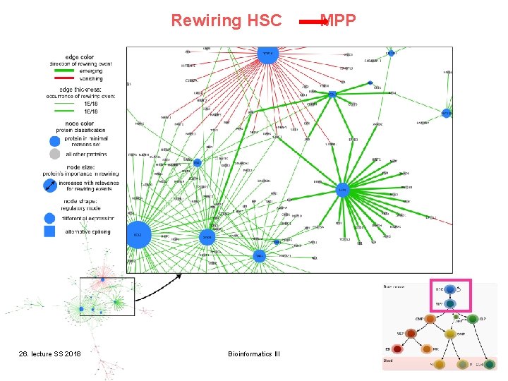
Rewiring HSC 26. lecture SS 2018 Bioinformatics III MPP 38
involvement of epigenetics in alternative splicing? V 21 – V 23 showed that chromatin state plays an essential role in regulating gene expression. Although epigenetic signatures are mainly found to be enriched in promoters, it has become increasingly clear that they are also present in exon regions, indicating a potential link of epigenetic regulation to splicing. 26. lecture SS 2018 Bioinformatics III 39
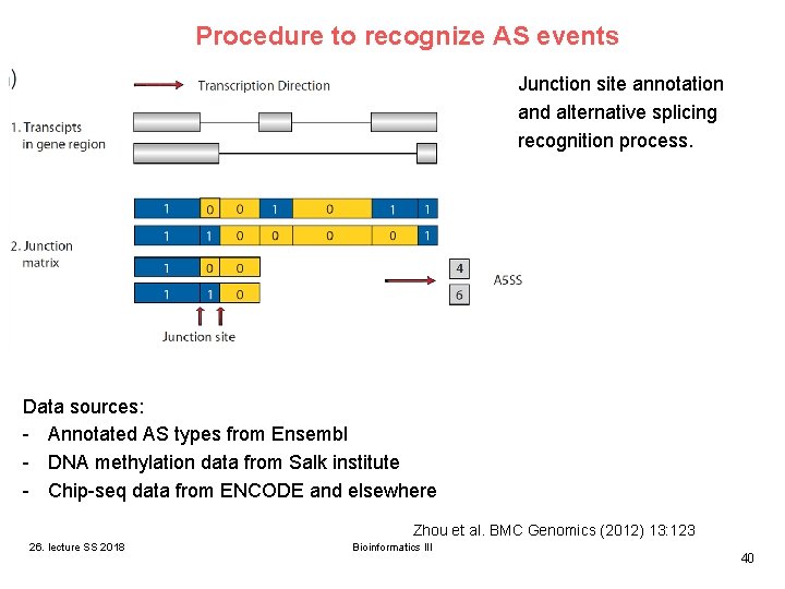
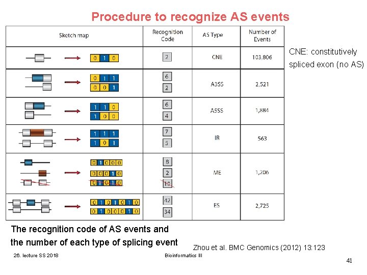
Procedure to recognize AS events Junction site annotation and alternative splicing recognition process. Data sources: - Annotated AS types from Ensembl - DNA methylation data from Salk institute - Chip-seq data from ENCODE and elsewhere Zhou et al. BMC Genomics (2012) 13: 123 26. lecture SS 2018 Bioinformatics III 40
Procedure to recognize AS events CNE: constitutively spliced exon (no AS) The recognition code of AS events and the number of each type of splicing event 26. lecture SS 2018 Zhou et al. BMC Genomics (2012) 13: 123 Bioinformatics III 41
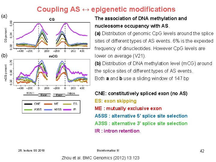
Coupling AS ↔ epigenetic modifications The association of DNA methylation and nucleosome occupancy with AS. (a) Distribution of genomic Cp. G levels around the splice sites of different types of AS events. 6% is the expexted frequency of dinucleotides. However Cp. G levels are lower on average (V 21). (b) Distribution of DNA methylation level (m. CG) around the splice sites of different types of AS events. Both a and b use a sliding window of 147 bp CNE: constitutively spliced exon (no AS) ES: exon skipping ME : mutually exclusive exon A 5 SS : alternative 5' splice site selection A 3 SS : alternative 3' splice site selection IR : intron retention. 26. lecture SS 2018 Bioinformatics III Zhou et al. BMC Genomics (2012) 13: 123 42
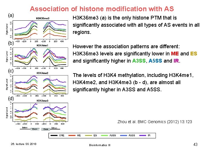
Association of histone modification with AS H 3 K 36 me 3 (a) is the only histone PTM that is significantly associated with all types of AS events in all regions. However the association patterns are different: H 3 K 36 me 3 levels are significantly lower in ME and ES and significantly higher in A 3 SS, A 5 SS and IR. The levels of H 3 K 4 methylation, including H 3 K 4 me 1, H 3 K 4 me 2, and H 3 K 4 me 3 (b - d), are almost all significantly higher in A 3 SS and A 5 SS. Zhou et al. BMC Genomics (2012) 13: 123 26. lecture SS 2018 Bioinformatics III 43
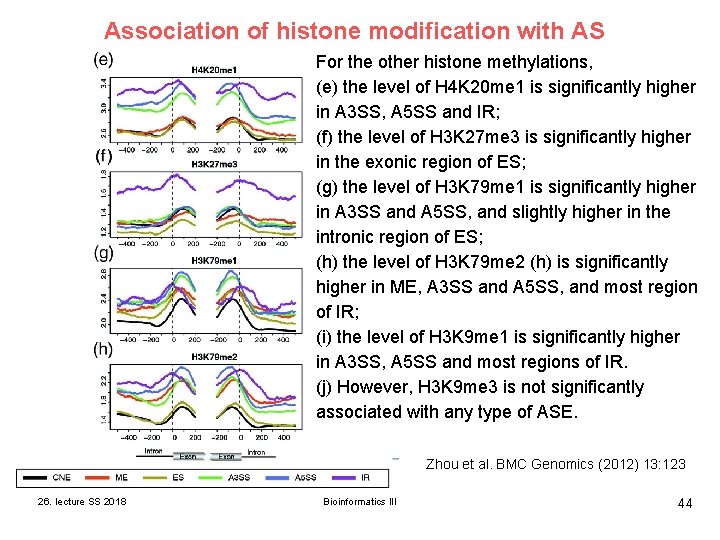
Association of histone modification with AS For the other histone methylations, (e) the level of H 4 K 20 me 1 is significantly higher in A 3 SS, A 5 SS and IR; (f) the level of H 3 K 27 me 3 is significantly higher in the exonic region of ES; (g) the level of H 3 K 79 me 1 is significantly higher in A 3 SS and A 5 SS, and slightly higher in the intronic region of ES; (h) the level of H 3 K 79 me 2 (h) is significantly higher in ME, A 3 SS and A 5 SS, and most region of IR; (i) the level of H 3 K 9 me 1 is significantly higher in A 3 SS, A 5 SS and most regions of IR. (j) However, H 3 K 9 me 3 is not significantly associated with any type of ASE. Zhou et al. BMC Genomics (2012) 13: 123 26. lecture SS 2018 Bioinformatics III 44
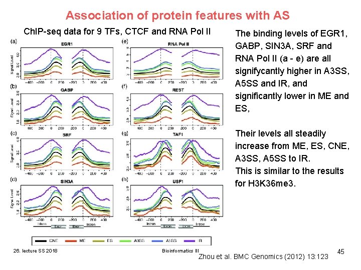
Association of protein features with AS Ch. IP-seq data for 9 TFs, CTCF and RNA Pol II The binding levels of EGR 1, GABP, SIN 3 A, SRF and RNA Pol II (a - e) are all signifycantly higher in A 3 SS, A 5 SS and IR, and significantly lower in ME and ES, Their levels all steadily increase from ME, ES, CNE, A 3 SS, A 5 SS to IR. This is similar to the results for H 3 K 36 me 3. 26. lecture SS 2018 Bioinformatics III Zhou et al. BMC Genomics (2012) 13: 123 45
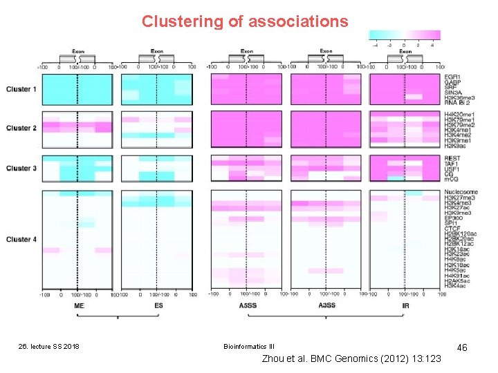
Clustering of associations Ch. IP-seq data for TF binding. 26. lecture SS 2018 Bioinformatics III Zhou et al. BMC Genomics (2012) 13: 123 46
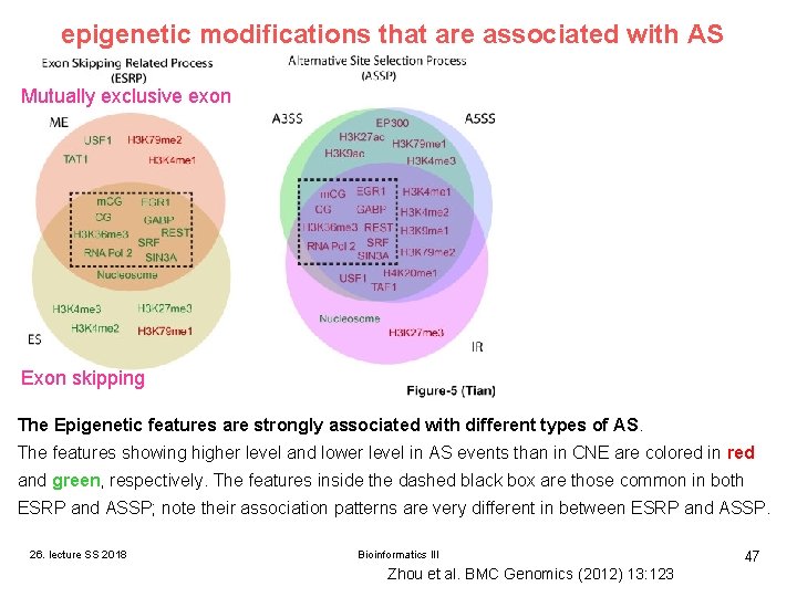
epigenetic modifications that are associated with AS Mutually exclusive exon Exon skipping The Epigenetic features are strongly associated with different types of AS. The features showing higher level and lower level in AS events than in CNE are colored in red and green, respectively. The features inside the dashed black box are those common in both ESRP and ASSP; note their association patterns are very different in between ESRP and ASSP. 26. lecture SS 2018 Bioinformatics III Zhou et al. BMC Genomics (2012) 13: 123 47
Coupling AS ↔ epigenetic modifications Epigenetic features are strongly associated with AS. This suggests that epigenetic regulation may be involved in AS. Clustering yielded 4 tight clusters of epigenetic features that are associated with AS. The AS events may be grouped into 2 classes on the basis of their association patterns with epigenetic features. - the exon skipping related process (ESRP) (including ME and ES) and - the alternative splice site selection process (ASSP) (including A 3 SS, A 5 SS and IR) → these 2 processes may involve different mechanisms of epigenetic regulation. 26. lecture SS 2018 Bioinformatics III Zhou et al. BMC Genomics (2012) 13: 123 48