Uveitis Prof Faiz Shakarchi Uveitis Uvea Iris Ciliary


 Toxoplasmosis 52 (16. 4%) Presumed Ocular Tuberculosis 28 (8.")
,")

 Endothelium dusting -Granulomatous uveitis:")
 Ocular 1 - Laterality")

 - Circum-corneal congestion -")
")


 -hypopyon (aggregation")
 Irregular, posterior synechiae (adhesion between the iris and the lens),")
. Iris nodules granuloma (granulomatous uveitis)")
: may be normal, elevated or depressed")


.")


 Multi-systemic auto-immune disorder affect pigmented cells in the")
 Systemic manifestations: Acute - Neurological and auditory manifestations: Headache, Tinnitus")
 - Bilateral, chronic, panuveitis Acute stage: non-granulomatous Multifocal serous detachment")


- Slides: 48
Uveitis Prof. Faiz Shakarchi
Uveitis
Uvea Iris Ciliary body Choroid
Iris • • Posterior pigment epithelium Stroma Sphincter pupillae muscle Dilator pupilae muscle • • Function: Regulates amount of light enters the eye
Ciliary body Anatomy • Pars Plicata- ciliary processes • Pars Plana Histology • Posterior epithelium • Stroma Ciliary muscles Functions: 1 - Secretion of aqueous humor 2 - Accommodation
Choroid Vascular layer Highest O 2 tension in the body • Supplies O 2 and nutrient to the photoreceptors • Thermo-regulator
Uveitis Inflammation of the uveal tract
Uveitis is a sight threatening disorder, and may be associated with life threatening diseases. Uveitis can be caused by various ocular or systemic infectious, immunological, and malignant diseases.
Etiology: - Infections: Viral; CMV, Herpes virus Bacteria; T. B. , T. pallidum, M. leprae Fungal; candidia Parasite; toxoplasmosis. toxocara -Non-infectious: Systemic: Arthrits; Ankylosing spondylitis, Skin diseases: VKH, Behjet disease, Psoriasis C. N. S. disorders: Multiple sclerosis Respiratory diseases; Sarcodosis: G. I. T. diseases: Ulcerative colitis Genitourinary diseases : Reiter’s disease Ocular: Specific ocular; Fuch’s hetrochromic iridiocyclitis Non-specific idiopathic
A- Infectious origin 92(28. 9%) Toxoplasmosis 52 (16. 4%) Presumed Ocular Tuberculosis 28 (8. 8%) Herpes simplex anterior uveitis 4 (1. 3%) Herpes zoster anterior uveitis 2 (0. 6%) Syphilis 2 (0. 6%) Acute retinal necrosis 2 (0. 6%) Presumed toxocarasis 1 (0. 3%) CMV retinitis 1 (0. 3 %) B-Non-infectious 118 (37. 1%) -Systemic diseases 74 (23. 3%) VKH 39 (12. 3%) Behçet's disease 26 (8. 2%) Ankylosing sponylitis 3 (0. 9%) Juvenile idiopathic arthritis 2 (0. 6%) Multiple sclerosis 2 (0. 6%) Antiphospholipid Syndrome 1 (0. 3%) Sarcodosis 1 (0. 3%) -Primary specific ocular disorders 44 (13. 8%) Pars planitis 18 (5. 7%) Punctate inner choroidopathy 5 (1. 6%) Fuchs heterochromic iridocyclitis 5 (1. 6%) Eales disease 4 (1. 3%) Bird shot retinochoroidopathy 3 (0. 9 %) Primary retinal vasculitis 3 (0. 9%) Serpiginous choroidopathy 2 (0. 6%) APMPPE* 2 (0. 6%) Antiphospholipid syndrome 1 (0. 3%) Sympathetic ophthalmitis 1 (0. 3%) Non-specific idiopathic 108 (34%) Total 318
• Anatomical: Classification: - Anterior uveitis 75% : inflammation of the iris (iritis), and anterior part of the ciliary body (iridocyclitis). - Intermediate uveitis: inflammation of the ciliary body (cyclitis, pars planatis). - Posterior uveitis : inflammation behind ora serrata; inflammation of the choroid and retina (choroiditis, retinitis, chorio-retinitis, retinal vasculitis). - Pan uveitis: inflammation of the entire uveal tract.
Classification • Clinical: - Acute: sudden, short duration - Chronic: insidious, long duration, sometimes with exacerbation and remission - Recurrent: repeated episodes of uveitis separated by periods of inactivity without treatment.
Classification • Pathological classification: -Non granulomatous Small keratic precipitate (KP) Endothelium dusting -Granulomatous uveitis: Large KP (muttan fat) or median size KP Iris nodules Iris granuloma
Diagnosis of uveitis depends on: 1 -Clinical features (Main stone) Ocular 1 - Laterality and Coarse; acute or chronic, recurrent 2 - Anatomical site; Anterior, intermediate, posterior and panuveitis 3 - Type; Granulomatous or non granulomatous Systemic manifestations 2 –Supported by selected lab investigations according to the probable causes.
Anterior uveitis Symptoms: - Pain - Photophobia - Redness - Lacrimation - Blurring of vision
Anterior uveitis Signs - Reduced visual acuity (V. A. ) - Circum-corneal congestion - Cornea: Keratic precipitate. KP: (aggregation of inflammatory cells on the posterior surface of the endothelium) small , endothelium dusting (non-granulomatous uveitis) Large (muttan fat) (chronic, granulomatous uveitis) - Ant. Chamber: cells and flare ( increase protein in the aqueous) hypopyon (aggregation of inflammatory cells at the bottom of anterior chamber with fluid level) - Pupil: Miosed (constricted) Irregular, posterior synechiae (adhesion between the iris and the lens), - Iris: Rubeosis ( iris neo-vascularization). Iris atrophy (chronic) Iris nodules and granuloma (chronic, granulomatous uveitis) - Intra-ocular pressure (IOP): may be normal, elevated or depressed
Anterior uveitis Signs - Reduced visual acuity (V. A. )
Signs -Circum-corneal congestion
Signs -Cornea: Keratic precipitate. KP: aggregation of inflammatory cells on the posterior surface of the endothelium) Small , endothelium dusting (non-granulomatous uveitis) Large (muttan fat) (chronic, granulomatous uveitis) Band keratopathy: deposition of calcium at Bowmann’s layer
Signs -Ant. Chamber: - cells -flare ( increase protein in the aqueous) -hypopyon (aggregation ofinflammatory cells at the bottom of anterior chamber with fluid level)
Signs -Pupil: Miosed (constricted) Irregular, posterior synechiae (adhesion between the iris and the lens),
Signs -Iris: Iris atrophy Rubeosis ( iris neovascularization). Iris nodules granuloma (granulomatous uveitis)
Signs -Intra-ocular pressure (IOP): may be normal, elevated or depressed
Ankylosing Spondylitits Idiopathic , chronic arthritis Young adults Male are affected more Arthritis; axial skeleton -sacroiliac joint and intervertebral joints 90% positive HLA-B 27 25% of patients with AS have uveitis Anterior Uveitis Acute, Recurrent, Non-granulomatous
Juvenile idiopathic arthritis Idiopathic, Chronic arthritis Children under 16 years. Females are affected more commonly Pauciarticular onset (less than 5 joints are involved) Polyarticular onset Still’s disease 80% positive for ANA sero-negative (for R. F. ) 20% of children with pauciarticulae type have uveitis Bilateral, Chronic, non-granulomatous Ant. Uveitis painless, Complications: 1 - Cataract 2 - Band keratopathy .
• Intermediate uveitis: inflammation of the ciliary body (cyclitis, pars planatis).
Intermediate uveitis • Symptoms - Floaters (moving shadows in the field of vision caused by vitreous opacities) - Blurring of vision
Intermediate Uveitis Signs: • Vitreous: cells, snow-balls snow banking
Intermediate Uveitis • Causes of intermidiate uveitis 1 - Pars planatis: Most common Idiopathic 2 - TB 3 - Multiple sclerosis
Posterior uveitis: inflammation behind ora serrata, inflammation of the choroid and retina - choroiditis, - retinitis, - chorio-retinitis, - retinal vasculitis .
Posterior Uveitis • Symptoms - Floaters (moving shadows in the field of vision caused by vitreous opacities) - Blurring of vision
Posterior uveitis Signs: • Vitreous: cells, flare and opacities • fundus lesions may be focal, multi-focal, or geographical lesions -Choroiditis; deep, yellow, well demarcated patches -Retinitis; superficial, white cloudy patches with indistinct margins -Old inactive lesion appears as white well defined area of chorio- retinal atrophy with pigmented borders -Vasculitis; acute: vascular cuffing: fluffy haziness surrounding blood columns chronic: vascular sheathening: fluffy haziness surrounding blood columns
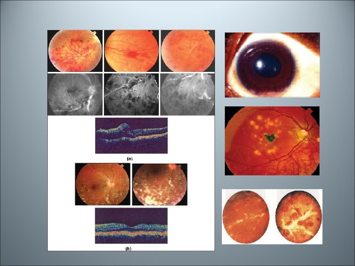
Toxoplasmosis • Toxoplasma gondii is an obligatory, intracellular protozoan parasite Acquired, congenital Vitritis. Retinitis; Active lesion: creamy-white lesion with indistinct margins Inactive lesion: white well defined area of chorio- retinal atrophy with pigmented borders
Toxoplasmosis Treatment: Antiprotozoal drugs; Clindamycin, sulphonamides Steroids
- Pan uveitis: inflammation of the entire uveal tract.
Behçet syndrome Idiopathic, multisystem disease characterized by recurrent episodes of oro -genital ulceration, uveitis and vasculitis
Behçet syndrome • Diagnosis criteia: According to the international study group for Behjet's syndrome • 1 - Recurrent oral ulceration characterized by painful aphthous lesions that have recurred at least three times in a 12 -month period. 2 - Plus at least two of the following: • Recurrent genital ulceration • Ocular inflammation. • Skin lesions include erythema nodosum, folliculitis, acneiform nodules • Positive pathergy test, cutaneous hypersensitivity, which is characterized by the formation of a pustule after 24– 48 hours at the site of a sterile needle prick
Behjet’s disease , Ocular features; Bilateral, chronic with exacerbation and remission, non- granulomatous pan-uveitis iridocyclitis Vitritis Retinitis Vasculitis; venous occlusion, neovascularization ,
Behjet’s disease
Pan uveitis Vogt Koyanagi Harada (VKH) Multi-systemic auto-immune disorder affect pigmented cells in the body. Involves CNS, eyes, and skin
Vogt Koyanagi Harada (VKH) Systemic manifestations: Acute - Neurological and auditory manifestations: Headache, Tinnitus Chronic- Integumentary findings: followed central nervous system and ocular manifestations alopecia, poliosis and vitiligo
Vogt Koyanagi Harada (VKH) - Bilateral, chronic, panuveitis Acute stage: non-granulomatous Multifocal serous detachment of sensory retina Disc swelling Bilateral exudative retinal detachment Chronic stage: granulomatous Depigmentation of the fundus; Vitilligo of the fundus Dalen Fuchs atrophic spots
Presumed Tuberculous Uveitis According to WHO: About one third of the world's population, are infected by tuberculosis 10% of infected people are symptomatic 90% have latent TB TB uveitis develops following hematogenous spread from a primary latent focus and usually occurs without evidence of systemic TB. TB is endemic in Iraq
Presumed TB uveitis • Clinical signs: • Chronic, Granulomatous, Panuveitis -Iris nodules or granuloma - Focal, multifocal choroiditis, -choroidal granuloma, - Retinal vasculitis.
Management of Uveitis: 1 - Investigations: Aimed for determining the etiology. Indications: chronic, recurrent, and granulomatous anterior uveitis Intermidiate, posterior and pan-uveitis 2 -Steriods: Topical, Side effects of corticosteroids eye-drops: • Flare up of pre-existing eye infection • Predispose for microbial keratitis, e. g. viral keratitis. • Inhibit collagen synthesis of the cornea, and predispose for corneal thinning • Cataract (chronic use) • Open angle glaucoma (chronic use) Periocular injections of steroids; used in severe ant. , Intermidiate, posterior uveitis Systemic steroids; used in; severe anterior, Intermidiate uveitis, posterior uveitis 3 - Mydriatics: -decrease pain by relieve ciliary muscles spasm - prevent synechiae, break down synechiae 4 - Anti-microbial drugs: in infectious types; Toxoplasmosis, Presumed TB uveitis. 5 - Immunomodulator and/or Immunoppressive agents: In bilateral, severe , vision threatening , steroid sparing, non-infectious -Cyclosporin; T-cell immunosuppressive agent -Methotrexate - Azathioprine
Red Eye Symptoms Conjunctivitis Keratitis or corneal foreign body Acute iritis Acute Glaucoma Vision Not affected depressed Redness + + Pain Foreign body sensation, itching Pain localized to the eye Severe pain radiating Secretion Watery, mucoid watery or purulent watery Photophobia absent marked mild Systemic Sometimes, e. g. noneadenovirus mild 50% associated Headache, with systemic nausea and disorders vomiting
Signs Conjunctivitis Keratitis or Acute iritis corneal foreign body Acute Glaucoma Congestion Conjunctival Cicumcorneal Cornea normal Ulceration or Suppuration Keratitic precipitates Oedema Anterior chamber normal Cells, Hypopyon Shallow Pupil normal Constricted, irregular Fixed, middilated Tension normal Normal High, Normal, or Low High