US Upper abdomen kidney spleen pancreas vessels bowel
 Suntharee moonrintah Radiologist NKP. Hospital 28")
US. Upper abdomen (kidney spleen pancreas vessels bowel) Suntharee moonrintah Radiologist NKP. Hospital 28 -6 -2060
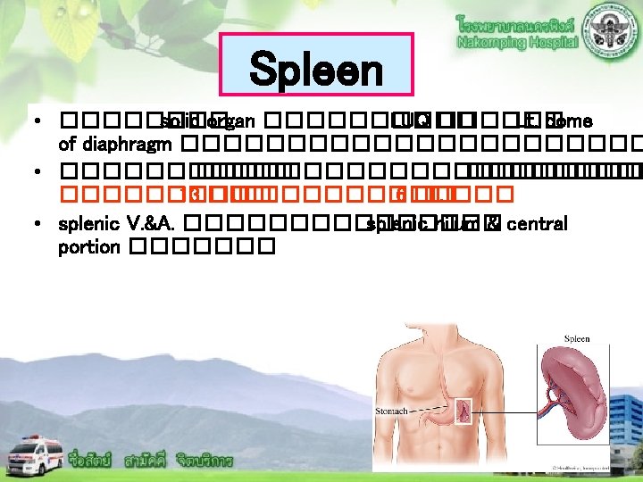

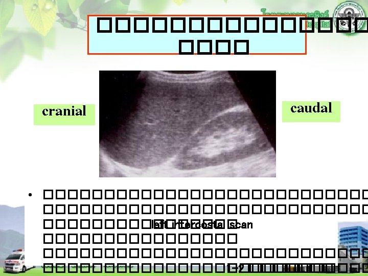
Spleen • ������� intercostal approach �������������� supine ���� Rt. Lateral decubitus ����� Lt. subcostal approach ����� splenomegaly

Spleen Splenomegaly Common cause v. Heart failure v. Portal hypertension v. Leukemia v. Lymphoma v. Hepatitis v. Mononucleosis v. Generalized infection v. Hemolytic anemia Uncommon cause v. Glycogen storage disease v. Malaria v. Myelofibrosis

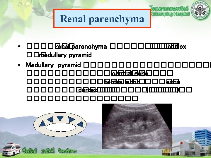
Splenomegaly Normal spleen: size ≤ 13 cm long, ≤ 6 cm thick
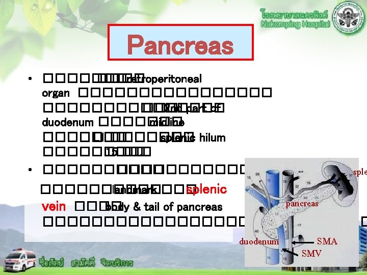

Pancreas • ������ head, neck, body, tail ��� uncinate process ���� head ������� IVC������ , body ������ vertebral body of L 1, tail ����������� spine ��� uncinate process ��������� head ����� inferomedial head body tail Splenic vein spine

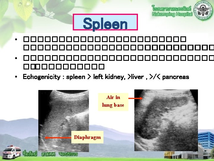
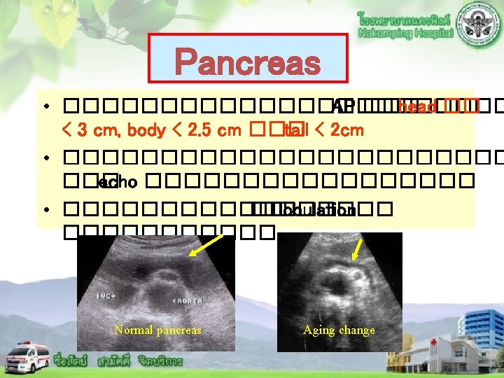
Pancreas • ������ ��echo ������������������ • ������ homogeneous ��echo ����������� echo. Normal ������� liver parenchymal echo Normal pancreatic parenchymal echo

Pancreatic duct ��������� body of pancreas ������� echogenic line ���� lucency between two echogenic lines ������������ 2 �� CBD

Acute pancreatitis v. More difficult to image , Sensitivity 3390% v. US for Ø evaluate biliary tract ; stone and coexistent liver disease Ø FU lesion or fluid collection Ø intervention procedure

Acute pancreatitis Most common cause : Alcohol abuse , gallstones US findings; v May be normal in mild case v Decreased or heterogeneous pancreatic echogenicity v Diffuse or focal enlargement of pancreas, ill-defined border v Peripancreatic fluid (in subtle pancreatic abnormality( v Perivascular fluid collection v Perirenal fluid collection v Right pleural effusion

Acute pancreatitis

Chronic pancreatitis

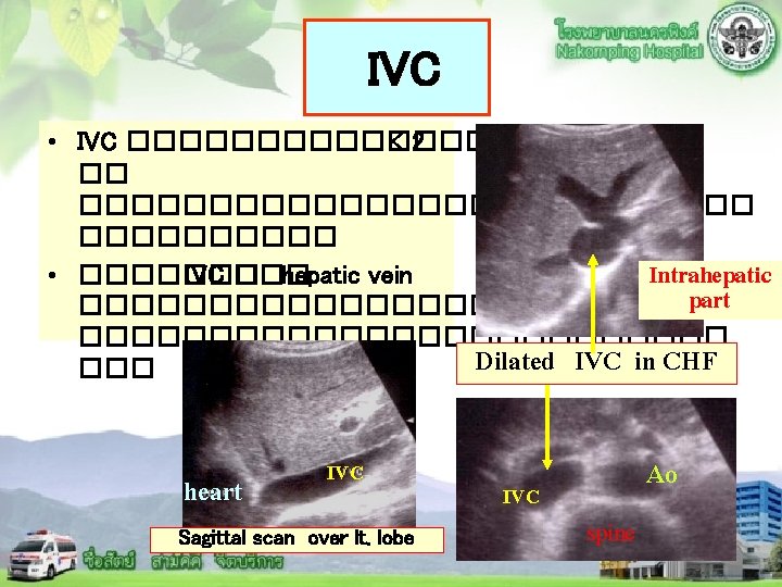
Abdominal vessels Hepatic v. IVC Aorta MPV Celiac axis Biliary tree Splenic v. SMV SMA
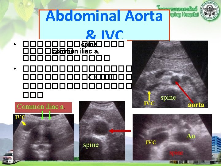

Abdominal Aorta ������������ spine ������ Distal part : ������ Proximal part

Abdominal vessels �������. 1 Celiac artery ��������������������� diaphragm 1 -2 ��. ������� common hepatic trunk ��� splenic artery ��������� Common hepatic a. Splenic a. Celiac trunk Ao Celiac origin Aorta

Abdominal vessels. 2 Superior mesenteric artery ������ aorta ������� celiac a. ������������� aorta Liver Lt. lobe SMA aorta Sagittal scan SMA aorta transverse scan

Abdominal vessels 3. Gastroduodenal artery ���� common hepatic trunk �������������� anterolateral Gastroduodenal a. Distal CBD pancreas Branches ���� aorta ���� IMA ��� renal artery ������

Abdominal vessels . 4 Splenic vessels �������� hilum ��� spleen splenic vein ��������� < 10 ��.

Abdominal vessels 5. Portal vein ��������� splenic vein ��� SMV �������������������� < 13 ��. Lt. PV MPV Oblique scan Rt. PV Rt. subcostal scan
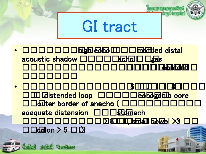

Stomach Liver : Lt. lobe stomach distended Stomach

Small Bowel in ascites Gas in bowel Fluid-filled bowel

Ascites

Lymphadenopathy v Discrete hypoechoic masses or anechoic masses without posterior enhancement, near the great vessels v nodal size criteria but normal nodal size could has disease

THANK YOU
- Slides: 38