URTICARIA ANGIOEDEMA Dr Neirita Hazarika Urticaria is characterized


 urticaria 2. Physical (inducible) urticarias")


 – transient, migratory 2. GI (20%)")

 of contact of urticant with")
 or in")


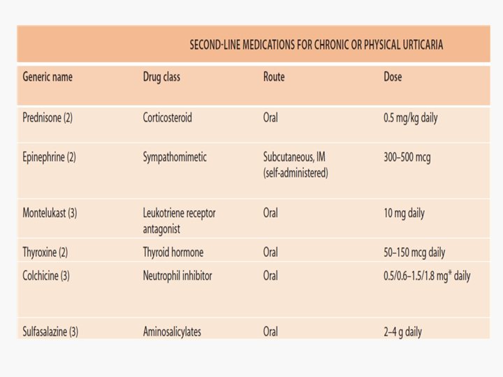
 • Oral corticosteroids • Leukotriene receptor antagonists •")

- Slides: 32
URTICARIA & ANGIOEDEMA Dr Neirita Hazarika
• Urticaria is characterized by transient skin or mucosal swellings due to plasma leakage. • Superficial dermal swellings are wheals • Deep swellings of the skin or mucosa are angioedema
WHEALS • Pruritic, pink/ red/pale swellings of the superficial dermis • ± Initial flare • Few millimeters to several centimeters • Number: few to numerous. • Hallmark : individual lesions come and go rapidly, by definition, in general within 24 hours.
ANGIOEDEMA • Swellings occur deeper in the dermis/subcutaneous/submucosal tissue. • May affect the mouth rarely, the bowel. • Involved areas : normal or faint pink in color, rather than red • Painful rather than itchy, • Larger and less well defined than wheals • Often last for 2 to 3 days
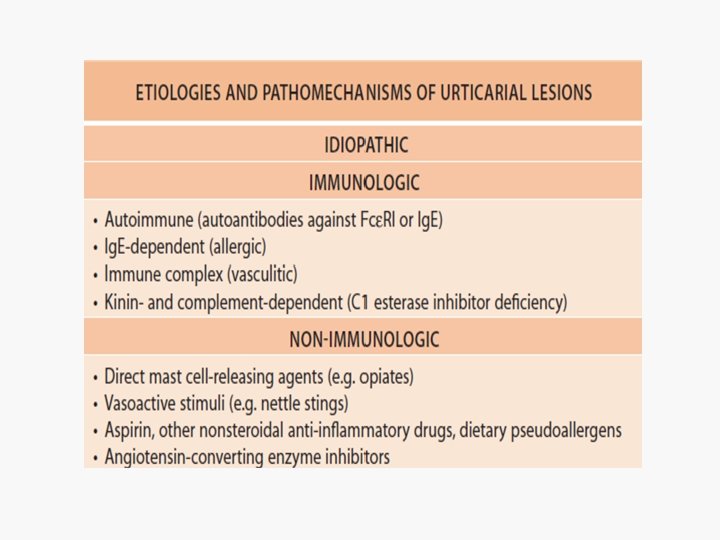
1. Classic immediate hypersensitivity binding of receptor-bound specific Ig. E by allergen. • Others: stimuli that act through the Ig. E receptor 2. anti-Ig. E and 3. anti-FcεRI antibodies 4. Non-immunologic stimuli: opiates, C 5 a anaphylatoxin, stem cell factor, some neuropeptides(e. g. substance P) • cause mast cell degranulation by binding specific receptors, independent of the FcεRI
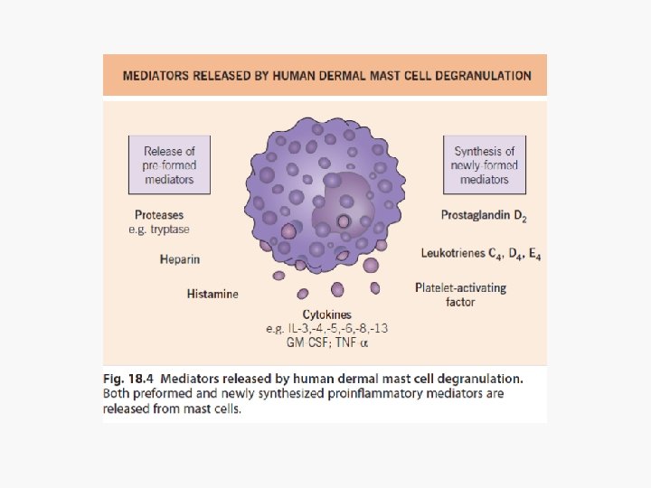
PATHOGENESIS • The mast cell is the primary effector cell of urticaria. • Degranulation: 1. Cross-linking of two or more adjacent FcεRI on the mast cell membrane 2. initiate a chain of calcium- and energy-dependent steps 3. fusion of storage granules with the cell membrane and externalization of their contents.
PATHOGENESIS • Basic pathology - ↑ capillary permeability, allowing proteins and fluids to extravasate to the dermis. 1. Histamine and other proinflammatory mediators released on degranulation » Bind receptors on postcapillary venules in the skin » Vasodilation and increased permeability to large plasma proteins (albumin and immunoglobulins). 2. Histamine, TNF-α and IL-8 upregulate adhesion molecules on endothelial cells, promoting the migration of inflammatory cells into the urticarial lesion.
CLINICAL CLASSIFICATION OF URTICARIA AND ANGIOEDEMA 1. “Ordinary” (spontaneous) urticaria 2. Physical (inducible) urticarias 3. Urticarial vasculitis (vasculitis on skin biopsy) 4. Contact urticaria (induced by percutaneous or mucosal penetration) 5. Angioedema without wheals 6. Distinctive urticarial syndromes
ACUTE vs CHRONIC URTICARIA • All urticarias are acute initially • “Chronic urticaria”: usually defined as 6 weeks or more. Applied to continuous urticaria occurring at least twice a week off treatment for ≥ 6 weeks • Urticaria occurring ≤ 6 weeks is called episodic / recurrent
ASSOCIATIONS OF CHRONIC URTICARIA • Autoimmune thyroid disorders • Vitiligo • Insulin dependent diabetes • Rheumatoid arthritis • Pernicious anemia • Helicobacter pylori gastritis • Intestinal strongyloidiasis(endemic countries) • ? ? ? Dental infections or gastrointestinal candidiasis
CLASSIFICATION OF PHYSICAL URTICARIA
CLASSIFICATION OF PHYSICAL URTICARIA
CLASSIFICATION OF PHYSICAL URTICARIA
URTICARIAL VASCULITIS 1. Favors middle-aged women 2. Urticarial lesions >24 hours in duration; painful & burning sensation as well as pruritus; 3. Residual purpura as they resolve 4. Sites: often occur at pressure points 5. Concurrent angioedema : up to 40% of pts 6. Disease course: average of 3 years
EXTRACUTANEOUS MANIFESTATIONS OF URTICARIAL VASCULITIS 1. Arthralgias (50%) – transient, migratory 2. GI (20%) – abdominal pain, nausea, vomiting, diarrhea 3. Pulmonary obstructive disease (20%) 4. Renal (5– 10%) – proteinuria, hematuria 5. Ocular (unusual) – conjunctivitis, episcleritis, uveitis 6. Others - Raynaud’s phenomenon, livedo reticularis, splenomegaly, lymphadenopathy, idiopathic intracranial HTN, pericardial or muscle involvement
ASSOCIATED DISORDERS OF URTICARIAL VASCULITIS • Systemic lupus erythematosus • Sjögren’s syndrome • Serum sickness • Cryoglobulinemia • Infections – hepatitis B or C virus, Epstein-Barr virus • Rarely, solar or cold urticaria, drugs, hypergammaglobulinemia
CONTACT URTICARIA • Development of urticaria at the site(s) of contact of urticant with skin or mucosa • Percutaneous or mucosal penetration of the urticant may have distant effects, including acute urticaria or even anaphylaxis • Immunologic and non-immunologic forms are recognized
CONTACT URTICARIA • Immunologic: sensitized to environmental allergens (grass, animals and foods) or in glove-wearers (latex). • Non immunologic: • Percutaneous microinjection of vasomediators (histamine, acetylcholine, serotonin) via nettle stings • contact with histamine liberators that degranulate mast cells (dimethylsulfoxide, cobalt chloride)
FOOD CONTACT HYPERSENSITIVITY SYNDROME • Itching and mild swelling of the mouth, tongue and soft palate within minutes of eating fresh fruits but not cooked fruit • apples, pears, peaches and cherries,
DIAGNOSIS • Comprehensive history • Duration of individual lesions, presence of purpura • Weals lasting more than 24– 48 h, particularly if painful or tender, suggest urticarial vasculitis • Frequency of attacks, duration of disease, previous treatment, known triggers • Past and family history, • Occupation and leisure activities,
DIAGNOSIS • Assessment of the impact of the disease on the patient’s quality of life. • Asso. angio‐oedema (eg. Oropharynx result in difficulty in swallowing or breathing) • Systemic symptoms • Recent acute infection, drugs, non‐prescription and prescription medicines, food
INVESTIGATIONS • Rule out Infections • Complete blood count • Stool for ova, cysts and parasites • Thyroid autoab, Thyroid function tests • C 4 complement (angio‐oedema without weals) • Non‐organ specific autoantibodies (eg ANA) • Basophil histamine release assay / basophil activation tests • Helicobacter pylori (stool antigen or urea breath test) • Chest X‐ray • 25‐hydroxycholecalciferol (vitamin D)
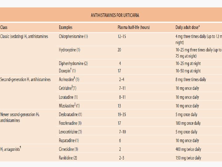
TREATMENT • Detection and avoidance of the cause. • First line therapies (H 1 antihistamines) minimal dosing which control episodes. H 1 – non sedating day time, sedating at night. A combination of an H 1 antihistamine with an H 2 antagonist may be more effective than H 1 antihistamines alone in some patients
TREATMENT Second line therapies (targeted therapy) • Oral corticosteroids • Leukotriene receptor antagonists • Doxepin, • Danazol • Sulphasalazine and dapsone
THANK YOU