Urogenital Tract 3 rd year Schistosomia hematobium Dr

Urogenital Tract / 3 rd year Schistosomia hematobium Dr Hamed Al-Zoubi MD, Ph. D Associate Prof. of Medical Microbiology

SCHISTOSOMIASIS BILHARIZIASIS

Is a human disease syndrome due to infestation by Schistosoma Most human schistosomiasis is caused by 1. Schistosoma haematobium discovered by Theodor Bilharz in Cairo in 1861 (mainly UTS). 2. Schistosoma mansoni (mainly GIT). 3. Schistosoma japonicum (mainly GIT).

EPIDEMIOLOGY

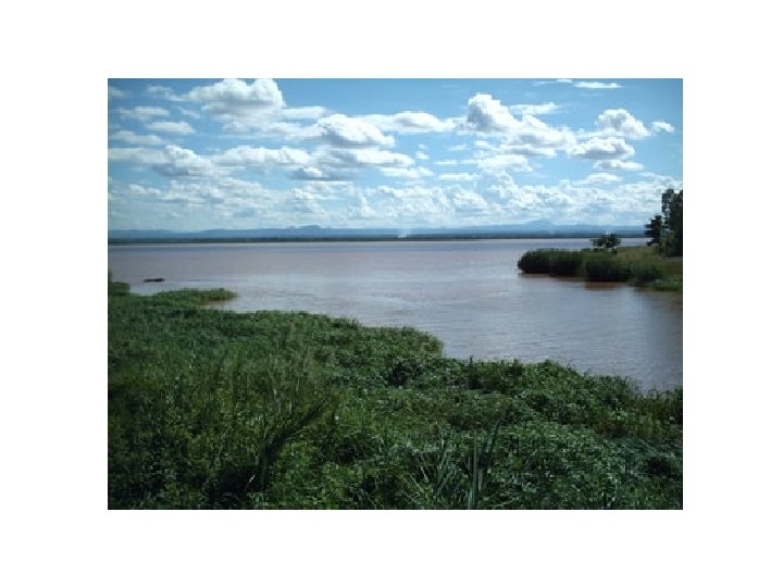
• Found in Africa in tropical & subtropical countries. • In Egypt it is prevalent all over the Nile valley. • In Asia, Syria, Palestine, Iraq& Yemen • Man is the only natural host for S haematobium • Habitat • Adult worm inhabit the bladder & pelvic venous plexuses rarely in the portal blood stream.

GLOBAL DISTRIBUTION

NILE RIVER

The spread continues because of a. water resource contamination by stool and urine in developing countries b. the migration of infected populations.

• It is estimated that than 200 million are infected all over the world & about 500 -600 million are exposed to infection. . • Disease prevalence tends to be worse in areas with poor sanitation, increased freshwater irrigation usage, and heavy schistosomal infestation of human and/or snail populations.

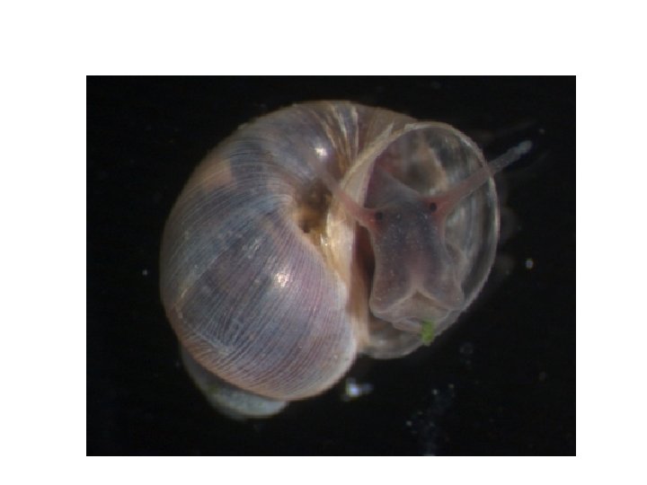
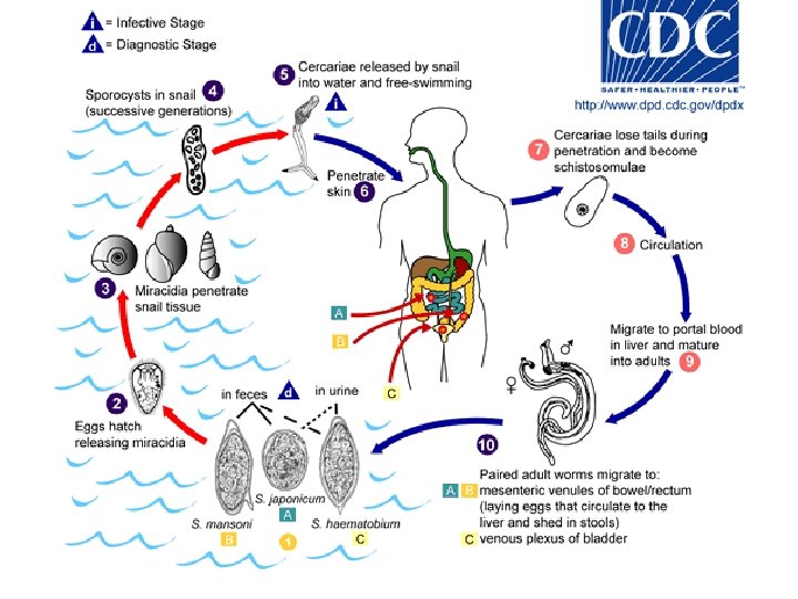
LIFE CYCLE • The ovum is passed in the urine or faeces of infected individuals and gains access to fresh water where the ciliated miracidium inside it is liberated; it enters its intermediate host, a species of freshwater snail, in which it multiplies. • Large numbers of fork-tailed cercariae are then liberated into the water, where they may survive for 2 -3 days.

• Cercariae can penetrate the skin or the mucous membrane of the mouth of their definitive host, humans. • They transform into schistosomulae and moult as they pass through the lungs and are carried by the blood stream to the liver and so to the portal vein where they mature. • Within 4 -6 weeks of infection they migrate to the venules draining the pelvic viscera, where the females deposit ova. •

LIFE CYCLE

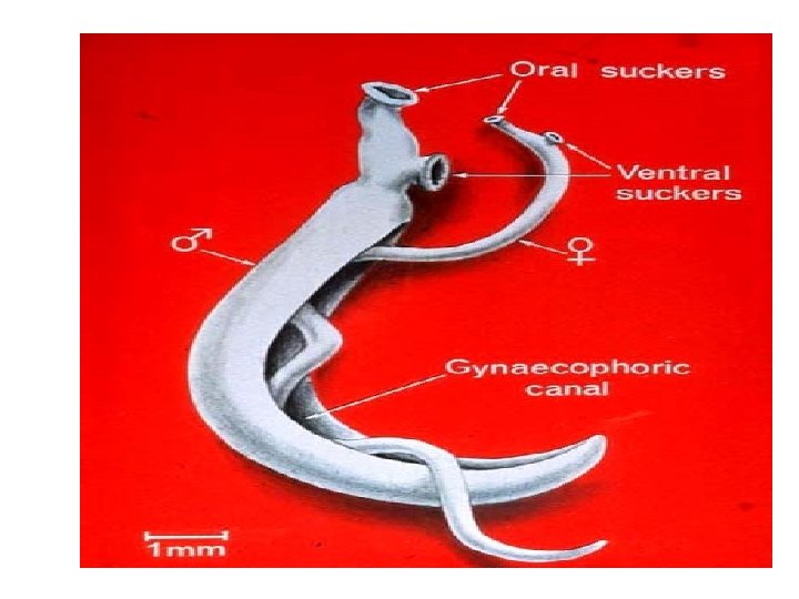
• Morphology • Adult male & female have oral sucker surrounding the mouth anteriorly & ventral Sucker on the ventral surface with which it attaches itself to the wall of the vessel in which it lives. • The male worm is flat, leaf like &folded to form the gynacophoric canal which enfolds the slender female for almost its entire length. • testes • ovary

PATHOLOGY • The pathological changes and symptoms depend on species and stage of infection. • Most of the disease is due to the passage of eggs through mucosa and to the granulomatous reaction to eggs deposited in tissues. • Granulomas are composed of macrophages, eosinophils, epithelioid and giant cells around an ovum. • The eggs of S. haematobium pass mainly through the wall of the bladder, but may also involve rectum, seminal vesicles, vagina, cervix and uterine tubes.

• Later there is fibrosis and eggs calcify, often in sufficient numbers to become radiologically visible. • Eggs of S. haematobium, and of the other two species after the development of portal hypertension, may reach the lungs.
 - During the early stages of infection")
Manifestation: Patients with acute schistosomiasis (Katayama fever) - During the early stages of infection there may be itching lasting 1 -2 days at the site of cercarial Penetration (swimmer itch). - After a symptom-free period of 3 -5 weeks, acute schistosomiasis (Katayama syndrome) may present with allergic manifestations such as urticaria, fever, muscle aches, abdominal pain, headaches, cough and sweating and HSM - Eosinophilia

- Other chronic symptoms may arise from 2 months to over two years after infection: - In light infection the symptoms are mild or non & the infection is detected on routine examination of urine. -Painless Terminal Haematuria is the first sign of infection. It may last for months or even years - Frequency & urgency of micturation are early symptoms. -Hydronephrosis & hydroureter and renal failure -Bladder changes -association of S. haematobium infection with squamous cell carcinoma of the bladder.

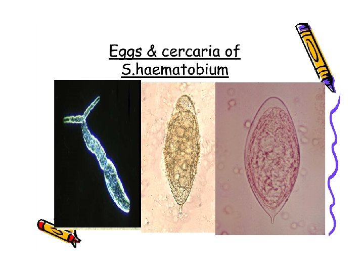
DIAGNOSIS 1. CLINICAL 2. HEMATOLOGICAL, BIOCHEMICAL 3. CONFIRMED BY Detection of ova in urine deposit of terminal stream (especially after exercise) or from biopsy from bladder wall. Ovum has sharp terminal spine in S. Hematobium 4. Serological tests ELISA for screening 5. Radiological and scope examination

SPECIFIC TREATMENT • Praziquantel 40 mg /kg for all types and as a single dose is treatment of choice. • Induce parasitological cure in 80% of people and over 90% reduction in egg count in the remaining individuals.

COMPLICATIONS OF SCHISTOSOMIASIS BILHARIZIASISIN GENERAL 1. Pulmonary hypertension 2. Cor pulmonale 3. Portal hypertension. 4. Obstructive uropathy. 5. Squamous cell carcinoma of the bladder. 6. Gastrointestinal bleeding 7. Neurological complications

PREVENTION Travelers to endemic areas should avoid exposure to freshwater that is likely to be contaminated. No accepted prophylactic regimens have been developed. No vaccines are currently available, although vaccine development is increasingly promising. Early treatment after high-risk exposures should minimize morbidity. Improved sanitation to decrease freshwater contamination with sewage should decrease disease prevalence.

The End Thank you
- Slides: 27