URINARY TRACT TRAUMA PROF DR METE KLCLER DEPARTMENT


 Ureter Injury Severity Scale Grade Injury")


- Slides: 84
URINARY TRACT TRAUMA PROF. DR. METE KİLCİLER DEPARTMENT OF UROLOGY, SCHOOL OF MEDICINE, BAHÇEŞEHİR UNIVERSITY
Renal Trauma
Renal Anatomy Retroperitoneal Upper poles protected by ribs so lower poles more commonly injured Right kidney inferior to left and more commonly injured
Renal Trauma Occurs in 8 -10% of patients with abdominal trauma Accounts for 50% of all GU trauma
Renal Trauma • 10% of abdominal injuries involve the kidneys • Renal trauma is the most common GU injury— 65% of GU injuries • Mechanism – 80 -95% due to blunt force— falls, assaults, sporting events
When are you concerned about renal injuries? • Mechanism of Injury – Penetrating injuries of abdomen, back or flank – Deceleration injuries • Physical exam – Tenderness of abdomen or flank – Echymosis of abdomen or flank • X-ray – Fractures of lower ribs, thoraco-lumbar spine
Mechanism of Injury - Blunt • Suspect some type of renal injury if fractures of the posterior ribs or lumbar vertebrae are present • Acceleration -Deceleration forces may cause damage to the renal vasculature
Mechanisms of Injury - Penetrating High velocity: Gun shot wound Low velocity: Knife wound
When are you concerned about renal injuries? • Hematuria—over 95% of patients with renal trauma DEGREE OF HEMATURIA DOES NOT CORRELATE WITH THE SEVERITY OF THE INJURY – 25% of patients with gross hematuria have minor injuries – 40% of the most serious renal injuries do not have any hematuria
Physical Assessment • • Inspection Palpation Gray Turner’s Sign Percussion
When is Imaging Indicated ? • • Penetrating trauma Pediatric trauma Deceleration injury Adult blunt trauma with hematuria
Imaging techniques • Contrast enhanced CT—the best test, up to 98% accurate, not great for renal vein injuries • IVP • Contrast Enhanced Ultrasound • MRI
Why is CT the Best Imaging Study? – Accurate staging – Non-invasive – Detects associated injuries – Rapid – Need contrast
AAST Kidney Injury Severity Scale American Association for the Surgery of Trauma
Grade I-Renal contusion
Grade I-Subcapsular Hematoma
Grade II-Small Cortical Laceration
Grade III-Major Renal Laceration
Grade IV-Major Laceration involving Collecting System
Grade IV- Multiple Renal Lacerations
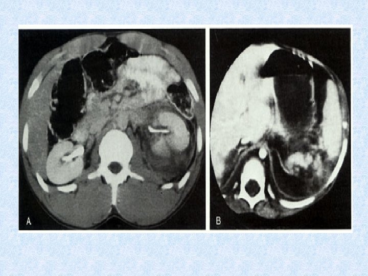
Grade IV-“Shattered” Kidney
Grade V- Avascular Left Kidney
Blunt Injury • Only 1 -2% of injuries involve the pedicle, • Renal injuries are more common, result from deceleration tend to be partial tears • Renal lacerations account for 2 -4% of all renal injuries, diagnosed by CT
Management of Renal Injuries • Grade I—home • Grade II-IV—admit, observe • Grade V—observe, vascular repair/stent, or nephrectomy absolute indications for surgery are: • persistent renal bleeding with hemodynamic instability, • active extravasation of IV contrast, • expanding or pulsatile perirenal hematoma • suggesting Grade V vascular injury
Blunt Injury • Surgical repair controversial • Minor renal lacerations/contusions managed expectantly
Penetrating Injuries • Hematuria is of no consequence as all patients need CT, most will need surgery
Complications of Renal Injuries • Mortality 3% • Complications – First six weeks • Hemorrhage/shock • Sepsis/abscess • ATN – Late • Renovascular HTN 4%
Renal Trauma Complications Major Trauma Abscess/urinomas Sepsis Decreased H/H Expanding perirenal mass Hemodynamic instability Sepsis Fistula Renal atrophy Rhabdomyolysis/myoglobinuria Renal HTN Renal Failure
Ureteral Trauma
Ureteral Anatomy • Thin, mobile tubes running between renal pelvis and posterior superior angle of bladder • Retroperitoneal in abdomen • Protected from injury by size and mobility
Ureteral Trauma • Accounts for 1% of urologic trauma • Penetrating (usually Gun Shot Wound) trauma to abd, back, flank • Most commonly iatrogenic following GU, gynecologic, vascular or colorectal surgery
Diagnosis/Treatment • Usually made by finding urine in surgical wounds/dressings or the development of a urinoma • Contrast CT or bolus IVP can Show the injury • Retrograde pyelography will aid in diagnosis • All injuries need surgical repair
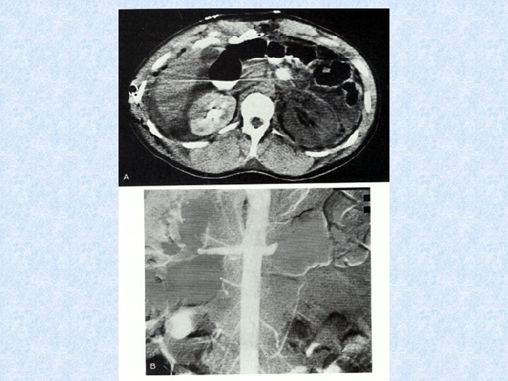
IMAGING FOR URETERAL INJURIES • Most injuries diagnosed during laparotomy • Contrast CT—most common findings are extravasation of contrast into medial perirenal space and absence of contrast in distal ureter if transected • IVP • Retrograde pyelogram
Delayed CT images showing extravasation of urine from ureteral injury
American Association for the Surgery of Trauma (AAST) Ureter Injury Severity Scale Grade Injury Description I Hematoma Contusion or hematoma without devascularization II Laceration < 50 % transection III Laceration > 50 % transection IV Laceration Complete transection with 2 cm devascularization V Laceration Avulsion of renal hilum which devascularizes kidney
Ureter Trauma Management • Ureterostomy • Irrigation and Drainage • Antibiotics • Stenting
Complications of Ureter Trauma Missed injuries usually manifest by Fever Flank mass or discomfort Ileus Leukocytosis Lethargy Urinary fistula to skin or vagina – Sepsis – Wound infection – – – Complications – Fistula – Stricture or ureteral obstruction – Retroperitoneal urinoma – Infection – Obstructive hydronephrosis
Urinary Bladder Trauma
Bladder Anatomy • Lies within pelvis when empty, can reach umbilicus when full • Consists of 3 muscle layers • Blood supplied from int. iliac artery, nerve supply from lumbar and sacral plexus • Bladder trauma usually associated with severe injuries, mortality 22 -44%
Urinary Bladder Anatomy • Empty bladder is a pelvic organ and protected by pelvic bones • With distention, becomes an abdominal organ and more prone to injury due to direct trauma • Peritoneum covers superior surface of bladder
Pathophysiology • Intraperitoneal rupture usually from blunt trauma in patients with a full bladder • Clinically will see lower abdominal pain, inability to urinate, blood at meatus
Pathophysiology • Can rupture in or outside of peritoneum, or both • Extraperitoneal rupture usually from pelvic fracture with laceration of bladder, but may occur with blunt trauma
Urinary Bladder Trauma • Mechanisms of Injury – Blunt—up to 85% of cases • 10% of patients with pelvic fractures will have bladder injuries – Penetrating—up to 15% of cases – Surgical/Cystoscopy
When are you concerned about a bladder injury? • Clinical Presentation – Suprapubic pain – Difficulty voiding • Hematuria • X-ray – Widened symphysis pubis is stongest predictor – Pelvic, sacrum, iliac, ramus fractures
Lab • Gross hematuria indicative of urologic injury • Clear urine and no pelvic fracture virtually eliminates possibility of bladder rupture • 98% of patients with bladder rupture have gross hematuria
Diagnostic Studies • Retrograde cystogram • Retrograde CT cystogram • Either one follows urethogram if concern for urethral injury exists
Indications for Cystography • Blunt Trauma in close proximity to bladder with gross hematuria • Pelvic fractures from blunt mechanism with any degree of hematuria • Penetrating Trauma in proximity to the bladder • Penetrating trauma with any degree of hematuria
Retrograde Cystogram • Place a foley catheter, fill the bladder with the contrast and than clamped and AP film taken • Then empty bladder and take post-evacuation film • If extraperitoneal perforation, will see contrast in area of pubic symphysis, intraperitoneal perforation will outline abdominal contents
Retrograde Cystogram--Normal
Retrograde Cystogram—Post-Void, Normal
Extraperitoneal Bladder Rupture • 50 -90% of bladder ruptures • Usually associated with pelvic fracture • Usually treated with urethral/suprapubic catheter
Retrograde Cystogram—Extraperitoneal Rupture
Retrograde Cystogram—Extraperitoneal Rupture
CT Cystogram—Extraperitoneal Rupture
CT Cystogram with Extraperitoneal Rupture
CT Cystogram with Extraperitoneal Rupture with Sagittal View
Intraperitoneal Bladder Rupture • 15 -35% of bladder ruptures • Bladder usually distended at time of trauma • Historically treated surgically • Conservative management possible
Retrograde Cystogram—Intraperitoneal Rupture
Retrograde Cystogram—Intraperitoneal Rupture
Bladder Injury Scale American Association for the Surgery of Trauma Grade I Injury Description Hematoma Contusion, intramural hematoma Laceration Partial thickness II Laceration Extraperitoneal bladder wall laceration < 2 cm III Laceration Extraperitoneal (> 2 cm) or intraperitoneal ( < 2 cm) bladder wall lacerations IV Laceration Intraperitoneal (> 2 cm) bladder wall lacerations V Laceration Intra or extraperitoneal bladder wall laceration extending into the bladder neck or urethral orifice (trigone)
Treatment • If no extravasation treat with or without Foley drainage • Extraperitoneal ruptures treated with Foley drainage for 7 to 15 days with 20 Fr. or greater sized catheter
Treatment • Surgical repair if rupture involves bladder neck or proximal urethra • Intraperitoneal ruptures always require surgical repair – Children 77% – Increased Bun/Cr – Potentially lethal
Complications of Bladder Trauma • Mortality associated with bladder injury is reported to be 11 -44%. Higher mortality associated with intraperitoneal rupture. • Death from a bladder injury is usually attributed to hemorrhage, sepsis or anorectal injury.
Urethral Injuries
Urethral Anatomy • Anatomy based on relation to urogenital diaphragm – Posterior • Prostatic • Membranous – Anterior • Bulbous • Penile
Urethral Injuries • 10% of all injuries to GU system • Potentially most debilitating GU injury due to complications • Rare in women • Mechanism of Injury – Blunt trauma such as mvc, bike accidents, straddle mechanisms – Often associated with pelvic fractures – Rarely penetrating trauma – Occasionally iatrogenic
Urethral Trauma Mechanism of Injury • Posterior injury usually accompanies pelvic fractures • Trauma to anterior urethra usually isolated • Trauma to posterior urethra usually co-exists with damage to other structures • Sudden deceleration injuries (bladder shears off urethra)
Signs and Symptoms of Urethral Trauma • Suprapubic pain • Urge to urinate but are unable • Hematuria (may be microscopic) • Blood at external meatus • Perineal butterfly eritem • • Scrotal Hematoma Rebound tenderness Abdominal wall muscle rigidity Displaced prostate gland during rectal exam
Physical Assessment • Perineal area – Bleeding from urinary meatus – Butterfly pattern ecchymosis – Scrotal edema
Posterior Urethral Injuries • 80 -90% occur in combination with pelvic fracture • 10 -25% of pelvic ring fractures disrupt posterior urethra as puboprostatic ligaments are torn or stretched • Associated with bladder injuries and vaginal lacerations
Anterior Urethral Disruption Usually due to direct blunt force trauma such as saddle injury Does not cause high riding prostate as injury is below the urogenital diaphragm Ureteral injury present in 1038% of penile fractures
Urethral Injury Scale American Association for the Surgery of Trauma Grade Injury Description I Contusion Blood at urinary meatus, urethrography normal II Stretch Injury Elongation of urethra without extravasation on urethrography III Partial Disruption Extravasation of urethrographic contrast medium at injury site, with contrast visualized in the bladder IV Complete Disruption Extravasation of urethrographic contrast medium at injury site without visualization in the bladder, < 2 cm of urethral separation V Complete Disruption Complete transection with > 2 cm urethral separation or extension into the prostrate or vagina
Retrograde Urethrogram • Complete disruption—contrast extravasates and none reaches bladder • Partial disruption—contrast extravasates and some reaches bladder
Normal Urethrogram
Grade III-Partial Urethral Disruption
Grade III Partial Urethral Disruption
Grade IV or V Complete Urethral Disruption
Grade V Complete Urethral Disruption
Urethral Trauma Female • Female urethral trauma usually coexists with vaginal lacerations resulting in a urethrovaginal communication • Delay in diagnosis may result in: – Incontinence Necrotizing fasciitis, sepsis – Uretero-vaginal fistula – Dyspareunia, recurrent urethritis – Hematuria, cystitis
Urethral Trauma Complications • Erectile dysfunction – 13 -30% of patients with pelvic fracture and urethral distraction injury • Incontinence – Most with significant urethral distraction injury have injury to the external (striated) sphincter, continence is then provided by the bladder neck. • Stricture 83
Thank You