URINARY SYSTEM Urinary system Comprises Two kidneys Two


- Slides: 25
URINARY SYSTEM
Urinary system • Comprises – Two kidneys – Two ureters – A urinary bladder – A urethra
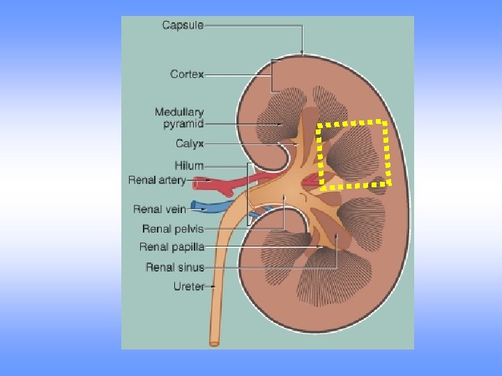
KIDNEY
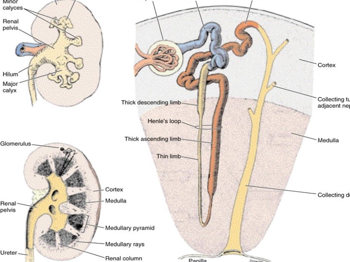
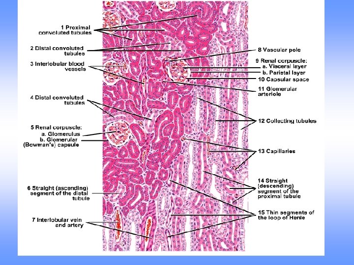
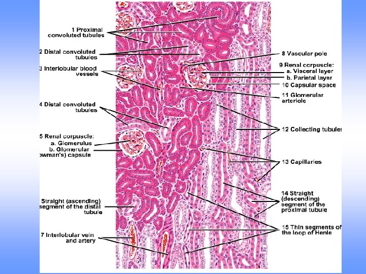
Microscopic structure of kidney § Covered by dense capsule § Substance is divided into: outer cortex and inner medulla § Medulla is arranged into units – renal pyramids § Medullary pyramids are separated by extensions of cortical tissue, called renal columns § Functional unit of the kidney – uriniferous tubule § Each tubule consist of nephron and collecting duct § Each nephron consist of renal corpuscle and renal tubule
CORTEX MEDULLA
CORTEX: • Darker outer portion • Contains renal corpuscles, proximal and distal convoluted tubles and medullary rays • Also contains interlobular arteries and veins • Pale stained medullary rays formed by straight portions of proximal and distal convoluted tubules and collecting tubules
GLOMERULI MEDULLARY RAYS
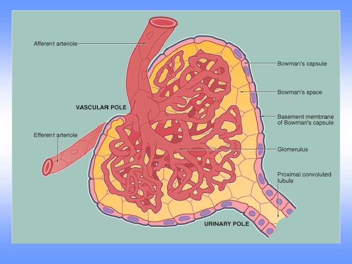
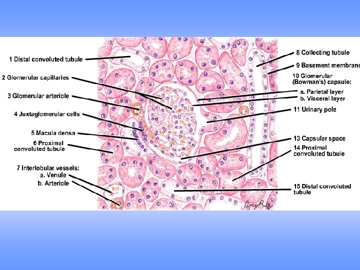
Renal corpuscle: • Dense rounded structures • Responsible for the filtration of plasma • Comprises: • Glomerulus is formed of a tight anastomosing network of capillaries • Bowman’s capsule
Bowman’s capsule • Outer paietal layer is lined by simple sqamous epithelium • Inner visceral layer consist of branching epithelial cells podocytes
CAPSULE GLOMERULUS
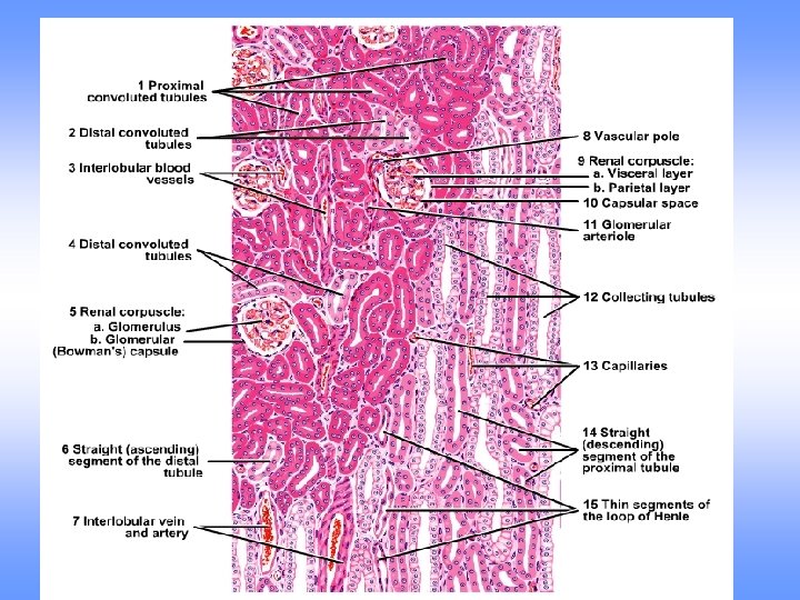
Proximal convoluted tubule: • Lined by simple cuboidal epithelium • Shows prominent brush border • Cytoplasm takes intense eosinophilic staining • Small or uneven lumen
Distal convoluted tubule: • Lined by simple cuboidal epithelium • No brush border • Clear distinct lumen • Less intensely stains • Less in number
Collecting tubules: • Lined by cuboidal epithelium • Becomes progressively taller • No brush border Collecting ducts: • Formed by the fusion of collecting tubules • Converge to form larger ducts (ducts of Bellinior papillary ducts) • Lined by tall columnar epithelium • Pale stained cytoplasm • No brush border
Medulla : • Contains the segments of the loops of Henle, straight portions of tubules, vasa recta and collecting ducts • Thin segments of Henle lined by simple squamous epithelium • Straight portions of tubules are lined by simple cuboidal epithelium • Collecting ducts (ducts of Bellini/papillary ducts) are lined by simple columnar epithelium
Juxtaglomerular apparatus • Juxtaglomerular cells: modified Smooth muscle cells in the wall of afferent arteriole • Macula densa: Modified cells of DCT adjacent to afferent arteriole
URETER • Muscular tube • Mucosa: transitional epithelium § Shows numerous folds § Wide lamina propria • Muscularis layer: arranged in two layers – inner longitudinal and outer circular • Outer coat: Adventitia – loose connective tissue with blood vessels, nerves and lymphatics
URETER
Thank you