Urinary System Presented by Yvonne Tsitsiou 210519 Lecture

Urinary System Presented by Yvonne Tsitsiou 21/05/19

Lecture 1 – The Urinary System

Learning Objectives • Urinary system: summarise the gross anatomy of the urinary system • Ureters: recall the functional anatomy of the ureters and mechanisms preventing reflux of urine • Bladder: recall the structural and functional anatomy, and histological features of the bladder; recall the mechanisms involved in reflex contraction in response to distension • Urinary sphincters: compare the sphincter urethrae and sphincter vesicae muscles and their nerve supplies • Kidney: recall the macroscopic structure of the kidney and be able to identify theses structures in histological sections • Renal vessels: explain the anatomy of blood vessels in the kidney and their functional significance (including filtration, reabsorption and countercurrent mechanism)

Kidney • Retroperitoneal organ • Surrounded by a dense fibrous capsule • Surrounding that is a fascial pouch which is made of adipose tissue protects against trauma • Right kidney is 11 th intercostal space – due to liver • Left Kidney is at 11 th rib • Hilum is at L 1 for both Urinary system: summarise the gross anatomy of the urinary system

Posterior relations of the kidney Urinary system: summarise the gross anatomy of the urinary

Anterior relations of kidney • Right: under liver and hepatic flexure of colon • Left: surrounded by stomach, pancreas, spleen, and splenic flexure Urinary system: summarise the gross anatomy of the urinary

Ureters • Run vertically down posterior abdominal wall • Run along vertical plane of transverse processes of lumbar vertebrae • They cross the pelvic brim anterior to the sacro-iliac joint & bifurcation of the common iliac arteries • The bladder is anterior so the ureters must come forward around the rectum (and uterus) to reach the bladder • Enter bladder at level of ischial spine Ureters: recall the functional anatomy of the ureters and mechanisms preventing reflux

Macroscopic structure of the kidney • Each kidney has superior and inferior pole • Granular cortex and an inner striated medulla because of the regular arrangement of the tubules and vessels • Human kidney is multilobular • Pyramids papilla minor calyx major calyx renal pelvis ureter bladder Kidney: recall the macroscopic structure of the kidney and be able to identify theses structures in histological sections

Mechanisms preventing reflux of urine • The muscles of the bladder run obliquely so they put pressure on the anterior wall of the ureter preventing reflux • A valve at the vesicoureteral (ureter-bladder) junction prevents backflow of urine Ureters: recall the functional anatomy of the ureters and mechanisms preventing reflux

Bladder • Triangular pyramid with the apex anterior and the base posterior • Apex is behind the pubic symphysis • Lined by urothelium (transitional epithelium) • 3 layered epithelium - slow cell turnover • Large luminal cells have highly specialised lowpermeability luminal membrane - prevents dissipation of urine-plasma gradients Posterior Anterior • Bladder is folded so that it can expand • Urethra passes through peroneal membrane • Female urethra passes straight into the perineum Bladder: recall • Male crosses the prostate glandthe structural and functional anatomy, and histological features of the bladder; recall the mechanisms involved in reflex contraction in

Bladder contraction • Occurs in response to distension Bladder: recall the structural and functional anatomy, and histological features of the bladder; recall the mechanisms involved in reflex contraction in
 • • At neck")
Urinary Sphincters • Sphincter vesicae (internal sphincter – smooth muscle) • • At neck of bladder Bladder wall tension causes reflex opening Relaxed by PNS Contracts by SNS • Sphincter urethrae (external sphincter – striated muscle) • In perineum • Tone maintained by pudendal nerve • Opened by voluntary inhibition of nerves Urinary sphincters: compare the sphincter urethrae and sphincter vesicae muscles and their nerve

Lymph drainage • Follows renal vessels to the renal lymph nodes • Ureter lymph vessels drain into deep iliac and common iliac nodes and you can palpate them in the groin

Q: What are the differences between male and female urethras?

ARQ Assertion: The left kidney is slightly lower than the right Reason: The liver pushes the kidney down A) B) C) D) E) True – reason is correct explanation True – reason is NOT correct explanation True False
 B) C) D) E)")
SBA What does bladder contraction occur in response to? A) B) C) D) E) Sympathetic stimulation Parasympathetic stimulation Distension from bladder filling Voluntary muscle contraction Endocrine signalling

Lecture 2 – Structural Basis of Kidney Function

Learning Objectives • Renal mechanisms: identify the mechanisms by which solutes enter and leave the tubular fluid and define the role that these mechanisms play in excretion of waste, define reabsorption and secretion; explain the meaning of transcellular and paracellular transport • Renal physiological functions: recall the physiological functions of the kidney including role in homeostasis, excretory function and endocrine function • Nephron: recall the constituent parts of a nephron and which compounds are absorbed in each area, explain the microscopic anatomy of the Bowman's capsule and list the regional features of tubular cells aiding urinary concentration, identify different sections and cell types of the nephron in light and electron microscopic images. • Renin-Angiotensin-Aldosterone Axis: recall the site of secretion of aldosterone and explain influences on rate of production, recall the physiological action of aldosterone and effect on sodium handling; list the effects of angiotensin II on renal function; recall stimuli for renin release; explain the effects of hyper- and hypo-aldosteronism

The body needs 2 litres of water a day –from food and drink Function of kidney: • Selective reabsorption occurs in the nephron to keep useful products • Tubular secretion of some components • Concentration of urine as necessary Renal physiological functions: recall the physiological functions of th kidney including role in homeostasis, excretory function and endocrin

Integrated functions of kidney Renal physiological functions: recall the physiological functions of th kidney including role in homeostasis, excretory function and endocrin

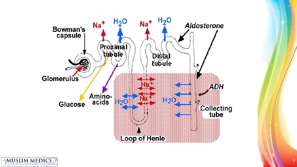
Production of urine • Filtration: Blood passing through the glomerulus is filtered and then everything lower than 50 000 molecular weight is filtered • Reabsorption: mainly occurs in the PCT includes ions, glucose, aa, small proteins, water etc. • Creation of Hyper-osmotic ECF: main function of the loop of Henle and vasa recta (blood vessels) – via Countercurrent mechanism • Adjustment of ion content of urine: DCT • Concentration of urine: Collecting duct = ADH Renal mechanisms: identify the mechanisms by which solutes enter and leave the tubular fluid and define the role that these mechanisms play in excretion of waste, define reabsorption and secretion; explain the meaning of transcellular and

Filtration – 3 layers • In the glomerulus, afferent arterioles are wider than efferent which allows an increase in BP (narrowing generates pressure) allowing ultrafiltration • 3 layers form a mesh which filters the blood 1. Fenestrated endothelium 2. Modified basement membrane 3. Podocytes • Filtrate passes through the Bowman’s capsule to the PCT isotonic at this point Nephron: recall the constituent parts of a nephron and which compounds are absorbed in each area, explain the microscopic anatomy of the Bowman's capsule and list the regional features of tubular cells aiding urinary concentration, identify different sections and cell types of the nephron in light and electron microscopic

Reabsorption PCT • 70% of fluid reabsorbed • Na+ uptake by basolateral Na+ pump water follows via aquaporins • Glucose reabsorbed by SGLT channel (Na+/glucose co-transporter) • Amino acids by Na+/aa co transporter • Proteins taken up by endocytosis • Lots of mitochondria due to high energy requirement Nephron: recall the constituent parts of a nephron and which compounds are absorbed in each area, explain the microscopic anatomy of the Bowman's capsule and list the regional features of tubular cells aiding urinary concentration, identify different sections and cell types of the nephron in light and electron microscopic

Hyper-osmotic Interstitium Loop of Henle • Descending limb is thin and permeable to water • Ascending limb is thick and impermeable to water • Na+/2 Cl-/K+ triple transporter pumps ions out of tubular fluid • Lots of mitochondria • Blood vessels (vasa recta) are also arranged in a loop • Creates hyper-osmotic gradient Nephron: recall the constituent parts of a nephron and which compounds are absorbed in each area, explain the microscopic anatomy of the Bowman's capsule and list the regional features of tubular cells aiding urinary concentration, identify different sections and cell types of the nephron in light and electron microscopic

Adjustment of ion content Mainly DCT • Under the control of aldosterone which alters sodium potassium balance • Macula densa detects changes in tubular fluid sodium - this is how BP is modified Nephron: recall the constituent parts of a nephron and which compounds are absorbed in each area, explain the microscopic anatomy of the Bowman's capsule and list the regional features of tubular cells aiding urinary concentration, identify different sections and cell types of the nephron in light and electron microscopic

Concentration of urine • Collecting duct • Water reabsorption under ADH control • Rate of water movement depends on aquaporin-2 in apical membrane • Basolateral membrane has aquaporin-3, not under control • See endo ppt for aquaporin detail Nephron: recall the constituent parts of a nephron and which compounds are absorbed in each area, explain the microscopic anatomy of the Bowman's capsule and list the regional features of tubular cells aiding urinary concentration, identify different sections and cell types of the nephron in light and electron microscopic

Juxtaglomerular apparatus • Endocrine cells • macula densa of DCT – senses conc of Na. Cl in tubular fluid • juxtaglomerular cells of afferent arteriole - senses stretch in arteriole wall • Secretes renin to control blood pressure via angiotensin • LOW BP sensed by JGA makes renin converts angiotensinogen to AT 1 ACE converts to AT 2 increases aldosterone production, increases sodium and water reabsorption and causes vasoconstriction Renin-Angiotensin-Aldosterone Axis: recall the site of secretion of aldosterone and explain influences on rate of production, recall the physiological action of aldosterone and effect on sodium handling; list the effects of angiotensin II on renal function; recall stimuli for renin release; explain the effects of hyper- and hypo-aldosteronism

• Aldosterone is secreted from the zona glomerulosa of the adrenal cortex • Stimuli for renin release: • • Low Na+ (low Na=low GFR=low BP) Low BP – juxtaglomerular cells act like baroreceptors SNS – beta 1 receptors on juxtaglomerular cells (high K+) Renin-Angiotensin-Aldosterone Axis: recall the site of secretion of aldosterone and explain influences on rate of production, recall the physiological action of aldosterone and effect on sodium handling; list the effects of angiotensin II on renal function; recall stimuli for renin release; explain the effects of hyper- and hypo-aldosteronism

ARQ Assertion: The afferent arteriole is wider than the efferent arteriole in the glomerulus Reason: This allows greater blood flow to supply very active cells of the glomerulus A) True – reason is correct explanation B) True – reason is NOT correct explanation C) True False D) False True E) False
 B) C) D) E)")
SBA Where is the majority of the water reabsorbed? A) B) C) D) E) PCT Descending limb of loop of Henle Ascending limb of loop of Henle DCT Collecting duct

Lecture 3 – Renal Blood Flow and Glomerular Filtration

Learning Objectives • Renal blood flow: recall what proportion of cardiac output normally perfuses the kidney, explain the effect of changes in renal blood flow on glomerular filtration rate (GFR) and be able to calculate renal plasma flow rate given appropriate values. • Glomerular filtration: define the term freely filtered, recall the factors affecting filtration of substances in the glomerulus, compare the composition of the glomerular filtrate and the plasma, define glomerular filtration rate and filtration fraction and recall normal values. • Filtration pressure: explain net filtration pressure (hydrostatic and oncotic pressures), explain how net filtration pressure may be affected by changes to either of these components (including arterial blood pressure, plasma protein concentration and ureteral obstruction). • Renal clearance: define renal clearance, explain the use of renal clearance in assessing renal function, and be able to perform the appropriate calculation given appropriate values.

Glomerular Filtration • Definition: formation of an ultrafiltrate of plasma in the glomerulus • Kidney failure is an abrupt fall in GF • Passive process - driven by by hydrostatic pressure • Small solutes are freely filtered - same concentration in the filtrate as the plasma

Basic Renal Process Renal input = renal artery Renal output = renal vein and ureter Primary urine Amount = Amount + Amount - Amount excreted secreted absorbed filtered Not all substances undergo all processes
 - hydrostatic pressure due")
Glomerular Filtration Pressure Driving force = glomerular capillaries pressure (Pgc) - hydrostatic pressure due to BP Opposing pressures: hydrostatic pressure of tubule (Pt) osmotic pressure of plasma proteins in glomerular capillaries (πgc) Together= net ultrafiltration pressure (Puf) Puf = Pgc - Pt - πgc Net filtration = 10 -20 mm. Hg Filtration pressure: explain net filtration pressure (hydrostatic and oncotic pressures), explain how net filtration pressure may be affected by changes to either of these components (including arterial blood pressure, plasma protein concentration

Glomerular Filtration rate GFR = Puf x Kf Where Kf is an ultrafiltration coefficient (membrane permeability and area available for filtration). • Any changes in filtration forces or Kf will result in GFR imbalances. • Kidney diseases may reduce number of functioning glomeruli = reduced surface area = fall in Kf so lower GFR • Dilation of glomerular arterioles by drugs/hormones will increase Kf Glomerular filtration: define the term freely filtered, recall the factors affecting filtration of substances in the glomerulus, compare the composition of the glomerular filtrate and the plasma, define glomerular filtration rate and filtration

Renal Blood Flow • RBF is 1 L/min which is 1/5 of cardiac output • Renal plasma flow is 0. 6 L/min • Filtration Fraction (FF) = 0. 2 because 20% is flitered • Normal GFR is 120 m. L/min • Glomerular filtration rate (GFR) = RPF x FF 80% 100% 20% Renal blood flow: recall what proportion of cardiac output normally perfuses the kidney, explain the effect of changes in renal blood flow on glomerular filtration rate (GFR) and be able to calculate renal plasma flow rate given

Autoregulation of GFR • DECREASED GFR • Not filtering enough • Constrict afferent arteriole, dilate efferent arteriole glomerular capillary pressure decreases • More time for kidneys to filter plasma • INCREASED GFR • Filtering too much (too many ions/ solutes lost in tubular fluid) • Dilate afferent arteriole, constrict efferent arteriole glomerular capillary pressure increases • Plasma passes through quickly, so less time for filtration Renal sympathetic nervous system: recall the effect of the sympathetic nervous system on the renal vasculature and renin

Mechanisms of Autoregulation 1. Myogenic mechanism • Vascular smooth muscle constricts when stretched • Keeps GFR constant when BP • Arterial pressure afferent arteriole stretched arteriole contracts (vessel resistance ) blood flow , GFR constant 2. Tubuloglomerular feedback • Where Na. Cl in fluid sensed by macula densa in JGA signal to afferent arteriole constriction decreased filtration Renal sympathetic nervous system: recall the effect of the sympathetic nervous system on the renal vasculature and renin release

Clearance https: //www. youtube. com/watch? v=zur. ZSXCO-Tw • Clearance is the number of litres of plasma that are completely cleared of the substance per unit time • C=Ux. V P ml/min U = concentration of substance in urine V = rate of urine production P = concentration of substance in plasma If a molecule is freely filtered and not reabsorbed/ secreted in the nephron the amount filtered = amount excreted GFR is measured by measuring the clearance of a freely filtered molecule e. g. insulin and creatinine Build up of creatinine = fall in GFR = renal disease Renal clearance: define renal clearance, explain the use of renal clearance in assessing renal function, and be able to perform the appropriate calculation given appropriate values.

Renal diagnostics • GFR = cardinal feature of renal disease • If GFR , excretory products build up in plasma • important with drugs- could accidently overdose as it isn’t excreted • plasma conc of creatinine is diagnostic of renal disease • Excretion of many other substances also impaired in renal failure- including some drugs • needs to be taken into account when calculating drug doses Measurement of renal function: recall methods of estimating global renal function and compare advantages and

ARQ Assertion: When there is a high GFR, the afferent arteriole is constricted and the efferent is dilated Reason: This decreases glomerular capillary pressure allowing less time for filtration A) B) C) D) E) True – reason is correct explanation True – reason is NOT correct explanation True False
 50% B)")
SBA What proportion of the cardiac output normally perfuses the kidney? A) 50% B) 30% C) 20% D) 10% E) 5%

Lecture 4 – Basic Tubular Function

Learning Objectives • Renal Mechanisms: identify the mechanisms by which solutes enter and leave the tubular fluid and define the role that these mechanisms play in excretion of waster, define reabsorption and secretion; explain the meaning of transcellular and paracellular transport. • Proximal tubule: recall the microscopic structure of the early proximal tubule (including tubular fluid, luminal membrane, basolateral membrane, peritubular capillary, tight junction, Na+/K+ pump); recall examples of ion transport (including ion-selective channel, co-transport of two solutes, counter-transport of two solutes) and explain the role of mutations to identifies transporters in causing renal dysfunction, recall the proportion of solute reabsorbed in this region and contrast this with the distal nephron. • Osmolarity: define osmolarity, define the minimum and maximum urine osmolarity in humans, explain how the kidney produces dilute and concentrated urine and explain why this is dependent on the osmolarity of the medullary and papillary interstitium, and permeability of the collecting ducts; explain how changes in plasma osmolarity and volume affect thirst, explain how and why osmolarity varies along the nephron • Vasopressin: explain the mechanisms controlling vasopressin release (including hypothalamic osmoreceptors) and recall the physiological action of vasopressin on the renal tubule

Osmolarity • The kidney is a central regulator of homeostasis OSMOLARITY: a measure of the osmotic pressure exerted by a solution across a perfect semi-permeable membrane. dependent on the number of particles in the solution all the concentrations of the different solutes added together normal plasma osmolarity is tightly controlled 285 -295 mosm/kg Osmolarity: define osmolarity, define the minimum and maximum urine osmolarity in

Renal Tubular Wall • Single layer of cells • Between the cells are tight junctions that vary in their tightness depending on where you are in the system • Secretion and reabsorption both occur either by transcellular or paracellular transport • Through cells- transcellular • Between cells- paracellular Renal Mechanisms: identify the mechanisms by which solutes enter and leave the tubular fluid and define the role that these mechanisms play in excretion of waster, define reabsorption and secretion; explain the meaning of transcellular and paracellular

Passive movement rate o Linear o non saturable- hydrophobic molecules Solute concentration rate Protein independent transport (lipophilic molecules) Protein dependent transport (hydrophilic molecules) o Saturable o Non linear Solute concentration Renal Mechanisms: identify the mechanisms by which solutes enter and leave the tubular fluid and define the role that these mechanisms play in excretion of waster, define reabsorption and secretion; explain the meaning of transcellular

Active movement – Cellular Energy dependent rate ATP ADP + Pi Solute concentration Directly coupled to ATP hydrolysis Na+ Glucose Na+ K+ rate ATP ADP + Pi Solute concentration Indirectly coupled to ATP hydrolysis

Transport maxima • It is the point at which a rise in solute concentration does not yield a rise in rate. • Limit to what can enter/exit cells • Applies to whole system, not just individual cells • Can vary depending on circumstances, stimulated maximum higher than basal max • Normally exceeds requirement • Amount of glucose filtered plasma glucose concentration • Up to certain plasma glucose concentration, all glucose is reabsorbed • Above certain plasma glucose concentration, can’t absorb any more so it is excreted diabetes mellitus

Secretion • Substances move from peritubular capillaries tubular lumen • Occurs either by diffusion or transcellular mediated transport • Most important substances secreted are H+ and K+ • Choline, creatinine, penicillin & other drugs also secreted • Active secretion from blood into tubule cells (basolateral membrane) and from cell into the lumen (luminal membrane) Renal Mechanisms: identify the mechanisms by which solutes enter and leave the tubular fluid and define the role that these mechanisms play in excretion of waster, define reabsorption and secretion; explain the meaning of transcellular and paracellular transport.

Reabsorption • Small molecules such as glucose, ions and amino acids are reabsorbed • Specialized proteins called transporters are located on the various cells of the nephron and trap molecules as they flow by • Water gets reabsorbed passively in response to build up of Na+/ions • Transporters are located in different parts of the nephron Renal Mechanisms: identify the mechanisms by which solutes enter and leave the tubular fluid and define the role that these mechanisms play in excretion of waster, define reabsorption and secretion; explain the meaning of transcellular and paracellular transport.

Proximal Convoluted Tubule • Reabsorbs most things: 60 -70% of all solute, all glucose, 90% bicarbonate ions, 65% Na+ and water • Water and anions follow Na • Na+K+ ATPase • needed to generate low intracellular sodium conc so that sodium is driven into the cell • most of the ATP is used for this • Passive reabsorption of urea and water • Active reabsorption of glucose, aa, Na+, K+, Ca 2+ and uric acid Proximal tubule: recall the microscopic structure of the early proximal tubule (including tubular fluid, luminal membrane, basolateral membrane, peritubular capillary, tight junction, Na+/K+ pump); recall examples of ion transport (including ion-selective channel, co-transport of two solutes, counter-transport of two solutes) and explain the role of mutations to identifies transporters in causing renal dysfunction, recall the proportion of solute reabsorbed in this region and contrast this with the distal nephron

Loop of Henle • Descending Limb relatively inert in terms of pumping, water is passively reabsorbed due to the high osmotic potential created by the ascending limb – draws in sodium and potassium • Ascending limb chloride actively reabsorbed, sodium passively reabsorbed and with it comes bicarbonate. Impermeable to water. Cuboidal epithelium, few microvilli. High energy requirement – prominent mitochondria. • Triple transporter Na+/K+/Cl. Nephron: recall the constituent parts of a nephron and which compounds are absorbed in each area, explain the microscopic anatomy of the Bowman's capsule and list the regional features of tubular cells aiding urinary concentration, identify different sections and cell types of the nephron in light and electron microscopic

Distal Convoluted Tubule • Proximal DCT has cuboidal epithelium, few microvilli • Complex lateral membrane interdigitations with Na+ pumps • Many large mitochondria • Na+ and Cl- cotransporter linked to Ca 2+ reabsorption • If Na+/Cl- channel blocked with thiazide, there is a rise in plasma Ca 2+ concentration • Macula densa detects changes in the amount of sodium in filtrate • Important in knowing how much sodium we need to conserve Nephron: recall the constituent parts of a nephron and which compounds are absorbed in each area, explain the microscopic anatomy of the Bowman's capsule and list the regional features of tubular cells aiding urinary concentration, identify different sections and cell types of the nephron in light and electron microscopic
 • Fine tuning of the filtrate to maintain homeostasis")
Collecting Duct (+ distal DCT) • Fine tuning of the filtrate to maintain homeostasis • DCT + CD– Sodium reabsorbed (dependent on aldosterone) • Water is reabsorbed under the control of ADH • Impermeable to water in the absence of ADH 2 main cells: • Principal cell: important in sodium, potassium and water balance (mediated via Na/K ATP pump) • Also has very tight epithelium allowing little paracellular transport • Allows you to regulate water transport across the membrane by changing the number of transporters in the system • Intercalated cells • important in acid-base balance (mediated via H-ATP pump) Nephron: recall the constituent parts of a nephron and which compounds are absorbed in each area, explain the microscopic anatomy of the Bowman's capsule and list the regional features of tubular cells aiding urinary concentration, identify different sections and cell types of the nephron in light and electron microscopic

PCT Cuboidal epithelium Tight junctions - water impermeable Contains aquaporins Brush border - SA to maximise rate of absorption energy, mitochondria vesicles from absorbing things Ascending thick limb • Very water-impermeable tight junctions • No aquaporins • Cuboidal epithelium, few microvilli • High energy requirement prominent mitochondria Descending thin tubule • Simple squamous epithelium • Contains aquaporins DCT • Cuboidal epithelium, microvilli • Complex lateral membrane interdigitations with Na+ pumps • large mitochondria Length of PCT > DCT so on a cross section there appears to be more proximal tubules

ARQ Assertion: The descending limb of the loop of Henle actively reabsorbs water Reason: There is a high osmotic gradient created by the ascending limb A) B) C) D) E) True – reason is correct explanation True – reason is NOT correct explanation True False
 side of the ascending")
SBA Which ion transporter is found on the apical (luminal) side of the ascending limb of the loop of Henle? A) B) C) D) E) SGLT transporter Na+/2 Cl-/K+ triple transporter Na+/K+ ATPase transporter K+/H+ exchanger Na+/Ca 2+ transporter

Lecture 5 – Control of Water Balance

Learning Objectives • Countercurrent multiplier: explain the mechanisms that lead to the development of the countercurrent multiplier and explain its physiological function • Vasopressin: explain the mechanisms controlling vasopressin release (including hypothalamic osmoreceptors) and recall the physiological action of vasopressin on the renal tubule • Extracellular fluid: explain the determinants and control of extracellular fluid volume • Renal sympathetic nervous system: recall the effect of the sympathetic nervous system on the renal vasculature and renin release

Water Balance • Osmolarity measures the solute conc in a solution • 1 osm = 1 mole of dissolved solutes/L • Plasma osmolarity is tightly controlled • Urine osmolarity varies depending on the amount of water reabsorbed • Water flows from low osmolarity high osmolarity through a partially permeable membrane Extracellular fluid: explain the determinants and control of extracellular fluid volume

Extracellular fluid: explain the determinants and control of extracellular fluid

• Must get rid of the excess volume • Or you will become oedematous and your blood pressure will increase • Must get rid of any excess water • or you will dilute the salt in your body • Cells will swell • Must get rid of any excess salt • or you will have too high a level of salt • Cells will shrink or you will have retain more water and you blood pressure will increase Extracellular fluid: explain the determinants and control of extracellular fluid

Fluid Compartments • Intracellular fluid compartment • Extracellular fluid compartment • • Interstitial fluid Plasma Transcellular fluid (CSF, synovial fluid) Lymph • Normal plasma osmolarity = 295 m. Osm/kg • Water is the main component of plasma and ECF • Na+ is the main solute of plasma and ECF Extracellular fluid: explain the determinants and control of extracellular fluid

How do we get rid of water? • Skin and Sweat • Fever, climate activity • Variable uncontrollable • Faeces • Normal or diarrhoea • Uncontrollable • Respiration • Depends on level of activity • Uncontrollable • Urine Output = 1500 ml/day • Variable and controllable Extracellular fluid: explain the determinants and control of extracellular fluid

Countercurrent multiplier: explain the mechanisms that lead to the development of the countercurrent multiplier and explain its physiological

Establishing the Gradient This by itself is not enough to generate 1200 osmoles/litre seen in medulla Countercurrent multiplier: explain the mechanisms that lead to the development of the countercurrent multiplier and explain its physiological

• Water is leaving collecting duct so conc of urea is greater than in base of loop of Henle • Generates gradient, so urea leaves collecting duct and moves into loop of H – circles round and round • Done via urea transporters • Descending limb • UT-A 2 receptors – let urea into tubule • Collecting duct • UT-A 1 – let urea into cell from tubule (apical) • UT-A 3 – let urea out of cell into interstitium (basolateral) Countercurrent multiplier: explain the mechanisms that lead to the development of the countercurrent multiplier and explain its physiological

https: //www. youtube. com/watch? v=Xb. I 8 e Y-Be. XY Countercurrent multiplier: explain the mechanisms that lead to the development of the countercurrent multiplier and explain its physiological

Control of Plasma Osmolarity • Main hormone is Vasopressin/ ADH • Secreted by posterior pituitary – acts on CD – synthesis and insertion of AQP 2 in apical membrane for water reabsorption • ADH release is controlled by osmoreceptors in the hypothalamus • Triggers release if: • osmolarity rises above 300 m. Os • marked fall in blood volume/pressure (baroreceptors/stretch receptors) • Ethanol inhibits ADH dehydration and polyuria Vasopressin: explain the mechanisms controlling vasopressin release (including hypothalamic osmoreceptors) and recall the physiological action of vasopressin on
 and recall")
Water Load Vasopressin: explain the mechanisms controlling vasopressin release (including hypothalamic osmoreceptors) and recall the physiological action of vasopressin on
 and recall the")
Dehydration Vasopressin: explain the mechanisms controlling vasopressin release (including hypothalamic osmoreceptors) and recall the physiological action of vasopressin on

Disorders of Water Balance • No/ insufficient ADH • Mutation in ADH receptor • No response to ADH signal (mutant AQP) • DIABETES INSIPIDUS • Symptoms thirst, dilute urine, polyuric Vasopressin: explain the mechanisms controlling vasopressin release (including hypothalamic osmoreceptors) and recall the physiological action of vasopressin on

ARQ Assertion: Urea leaves the collecting duct and moves into the loop of Henle Reason: The water leaving the collecting duct generates a gradient as the concentration of urea in the collecting duct is greater than in the base of the loop of Henle A) B) C) D) E) True – reason is correct explanation True – reason is NOT correct explanation True False
 B) C) D) E) Sympathetic stimulation Osmolarity")
SBA ADH release is triggered by: A) B) C) D) E) Sympathetic stimulation Osmolarity rising above 200 m. Os Renin Thirst A marked fall in blood volume/pressure

Lecture 6 – Control of Sodium and Potassium Balance

Learning Objectives • Sodium handling: recall the daily amounts of filtered and excreted sodium; explain the renal handling of sodium ions; explain how active sodium transport acts as the driving force for the reabsorption of water and other ions and molecules, explain the effects of changes in dietary sodium intake and sodium balance on sodium handling in the kidney and on fluid volume • Potassium handling: recall the daily amounts of filtered and excreted potassium; explain the renal handling of potassium ions; recall the cellular mechanism of potassium secretion and how this is influenced by potassium concentration, aldosterone, tubular flow rate and acid-base balance • Diuretics: explain the mechanism of action of diuretics and identify their site of action

ECF osmolarity and volume • Plasma osmolarity is tightly controlled between 285 -295 mosm/kg • Sodium = most abundant solute in ECF so determines ECF volume • Body does not allow changes in dietary sodium to affect osmolarity so: • Increase in dietary sodium: increase in ECF volume, blood volume and BP • Decrease in dietary sodium: decrease in ECF volume and define osmolarity, define the minimum and. BP maximum urine osmolarity in humans, hence. Osmolarity: a decrease in blood volume and explain how the kidney produces dilute and concentrated urine and explain why this is dependent on the osmolarity of the medullary and papillary interstitium, and permeability of the collecting ducts; explain how changes in plasma osmolarity and volume affect thirst, explain

Where is sodium reabsorbed? 65% Na reabsorbed in PCT because Na to reabsorb everything else e. g. glucose, aa and H+ excretion Sodium handling: recall the daily amounts of filtered and excreted sodium; explain the renal handling of sodium ions; explain how active sodium transport acts as the driving force for the reabsorption of water and other ions and molecules, explain the effects of changes in dietary sodium intake and sodium balance on

GFR and sodium excretion • If we increase the rate of GFR we increase the amount of sodium reabsorbed • If we decrease the rate of GFR we reduce the amount of sodium reabsorbed Sodium handling: recall the daily amounts of filtered and excreted sodium; explain the renal handling of sodium ions; explain how active sodium transport acts as the driving force for the reabsorption of water and other ions and molecules, explain the effects of changes in dietary sodium intake and sodium balance on sodium handling in the kidney and on fluid volume

Renin Angiotensin System • Liver produces Angiotensinogen Angiotensin 1 by Renin (produced in JGA of kidney) • Angiotensin 1 Angiotensin 2 by ACE produced in the lungs • Angiotensin 2 causes the production of aldosterone from the adrenal cortex (zona glomerulosa) Renin-Angiotensin-Aldosterone Axis: recall the site of secretion of aldosterone and explain influences on rate of production, recall the physiological action of aldosterone and effect on sodium handling; list the effects of angiotensin II on renal function; recall stimuli for renin release;

Effects of AII Increased ECF Increased blood pressure Increased water reabsorption vasoconstriciton Increased sodium uptake Vascular system Proximal tubule Angiotensin II Adrenal gland Aldosterone synthesis Renin-Angiotensin-Aldosterone Axis: recall the site of secretion of aldosterone and explain influences on rate of production, recall the physiological action of aldosterone and effect on sodium handling; list the effects of angiotensin II on renal function; recall stimuli for renin release; explain the effects of hyper- and hypo-
 •")
Aldosterone Stimulates: • Increased Sodium reabsorption • (controls reabsorption of 35 g Na/day) • Increased Potassium secretion • Increased hydrogen ion secretion • Aldosterone excess: • leads to hypokalaemic alkalosis Renin-Angiotensin-Aldosterone Axis: recall the site of secretion of aldosterone and explain influences on rate of production, recall the physiological action of aldosterone and effect on sodium handling; list the effects of angiotensin II on renal function; recall stimuli for renin release; explain the effects of

Hypoaldosteronism - Reabsorption of sodium in the distal nephron is reduced - Increased urinary loss of sodium - So ECF volume falls - Increased renin, Ang II and ADH release - Symptoms include: dizziness, low BP, salt craving and palpitations Renin-Angiotensin-Aldosterone Axis: recall the site of secretion of aldosterone and explain influences on rate of production, recall the physiological action of aldosterone and effect on sodium handling; list the effects of angiotensin II on renal function; recall stimuli for renin release; explain the effects of

Hyperaldosteronism - Reabsorption of sodium in the distal nephron is increased - Reduced urinary loss of sodium - So ECF volume increases (hypertension) - Reduced renin, ang II and ADH release - Increased ANP and BNP - Symptoms include: high BP, muscle weakness, polyuria, thirst Renin-Angiotensin-Aldosterone Axis: recall the site of secretion of aldosterone and explain influences on rate of production, recall the physiological action of aldosterone and effect on sodium handling; list the effects of angiotensin II on renal function; recall stimuli for renin release; explain the effects of

Diuretic Drugs • Osmotic Diuretics: Mannitol – increases osmolarity of tubular fluid thus decreasing water reabsorption • Carbonic Anhydrase Inhibitors – reduces sodium reabsorption by blocking conversion of bicarbonate into CO 2 and H 2 O in lumen • Loop Diuretics: Frusemide – blocks triple co-transporter Na+/K+/Cl- in the ascending limb of LOH • Thiazides: block Na/Cl co-transport in the DCT • K+ Sparing Diuretics: • amiloride – blocks Na channels in DCT • Spironolactone – aldosterone antagonist Diuretics: explain the mechanism of action of diuretics and identify their
, extracellular = 3")
Potassium Regulation • K+ is the main intracellular ion (150 mmol/l), extracellular = 3 -5 mmol/L • Extracellular K+ has effects on excitable membranes (of nerve and muscle) • High K+; depolarises membranes causing action potentials and heart arrhythmias • Low K+ also leads to heart arrhythmias (and asystole) • Therefore the immediate response of dietary potassium is to pump it into cells via the Na+/K+ ATPase Potassium handling: recall the daily amounts of filtered and excreted potassium; explain the renal handling of potassium ions; recall the cellular mechanism of potassium secretion and how this is influenced by potassium concentration, aldosterone, tubular

Potassium filtration • 70% reabsorbed in the PCT • 20% in the ascending limb of LOH • K+ secretion is stimulated by • • Increase in plasma [K+] Increase in aldosterone Increase in tubular flow rate Increases in plasma p. H • K+ secretion is mainly carried out by the principal cells moving K+ from the blood into the cell and then out through the potassium channels into the lumen Potassium handling: recall the daily amounts of filtered and excreted potassium; explain the renal handling of potassium ions; recall the cellular mechanism of potassium secretion and how this is influenced by potassium concentration, aldosterone, tubular

Aldosterone and Potassium • Aldosterone stimulates the Na+/K+ ATPase on the blood side but also the Na+ and K+ channels on the lumen side • Causes loss of potassium and gain of sodium Potassium handling: recall the daily amounts of filtered and excreted potassium; explain the renal handling of potassium ions; recall the cellular mechanism of potassium secretion and how this is influenced by potassium concentration, aldosterone, tubular

Hypokalaemia • One of the most common electrolyte imbalances Causes: • Diuretics (due to increase tubular flow rates) • Surreptitious vomiting • Diarrhoea • Also caused by genetics (Gitelman’s syndrome; mutation in the Na/Cl transporter in the distal nephron) Potassium handling: recall the daily amounts of filtered and excreted potassium; explain the renal handling of potassium ions; recall the cellular mechanism of potassium secretion and how this is influenced by potassium concentration, aldosterone, tubular

Hyperkalaemia • Common electrolyte imbalance present in 1 -10% of hospitalised patients • Seen in response to K+ sparing diuretics and ACE inhibitors • Often seen in the elderly Potassium handling: recall the daily amounts of filtered and excreted potassium; explain the renal handling of potassium ions; recall the cellular mechanism of potassium secretion and how this is influenced by potassium concentration, aldosterone, tubular

ARQ Assertion: Increased GFR leads to increased sodium reabsorption Reason: Cells in the macula densa sense the high sodium so more sodium channels are inserted A) B) C) D) E) True – reason is correct explanation True – reason is NOT correct explanation True False
 B) C) D) E) Na/Cl co-transporter in")
SBA Where do Thiazide diuretics act? A) B) C) D) E) Na/Cl co-transporter in the DCT Na+/Ca 2+ co-transporter in the PCT Na+/K+ ATPase pump in the DCT Na+/K+/Cl- co-transporter in the ascending limb of Lo. H Na+/K+ ATPase pump in the PCT

Lecture 7 – Mechanisms of Acid/Base Balance

Learning Outcomes • Physiological p. H: recall the normal plasma p. H and the limits compatible with life • Buffering: explain the bicarbonate buffer system, recall the normal value for plasma bicarbonate, recall and apply the Henderson. Hasselbach equation, and explain renal mechanisms to alter/maintain serum bicarbonate concentration (reabsorption and secretion) • Phosphate, ammonium and protons: explain the mechanisms involved in the excretion of phosphate, ammonium salts and hydrogen ions • Acid-base disturbance: recall and explain disturbances in acid-base balance (including respiratory acidosis, metabolic acidosis, respiratory alkalosis and metabolic alkalosis), and explain respiratory and metabolic compensation

• James Moss’ lecture is really good and he explains it far better so well worth panopto-ing it • He also made this really good video about acid/base balance– it really helps clear things up (useful for resp module as well) • He also puts up a work sheet of acid/base case studies; make sure you can complete this

Acid – Base Summary • Normal arterial blood p. H = 7. 45, blood +/- this range is alkalaemic/ acidotic respectively • Physiological buffering systems provide immediate responses to fluctuations in p. H • The main buffer in ECF is bicarbonate system (hours – for H+ ions), main buffer of ICF is phosphate system • Proteins can be buffers in plasma and cells and buffer acids and bases instantaneous most powerful buffer
![• RESPIRATORY ACIDOSIS • Caused by hypercapnia (increased [CO 2]) and hypoventilation •](http://slidetodoc.com/presentation_image_h2/abff49b9d61a61121e152b0ebe29c555/image-100.jpg "• RESPIRATORY ACIDOSIS • Caused by hypercapnia (increased [CO 2]) and hypoventilation •")
• RESPIRATORY ACIDOSIS • Caused by hypercapnia (increased [CO 2]) and hypoventilation • p. H decrease • Increase bicarbonate and remove H+ • RESPIRATORY ALKALOSIS • Caused by hyperventilation (low [CO 2] due to increased elimination) • p. H increase • Compensate by removing bicarbonate and retaining H+ • METABOLIC ACIDOSIS • Decreased p. H (excess H+) and bicarbonate level • Not attributable to CO 2 – hence metabolic • Fast breathing – blow off excess CO 2 to compensate • METABOLIC ALKALOSIS • Increased p. H (low H+) and increased bicarbonate • Compensate by retaining H+ in kidneys and by breathing slower and retaining CO 2

Lecture 8 – Kidney Failure

Learning Outcomes • Renal failure: recall the consequences of renal failure including: salt and water imbalance, hyperkalaemia, loss of endocrine function, increase in cardiovascular events; recall the signs and symptoms of renal failure • Urine dipstick testing: explain how dipsticks can be used to diagnose basic disorders • Renal disease: recall the nutritional implications of renal disease and renal replacement therapy and dietary modifications required • Measurement of renal function: recall methods of estimating global renal function and compare advantages and disadvantages of each

What happens when kidneys fail? • Loss of excretory function • Accumulation of waste products • Loss of homeostatic function • Disturbance of electrolyte balance • Loss of acid-base control • Inability to control volume homeostasis • Loss of endocrine function • Loss of erythropoeitin production • Failure to 1 alpha hydroxylase vitamin D • Abnormality of glucose homeostasis • Decreased gluconeogenesis Renal failure: recall the consequences of renal failure including: salt and water imbalance, hyperkalaemia, loss of endocrine function, increase in cardiovascular events; recall the signs and symptoms of renal failure

Signs and symptoms • Symptoms vary with rate of deterioration • Chronic – can feel well with 5% kidney function • • “Very unwell” Pale, Hands cold Poor skin turgor Pulse rate low 50/minute Blood pressure low - 67/35 mm Hg Jugular Venous Pulse not visible Tachypnoeic (compensating for acidosis) Clear lungs on auscultation Renal failure: recall the consequences of renal failure including: salt and water imbalance, hyperkalaemia, loss of endocrine function, increase in cardiovascular events; recall the signs and symptoms of renal failure

Clinical Findings: • • Symptoms of extreme lethargy, weakness and anorexia Clinically volume depleted severe hypotension Elevated plasma urea and creatinine make diagnosis of renal failure. This is complicated by • • hyperkalaemia ( K+) hyponatraemia ( Na+) metabolic acidosis anaemia • ULTRASOUND - 2 small shrunken kidneys • (renal size unchanged in acute) Renal failure: recall the consequences of renal failure including: salt and water imbalance, hyperkalaemia, loss of endocrine function, increase in cardiovascular events; recall the signs and symptoms of renal failure

Causes of lethargy and anorexia • Uraemia: accumulation of nitrogenous waste products, hormones, peptides and other ‘middle-sized’ molecules (Mol Weight 2 -5000) • Long term uraemia can cause peripheral nerve damage • Acidosis - H+ ion excretion and retention of acid bases • Buffered by H+/K+ ion exchanger – aggravates hyperkalaemia • Compensation: CO 2 loss through lungs - Kussmahl respiration (air hunger) • Exacerbates anorexia and increases muscle catabolism • • Hyponatraemia, hyperkalaemia Volume depletion (low blood pressure) Anaemia – decreased erythropoietin production Chronic neurological damage – peripheral neuropathy Renal failure: recall the consequences of renal failure including: salt and water imbalance, hyperkalaemia, loss of endocrine function, increase in cardiovascular events; recall the signs and symptoms of renal failure

Salt and water imbalance • 2 phenotypes • Most common for CKD is tendency to retain • Hypertension • Oedema • Pulmonary oedema DO NOT CONFUSE SERUM SODIUM LEVELS WITH TOTAL BODY SODIUM – CKD AND AKI ARE OFTEN WITH sodium: ASSOCIATED HYPONATRAEMIA • Salt and water loss found in patients with tubulointerstitial disorders in which concentrating mechanisms have been damaged • Causes: • Inability to increase sodium reabsorption when sodium depleted. • Osmotic diuresis - high conc of small waste substances e. g. urea. • Inappropriately high loss of salt and water results in volume depletion which causes low blood pressure

Cardiovascular effect • Hyperkalaemia can cause cardiac arrhythmias (usually initial loss of p waves and also bradycardia) and cardiac arrest • Heart rate partially controlled by the membrane potential determined by relative K+ • Increased plasma K+ membrane depolarisation - can be sufficient for a portion of Na channels to inactivate not able to contribute towards AP upstroke • Impairs conduction and excitability so severe hyperkalaemia can cause heart block • Depolarisation may also be sufficient to reduce SAN upstroke therefore firing rate which will also cause bradycardia. Renal failure: recall the consequences of renal failure including: salt and water imbalance, hyperkalaemia, loss of endocrine function, increase in cardiovascular events; recall the signs and symptoms of renal failure

• Major outcome for patient with CKD is CARDIOVASCULAR DISEASE • Hypertension • Secondary cardiac effects • Endothelial effects • Lipid abnormabilities

Methods for assessing GFR • Urea - Poor indicator - confounded by diet, catabolic state, GI bleeding (bacterial breakdown of blood in gut), drugs, liver function etc • Creatinine - Affected by muscle mass, age, race, sex etc. Need to look at the patient when interpreting result • Creatinine clearance • Difficult for elderly patients to collect an accurate sample • Overestimates GFR at low GFR (small amount of creatinine secreted into urine) • Inulin clearance - Laborious - used for research purposes only • Radionuclide studies, EDTA clearance etc , Reliable but expensive Measurement of renal function: recall methods of estimating global renal function and compare advantages and

ARQ Assertion: Patients with chronic kidney disease are hyponatraemic Reason: They lose Na in urine so have low serum Na A) B) C) D) E) True – reason is correct explanation True – reason is NOT correct explanation True False
 B) C)")
SBA What is needed to make a diagnosis of renal failure? A) B) C) D) E) Decreased plasma urea and creatinine Elevated plasma urea and low sodium Elevated urine creatinine Elevated plasma urea and creatinine Decreased plasma creatinine and low sodium

Next tutorial info • Tutorial: Pelvis and Perineum • Date: Thursday 23 rd May • Location: CX 10 th Floor LT • Time: 6 pm

Feedback
- Slides: 114