
Urinary System Outcome I can describe the main
Urinary System: § Outcome: I can describe the main structures found in the kidney and their functions. § Drill: What are the four main structures/organs in the urinary system?
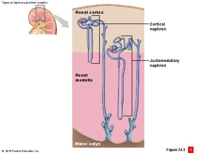
Types of nephrons and their location Renal cortex Cortical nephron Juxtamedullary nephron Renal medulla Minor calyx © 2015 Pearson Education, Inc. Figure 24. 3 3
Gross and Microscopic Anatomy of the Kidney Nephrons • Microscopic functional units of the kidney • Two types 1. Cortical nephrons • 85 percent of all nephrons • Located primarily in the cortex • Responsible for most regulatory functions 2. Juxtamedullary nephrons • 15 percent of all nephrons • Long nephron loop extending deep into medulla • Essential to producing concentrated urine © 2015 Pearson Education, Inc.
Segments of a Nephron components • Two components 1. Renal corpuscle • Blood pressure forces water and solutes out of the glomerular capillaries in a process called filtration • Produces filtrate (protein-free solution, similar to blood plasma) • Collected in the surrounding capsular space 2. Renal tubule • Tubular passageway up to 50 mm long • Receives filtrate and modifies it to create urine © 2015 Pearson Education, Inc.
Segments of a Nephron segments • Renal corpuscle • Glomerular capsule (cup-shaped chamber) • Capillary network (glomerulus) • Proximal convoluted tubule (PCT) • Reabsorbs nutrients from the filtrate (now called tubular fluid) © 2015 Pearson Education, Inc.
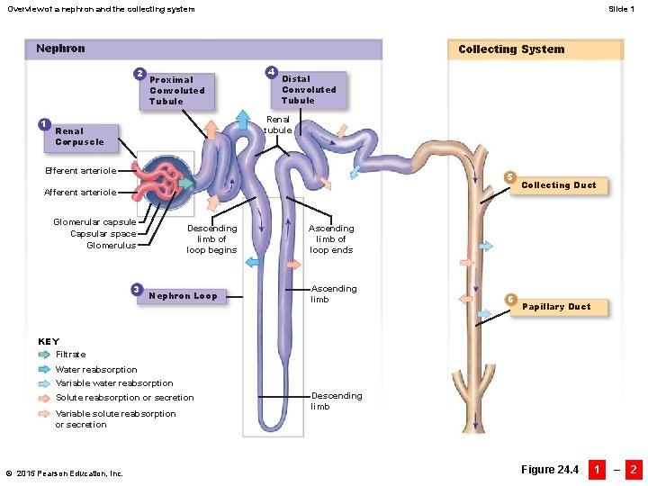
Overview of a nephron and the collecting system Slide 1 Nephron Collecting System 2 1 Proximal Convoluted Tubule 4 Distal Convoluted Tubule Renal tubule Renal Corpuscle Efferent arteriole 5 Afferent arteriole Glomerular capsule Capsular space Glomerulus 3 Descending limb of loop begins Nephron Loop Collecting Duct Ascending limb of loop ends Ascending limb 6 Papillary Duct KEY Filtrate Water reabsorption Variable water reabsorption Solute reabsorption or secretion Variable solute reabsorption or secretion © 2015 Pearson Education, Inc. Descending limb Figure 24. 4 1 – 2
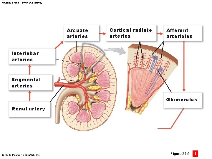
Arterial blood flow in the kidney Arcuate arteries Cortical radiate arteries Afferent arterioles Interlobar arteries Segmental arteries Glomerulus Renal artery © 2015 Pearson Education, Inc. Figure 24. 5 1
Circulation Patterns in the Kidney Arterial system • Renal artery delivers blood to kidney branching into: • Segmental arteries in the renal sinus, which branch into: • Interlobar arteries running within renal columns and branching into: • Arcuate arteries that arch along the boundary between the renal cortex and renal medulla and branch into: • Cortical radiate arteries that branch into: • Afferent arterioles that supply each nephron, specifically a capillary knot known as a glomerulus © 2015 Pearson Education, Inc.
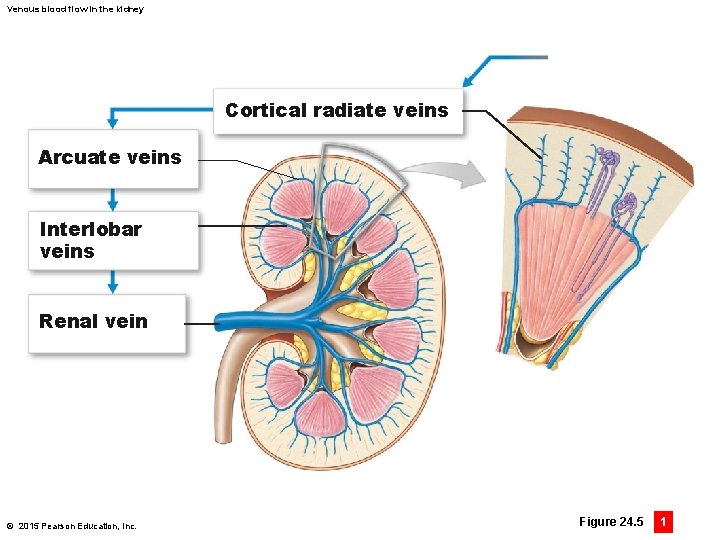
Venous blood flow in the kidney Cortical radiate veins Arcuate veins Interlobar veins Renal vein © 2015 Pearson Education, Inc. Figure 24. 5 1
Circulation Patterns in the Kidney Venous system • Cortical radiate veins collect blood from the capillaries of the nephron and drain into: • Arcuate veins, then to: • Interlobar veins, which drain into the: • Renal vein, which drains into the inferior vena cava © 2015 Pearson Education, Inc.
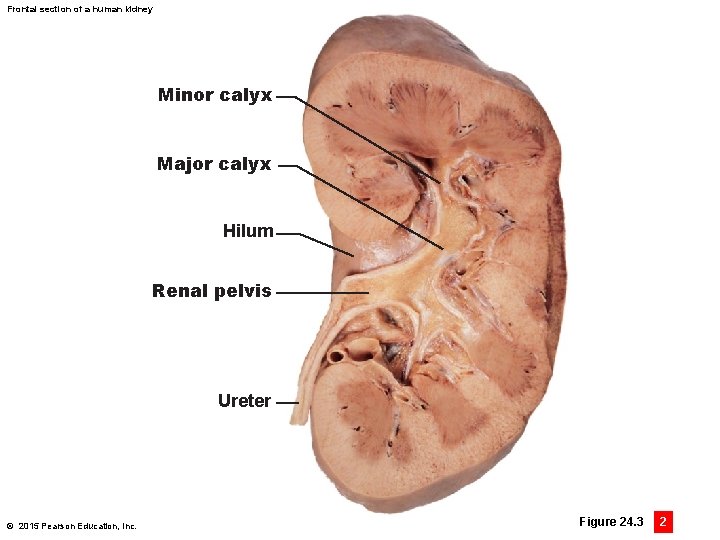
Frontal section of a human kidney Minor calyx Major calyx Hilum Renal pelvis Ureter © 2015 Pearson Education, Inc. Figure 24. 3 2
Urinary System: § Exit Ticket: § Describe the pathway of blood flow into the kidney. What arteries are being pass through (in order)?
- Slides: 12