URINARY SYSTEM Kidneys are large lobulated structure Right

URINARY SYSTEM

• Kidneys are large lobulated structure. • Right kidney is placed below the last rib upto 2 nd and 3 rd lumber transverse process. • Left kidney is placed more ventral under 2 nd or 3 rd to 5 th lumber vertebrae. • The ureters run and enters the dorsal wall of the urinary bladder. • The male urethra is divided into pelvis and extra pelvic part. • The urethra extends down from the pelvic part around the ischial arch down ventrally as a part of penis and gradually tapers down the preputial part of penis. • The penis of ram is similar to that of a bull except the free part and glans.

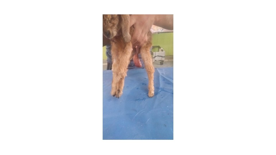
• In the terminal part urethra extends beyond the glans as the urethral process which is about 4 cm long. Obstructive Urolithiasis • Calculi form in both castrated and uncastrated males and also in females. • Obstructive urolithiasis is problem of castrated adult males. • Also recorded in uncastrated male calves of cows and buffaloes. • The obstruction in the flow of urine due to calculi may occur in any part of the urinary tract but mostly in urethra.

• Most common site in cattle is distal to sigmoid flexure. • Frequently the calculus gets lodged between the ischial arch and sigmoid flexure. • Sometimes may obstruct the neck of urinary bladder. • In few cases large calculus obstruct the urethra and multiple calculi recovered from the bladder. • Rare cases obstruct the ureters.

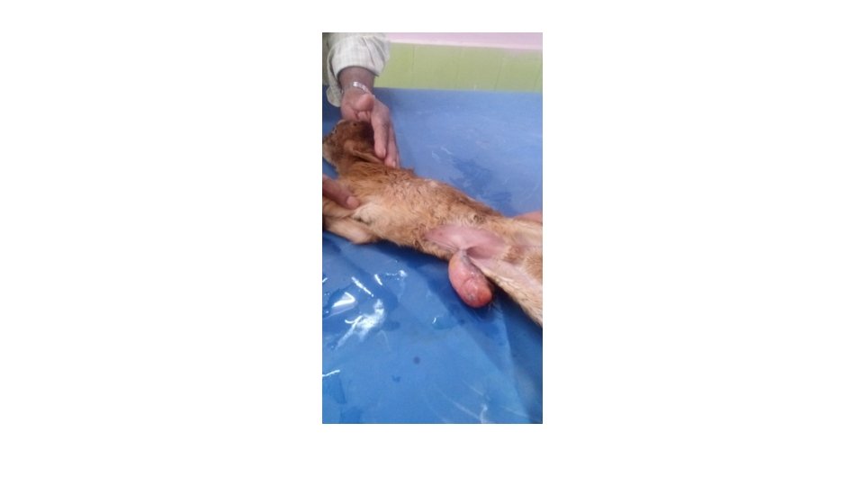
• In buffaloes, calculi are mostly lodged at the glans penis or at the sigmoid flexure and also obstruct neck of urinary bladder. • Most common site in sheep are urethral process at distal end of penis and region of sigmoid flexure. • Clinical Examination: • Obstruction in flow of urine leads to futile and painful attempt by the animal to void urine. • In delayed cases urinary bladder gets rupture (cystorrhexis) • In case of nephrolith – No relief from pain.

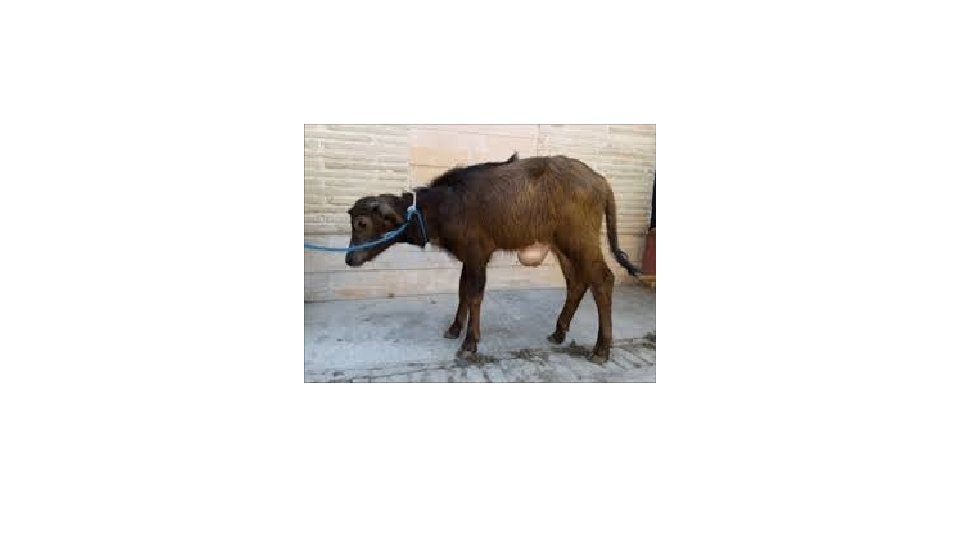
• Clinical signs of retention of urine – • Anuria and less severe in partial retention. • Dry muzzle, sunken and anaemic eyes, salivation, rough coat, turgid skin, anorexia and suspended rumination. • The animal passes dry mucous coated balls and variable degree of dehydration. • If bladder intact rectal palpation reveals a fully distended urinary bladder.

• UB crosses physiological limit gets ruptured. • Subserous or incomplete rupture usually causes atony of bladder such cases are more difficult to treat following complete rupture.

• Rectal exam reveals splashing sound and collapse bladder difficult to palpate. • Where tear as on dorsum, UB bladder usually contains some urine and hang down in abdominal cavity. • Fluid thrill on abdominal ballotment. • Water belly evident. • Paracentesis abdominis done for correction • In sharp edges of calculus have reptured urethra subcutaneous infiltration of urine occurs. • Cellulitis may over perineal and preputial regions • Necrosis of area involved.

• The abdominal fluid Creatinine concentration is 2 -3 times higher than plasma conc. • Severe degree of dehydration, laboured breathing , arched back, and protruded rectal mucosa indicate poor prognosis. • In nephritis and atonic bladder have little chance of recovery. • Location of calculous diagnosis is difficult due to sigmoid flexure prevent catheterisation. • Radiography is also difficult to locate.

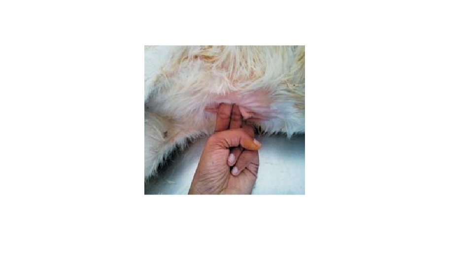
POST SCROTAL URETHROTOMY • Bovine restrained in a lateral recumbency • 20 -25 cm long incision site from scrotum to ischial arch prepared for emergency. • LA is given on midline about 20 cm length • Incise the skin and S/C tissue, muscle bluntly dissected. • Minimal haemorrhages in post scrotal area • Penis comes out by passing finger • Fascial covering removed by gentle incision • Penis can be exteriorized by strengthening the sigmoid flexure • Urethra lies n urethral groove ventrally. • Thoroughly examined to palpate the calculi.

• Nick given on site of calculi and pressed to remove the calculi • Firmly lodged calculi require Allies tissue or Artery forceps to hold and pull out. • Sterilised Polyethylene tube is passed inside urethra • Size of tube used to snuggly fit the lumen • The other end of catheter is passed down urethra and anchored with the prepucial sheath to prevent dislodgement • Snuggly fitted catheter prevent seepage of urine and allows healing of wound without suture also prevent narrowing of lumen. • Muscle are also left unsutured. • Skin and S/C suture is closed with Hz mattress or interrupted suture using non-absorbable suturing material. • About 2 -3 cm catheter protrude beyond the preputial opening.

• Catheter is kept for 3 -4 weeks.

ISCHIAL URETHROTOMY • Penis can not be pulled out in incision in the ischial arch • On linear incision on ischial arch • On exposing the penis directly faces the surgeon. • The catheter is passed through the urethral nick into the bladder and other end is anchored in the ischial region • Catheter is dislodged by movement of tail. • This technique is commonly used in Western country. • After a few days animal used for meat purpose.

CYSTORRHAPHY • Rupture of urinary bladder most common in complete destruction. • Dorsal tear sometimes heals spontaneously • Ventral tear required surgery. • Repair of urine bladder taken after removal of calculi • Cystography is done in standing position • Left laparotomy is performed after linear infiltration of xylocaine 2% below the external of the ileum. • Laparotomy incision should be large as much to introduce both hand into abdominal cavity. • Urine is siphoned out from ab cavity as much possible • Urinary bladder is located and presence of calculi and blood clot should carefully removed.

• Catheter is passed through tear in the bladder and also passed through post-scrotal urethrotomy upto entire length of urethra 2 -3 cm beyond the post- preputial sheath. • The upper layer of urinary bladder is weak and necrotic subtotal cystectomy required • Urinary bladder is sutured by continuous lock stitch suture using absorbable suture material. • Second row of continuous suture is also used to seal the wound • Remove the blood clots, cast cells, shreds etc. • Abdomen is closed in a routine manner.

• RUPTURE OF URETHRA : • It might occur due to irregular sharp edge calculus causes trauma leading to seepage urine in subcutaneous tissue. • Swelling observed in the urethral area. • Multiple stab incision in the affected area and sufficient hydrotherapy help to drain urine and lessen its irritant effect. • Magnesium sulphate – glycerine paste in the initial stage and bismuth –Iodoform paraffin paste (BIPP) and other antiseptic and fly repellent creams helps to remove necrotic tissue. • Antibiotic and analgesic should be given.

TRAUMETIC-RETICULIPERITONITIS • TRP is disease of gastric – intestinal tract in cattle. • Cattle and buffalo ingest foreign bodies due to their indiscriminate feeding habit. • Due to nutritional deficiency ingest various types of foreign bodies. • Sometime animal may consume ropes, plastic, sheets etc. • Metallic foreign bodies also recovered from reticulum. • Foreign bodies are swallowed straight into the reticulum where they inflict trauma in the reticulum and peritoneum causing traumatic reticulo-peritonitis.

• Higher incidence of TRP found in buffalo than cattle. • Buffalo above 6 yrs of age show very high incidence. • When foreign body is ingested get lodged into honey comb pattern at the reticulum. • Non potential foreign bodies like nuts, coins and stones are harmless and may passed out ultimately through faeces. • Potential foreign bodies like nails, needles and sharp wires may cause complications other than reticulitis. • Sequelae to potential foreign bodies may cause traumatic sternebrae, traumatic pericarditis, vagal indigestion, pyothorax, abscessation of liver and spleen, diaphragmatic hernia, traumatic pneumonia and pleurisy.

• Extensive adhesions of the reticulum with the diaphragm or other structure might interfere with the reticular contractions and eructations process. • Localised peritonitis may be developed. • Clinical Signs: - Reccurent tympany, complete or partial anorexia, retarded or suspended rumination, less milk yield. • In foreign body syndrome, chronic tympany may be absent. • In acute cases reduction in milk yield. • Stiffness of forelimb and abducted elbow also observed. • Grunt, nearly abnormal heart, distressed respiration and regurgitation may occur advanced cases of DH.

• Diagnosis: - History and clinical signs. • Neutrophilia with shift to left • Radiography showed nature and extent of damage caused by foreign body. • IB, s are located in the lower cranial, lower caudal and mid central part of the reticulum. • Treatment : • Rumenotomy done to remove foreign body. • Rumenotomy is indicated to remove impacted feed, trichobezors, phytobezors and other foreign body.

RUMENOTOMY • Incision parallel and lateral to last 5 cm below lumber transverse vertebrae. • It is easier to approach the hand upto reticulum. • Local infiltration is given at the site of incision. • Furious animal require light sedation for normal standing. • Operative area thoroughly cleaned and shaved. • After scrubbing drap the area for operation • Animal kept in trevis and left area facing the surgeon.

• The skin and muscle should be incised enough to enter hand of surgeon to explore the outer surface of rumen and reticulum. • Weingarth rumenotomy ring fitted at the site • The front of ring is fixed to the dorsal wound by thumb screw. • A part of rumen is brought out of the abdominal incision and one of rumen forcep is fixed to the dorsal part of the rumen wall. • The forceps is then hooked into the dorsal eye of the frame. • Similar procedure repeated at ventral part of rumen at a distance of 10 -12 cm from the dorsal forcep. • Exposed part of the rumen incised and hooked at the cut edges of rumen wall. • Scalpel should be discarded.

• After evacuating some contents of rumen, hand introduced into cavity to explore the foreign body in rumen and reticulum. • Penetrating foreign bodies are generally removed. • A magnet in hand used to remove loose foreign bodies. • Various types of foreign bodies are removed. • An Abscess in the reticulum can be incised by BP blade. • A cold and thick abscess should be cleaned before suturing. • Surgeon again rescrub the hand. • Rumen is sutured with catgut no. 2 using lambert pattern and reinforced with another layer cushing pattern. • Continuous lock stitch suture is using to close the muscle and peritoneum. • Skin suture closed with horizontal mattress pattern.

OMASAL IMPACTION • Secondary to rumen impaction and is result of poor quality food. • The omasum gets distended with stagnation of ingesta and absoprtion of fluid. • Signs: Anorectic, listless and dehydration shows stasis and absence of omasal sounds on auscultation at the level of right elbow at the 9 th intercostal space. • Treatment: 4 -5 litres liquid paraffin or mineral oil is administered through stomach to soften the content. • Rumen is massaged with fist and knee.

• Above treatment fails then rumenotomy. • Mineral oil is given directly into the omasum through tube inserted by rumen incision during rumenotomy to soften the content.

HERNIA • Protrusion of the contents of a body cavity through normal or abnormal opening either to lie beneath the skin or occupy another body cavity. • It may be congenital or acquired. • The primary predisposing cause of acquired hernia is mostly trauma or increased intra-abdominal pressure. • Contents of hernia: • It has tree parts • (i) Hernial ring (ii) Hernial Sac (iii) Hernial Contents

• The hernia sac made up of tissues that enclose the hernia content. • Hernial sac is usually skin, muscular fibres, fibrous tissue and parietal peritoneum. • No hernial sac in DH. • Classification: • Classified according to its location, Functional alteration, content and cause. • Location: • (i) External hernia: - Consists of hernial ring, hernia sac and contents of hernia. ex- Ventral hernia, umbilical hernia. • (ii) Internal hernia Which lacks of hernia sac, eg. DH, gut tie.
 Reducible hernia: Contents of hernia sac can be")
• Functional Alteration: • (i) Reducible hernia: Contents of hernia sac can be returned into original position through hernia opening • (ii) Irreducible hernia: Contents of hernia sac can not be returned to the original position through the hernial opening. • It could be of three types • (i) Hernia with adhesions: -adhesions between sac and its contents prevent reduction. • (ii) Incarcerated hernia: - Hernial content become too voluminous to be replaced through a narrow hernial ring.
 strangulated hernia: - When blood vessels supplying the hernial contents are")
• (iii) strangulated hernia: - When blood vessels supplying the hernial contents are affected to cause complication like necrosis and extensive adhesions. • Contents of sac: - Enterocele- (intestine), epiplocele (omentum), enteroepiplocele (intestine plus omentum), reticulocele (reticulum), Hysterocele (uterus) and vesiculocele (urinary bladder). • Cause- (i) Traumatic hernia : - Which occurs due to injury e. g. ventral hernia. • (ii) Infectious hernia: -which occurs following destruction of abdominal wall due to infections.

• Diagnosis: -External hernia has an external swelling. The swelling varies in shape and size and differentiated from other swellings. Such as abscess, cellulitis, haematoma, cyst and neoplasm. • In reducible hernia, the ring can be easily palpated. Incarcerated hernia ring is not felt easily because it is impossible to reduce the contents. • In strangulated hernia symtoms like abdominal pain, fever and evidence of bowel obstruction may coincide with the swelling • Radiography may be helpful to identify the contents of irreducible hernia in small ruminants and calves. • Exploratory puncture may also helpful to know the contents of swelling.

• UMBILICAL HERNIA • Generally seen in bovine calve, more frequent in female than male. • It can be congenital or acquired in nature. • Acquired hernia is noticed few weeks after birth. • It is hereditary. • The umbilical opening in the fetus allows the passage of the urachus and umbilical blood vessels. • After birth these structure are disrupted or severed and umbilical opening closes around the cord. The wound heals cicatrization and represents umbilicus. • Due to improper closure of the umbilicus opening at birth or from maldevelopment or hypoplasia of the abdominal muscles, a defect may remain in the mid ventral to form congenital opening


• Acquired umbilical hernia occurs primarily due to manual breaking or resecting of the cord to close the abdominal wall. • Excessive straining due diarrhea or constipation or infection of the cord preventing natural closure of the umbilical resulting to hernia. • Clinical signs: - Discrete spherical swelling seen on umbilicus. Contents are usually fat and omentum. Sometime contains loops of small intestines. The sac is formed by skin, fibrous tissue and peritoneum. • Treatment: - Treated by conservative and surgical methods. • Belly bandages reduce the hernia and allows the hernial ring to close by cicatrization. Previously hernial clamp is used to control the hernia

• Radical surgery is performed. • After fasting animal controlled in dorsal recumbency and liberal operation area is prepared for surgery. • Local anesthesia is infiltrated and adequate elliptical incision is made through skin or each side of swelling. • The skin incision are joined on the midline cranially and caudally. • The skin between the two elliptical incision is resected from the hernial sac and discarded. • The skin lateral to the incisions is then undermined to expose the hernial ring. • The sac is freed from the ring by blunt dissection and the intact sac is deposited into the abdominal cavity.

• A circular hernial ring should be converted into an oval shape for smooth closure by incising the ring caudally and cranially with Vshaped incision. • The ring is then closed by placing a series of overlapping sutures through its edges using heavy nonabsorbable suture material. • The Skin is closed by mattress with non-absorbable suture material. • Post-operative management should be done.

VENTRAL OR ABDOMINAL HERNIA Hernia through any part of the abdominal wall other than natural office • If hernia is ventral to the stifle skin fold. • Hernia is in the flank region are lateral abdominal hernia. • Common in ruminants and acquired in nature. • Trauma such kick, horn thrust or violent contact with blunt objects or abscess in the abdominal cavity. • Abdominal distention due to pregnancy or violent straining during parturition •

• Excessive long caudal incision for CS. • Commonly seen in along the costal arch, high or low in the flank, between the last few ribs or in the ventral abdominal wall near the midline. • It is difficult to palpate the ventral hernial ring during initial stage due to oedema or haematoma in the surrounding tissues. • Wait for inflammatory swelling to subside before examination is done to confirm the diagnosis. • this delay may also facilitate repair as recently traumatized tissues do not hold suture firmly. • The hernial sac formed by the skin and subcutaneous tissues may or may not be lined with the peritoneum.

• Lack of peritoneal covering favours the development of adhesions between the viscera and sac and finally may cause incarceration or strangulation. • The nature of hernial contents depend on the site of the herniation. • A hernia in the left flank may contain the rumen. • Treatment: • Herniorrhaphy is elective. • Delay the surgical repair for at least one week after injury. • Prolonged delay may cause complication. • If hernia is complicated due to incarceration or strangulation, immediate surgical intervention required.

• After linear incision skin is separated from swelling. • Hernial Sac is separated from ring. • Viscera separated from adhesion and returned into abdominal cavity. • The thick mass of fibrous tissue involving hernial swelling may be resected. • Ring edge are trimmed to provide raw surface. • The rent in the abdominal is then closed with sutures. • Hernioplasty may be required in some cases.

INGUINAL OR SCROTAL HERNIA • Protrusion of an abdominal organ through the inguinal canal is Bubonocele. • If contents coming up to scrotum is Oscheocele. • Contents may be omentum, intestine or both. • Sometime UB. • Generally acquired in rams and bulls. • Congenital inguinal hernia bovine calves and lambs. • Scrotal hernia may be traumatic in origin - Mounting.

• May be unilateral or bilateral. • The abdominal canal is slite like space between abdominal muscle. • CLINICAL SIGNS: - Swelling in the inguinal canal or neck of scrotum. • Bulls refuse to mount. • Enlarged scrotum touches ground in rare cases. • When hernial contents strangulated shows systemic signs. • Rectal examination may help in diagnosis. • TREATMENT: • The animal is tranquilized and kept in lateral recumbency with affected side up.

• The upper hind limb is abducted for maximum exposure. • Local anesthesia infiltrated. • Skin incision over swelling. • The hernial content returned into abdominal. • Ring sutured with overlapping suture. • The incision over sac is closed with simple continuous suture. • Another technique through paralumbar fossa

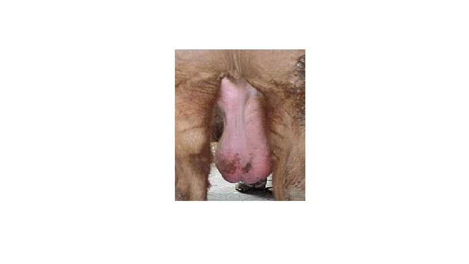
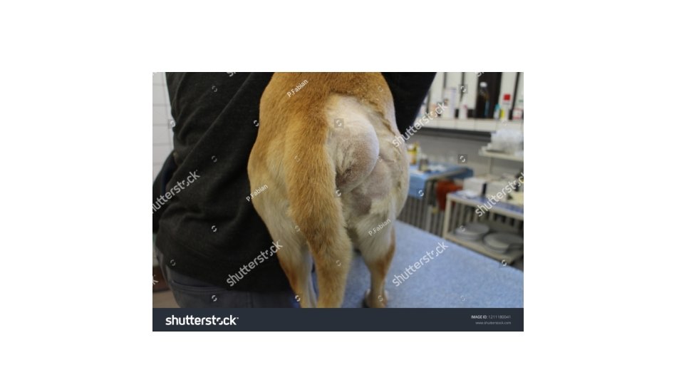
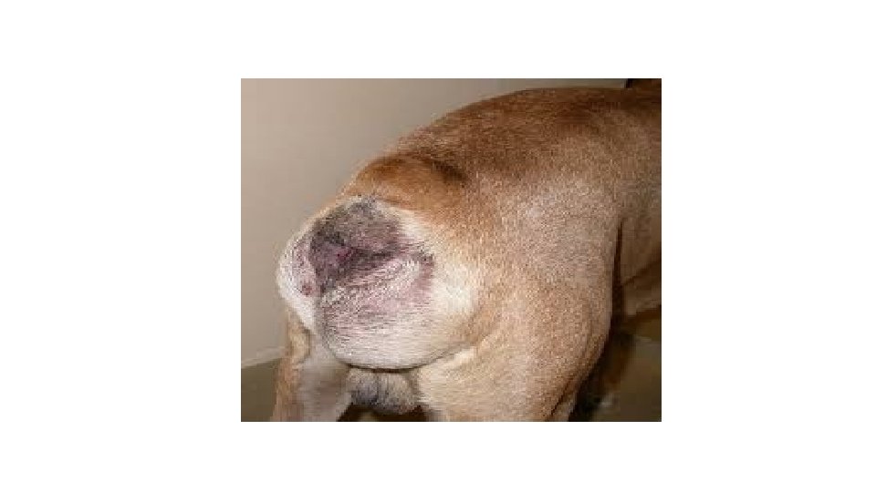
PERINEAL HERNIA • Protrusion of the abdominal or the pelvic viscera through the pelvic diaphragm which supports the rectal wall. • Common in dog. • The perineum is part of the body wall that covers the pelvic cavity outlet while surrounding the anal and urogenital canals. • Tearing of diaphragm muscles leads to herniation. • Reducible swelling observed along the side anus or vulval lips, • Contents are generally UB and retroperitoneal fat.

• Sedation of animal with GA kept in either lateral or sitting position. • A purse string suture placed around the anus after placing gauge plug in the anal opening. • A slightly curve incision given vertically. • Subcutaneous tissue separated expose the organs, • Contents retuned into original position sutured with two layer suture • Skin closed with mattress suture.

DIAGPHRAGMATIC HERNIA • Passage of abdominal viscera into Thoracic cavity. • Congenital or acquired • Commonly reticulum but sometime other organs may also involved. • Frequently in buffalo. • AETIOLOGY: • Weakening of diaphragm by TRP • Congenital weak point of diaphragm • During pregnancy or act of parturion.

• Presence of foreign body in the reticulum • Abscess formation in the diaphragm • Tympany, advance pregnancy, straining at the time of parturion. CLINICAL SIGNS: -Recurrent tympany, abrupt fall in milk yield, scant defecation or diarrhea with smell, regurgitation, aspiration pneumonia , brisket oedema with or without jugular pulsation, abduction of limb, chronic cough, weakness, dehydration. Pathology: - In buffalo DH occurs mostly in right hemidiaphragm -90% and rarely in left 7% or in centre 3%. Generally one hernial ring but occasionally multiple hernial ring. The extent of reticular herniation through diaphragmatic rupture varies in different animals.

• DIAGNOSIS: - History, clinical signs, recent parturition, Muffled heart sound, reticular sound heard cranial to 6 th rib, • Pain or contrast radiography, left flank laparotomy, Exploratory laparotomy and rumenotomy. • Laparo-rumenotomy is primary step. • TREATMENT: • Laparo-rumenotomy, Evacuate the content, Foreign body removed. • Two technique- Thoracic and abdominal. • Kept off feed 48 hours following rumen evacuation. • Corticosteroid given before operation • Animal is anaesthetized with 5% thiopentone and 2 -4% Halothane.

• After intubation endotracheal tube connected to positive pressure ventilator for intermittent positive pressure • Abdominal Approach: - Animal controlled in dorsal recumbency. • Right hemi diagram prepared for surgery. • The abdomen is entered through an incision of 25 to 35 cm in length. • Five cm caudal to xiphoid cartilage and running to costal arch. • Least haemorrhage. • The adhesion between reticulum and diaphragmatic ring are severed by blunt dissection. • The palm of hand is glided through the hernial ring to carefully break the adhesion between reticulum and thoracic organs.

• The reticulum is withdrawn into abdominal cavity and ring is closed with continuous lock stich suture using nonabsorbable suture material. • The suture starts from lower part of the ring. • Abdominal wall is closed with two to three layer. • Thoracic approach: • Right or left lateral thoracotomy. • A cutaneous incision 25 cm in length is made midway on the 7 th rib and extended ventrally to the costo-chondral junction. • The technique of rib resection to the thoracic cavity. • After pleura has been incised the herniated reticulum comes in view.

• Adhesions of the reticulum with the lungs, pericardium and pleura ae separated carefully by blunt dissection. • The reticulum is freed from ring by breaking the adhesions with a gentle blunt dissection. • The reticulum is then pushed into abdominal cavity. • Ring is closed as the same way. • Negative pressure in the chest cavity is created by suction of air. • The respirator is disconnected • The endotracheal tube removed after swallowing reflex. • The animal came into sternal recumbency.

• Post-operatively • Broad spectrum antibiotic 7 -10 days • Analgesic 3 to 5 days. • Adequate fluid therapy. • In brisket oedema diuretics may used. • Suture after 10 to 12 days.
- Slides: 58