URINARY SYSTEM ANATOMY BIO 139 ANATOMY PHYSIOLOGY II

URINARY SYSTEM ANATOMY BIO 139 ANATOMY & PHYSIOLOGY II

Urinary System Organs • Kidneys • Ureters • Urinary Bladder • Urethra
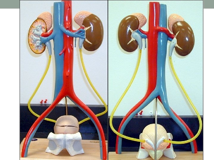

The Kidneys • Posterior abdominal cavity, slightly above waistline • Retroperitoneal • Right kidney lies below left kidney • Due to presence of liver • Supplied by renal artery and renal vein at the renal hilus • Functional unit is the nephron
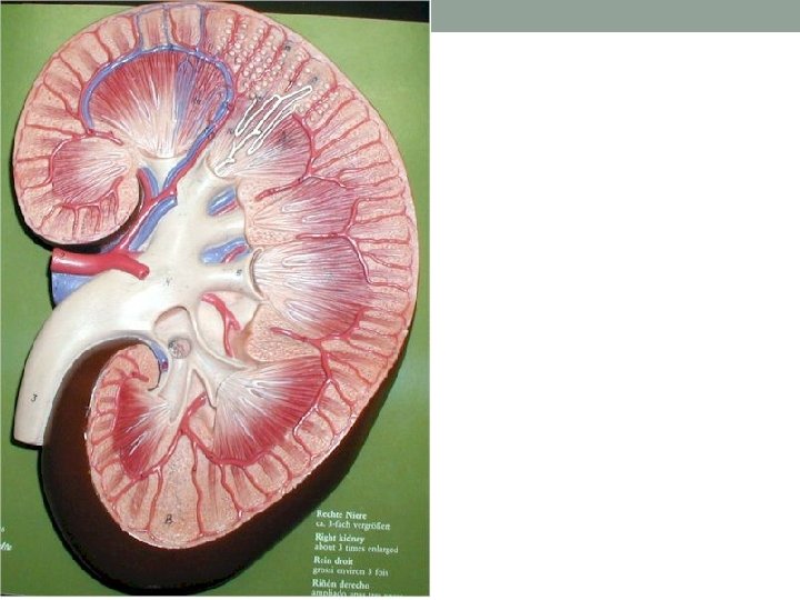
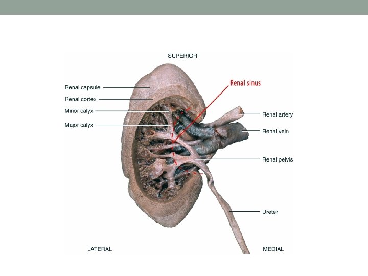
 • Renal Hilus – indention of kidney • Renal Cortex –")
Kidney Anatomy (Coronal) • Renal Hilus – indention of kidney • Renal Cortex – outermost layer, granular • Renal Medulla – striated, composed of renal pyramids (kidney tubules here) • Renal Pyramids • Minor Calyx – receive urine from renal pyramids • Major Calyx – receive urine from minor calyces • Renal Pelvis – funnel shaped portion of ureter; receives urine from major calyces • Ureter

Ureters • 25 cm long tubes • transport urine from the renal pelvis of the kidneys to the bladder through peristalsis

Urinary Bladder • Hollow, distensible organ that sits on the pelvic floor • Has a capacity that averages 700 -800 m. L • Urine exits through the urethra

Location of Male and Female Urinary Bladders Contains Transitional Epithelium • Bladder sits on top of the prostate in males

Male Urinary Bladder Longitudinal section and posterior view of male urinary bladder n. Why does prostatitis affect urinary output in men?
 • Urge to micturate (urinate) occurs when the bladder contains 150 -200")
Urination (Micturition) • Urge to micturate (urinate) occurs when the bladder contains 150 -200 m. L • When volume increases, stretch receptors send signals to a micturition center in the spinal cord triggering a spinal reflex – the micturition reflex. • In early childhood, we learn to initiate and stop the reflex voluntarily. • This then encourages the bladder to expel urine through the external urethral orifice

Histology of Ureter and Urinary Bladder • Inner layer is transitional ET – Distensibility • Smooth muscular layer surrounds the ET Ureter

Urethra • Tube that connects the bladder to the outside of the body • Excretory function in males and females • Also has a reproductive function in males • Passage of sperm • 1 -2 inches long in females • About 7 inches in males

Male and Female Urethras 20 -35

Urinary Anatomy • Kidneys • Ureters • Urinary Bladder • Urethra • External urethral orifice

THE FUNCTIONAL UNIT OF THE KIDNEY IS THE NEPHRON. A nephron is the smallest unit that can make urine.

The Nephron • Functional unit of kidney • 1 million per kidney • Consist of 2 components: • Vascular Component • Tubular Component

Tubular Component of Nephron • Bowman’s Capsule - cortex • Surrounds a glomerulus (capillaries where blood is filtered, 1 st step in urine formation) • Bowman’s capsule receives the glomerular filtrate • Afferent arteriole leads to glomerulus • Efferent arteriole leads away from glomerulus
 –cortex • Receives from Bowman’s")
Tubular Component of Nephron • Proximal Convoluted Tubule (PCT) –cortex • Receives from Bowman’s capsule • Loop of Henle – dips down into the medulla • Descending • Ascending • Distal Convoluted Tubule (DCT) - cortex • Collecting Duct • DCT fluid drains into here and then to the renal pelvis and ureter • Not an actual nephron structure

Tubular Component of Nephron

Histology of Tubular Component • Bowman’s Capsule • Lined by simple squamous epithelium • Filtration • Kidney Tubules • Lined by simple cuboidal epithelium • Secretion and reabsorption

Vascular Component of Nephron • Afferent Arteriole - Branch of renal artery • Sends blood to glomerulus • Glomerulus - Tuft of capillaries in Bowman’s capsule • Filtration of plasma occurs here • Efferent Arteriole • Receives unfiltered blood from glomerulus • Peritubular Capillaries • Form from efferent arteriole • Surrounds tubular component • Eventually drains back into renal vein

Vascular Component of Nephron

Juxtaglomerular Apparatus • Formed where DCT comes in close contact with Bowman’s capsule, between the afferent and efferent arteriole • (see picture from H/O) • Important in regulating kidney function • JG apparatus releases the hormone renin

Nephron Types • Bowman’s capsule & PCT always in renal cortex • Loop of Henle dips into renal medulla • Juxtamedullary • Cortical Nephrons • BC near outer edge of cortex • Loop of Henle only dips slightly into medulla Nephrons • BC in cortex near medulla • Loop of Henle deep through entire length of medulla • Vasa Recta – branches of peritubular capillaries surrounding juxtamedullary nephrons

Nephron Types
- Slides: 27