Urinary bladder nd 2 year Mbbsmc Dr muhammad

Urinary bladder nd 2 year Mbbs-mc Dr muhammad zubair Dr hifza kayani

URINARY BLADDER • IT IS A HOLLOW MUSCULOMEMBRANOUS SAC WHICH ACTS AS A RESORVOIR FOR THE URINE. • IT IS THE MOST ANTERIOR ELEMENT OF THE PELVIC VISCERA. • IT IS A SUBPERITONEAL ORGAN AND HAS PARIETAL PERITONEUM ONLY ON ITS SUPERIOR SURFACE. • URINE ENTERS THE BLADDER VIA URETERS AND EXITS VIA THE URETHRA.

ANATOMICAL LOCATION ▸ When "Empty" , the adult urinary bladder is located in the "Lesser pelvis" lying partially superior to and partially postetior to the pubic Bones. ▸ As the bladder fills it enters the "Greater Pelvis". ▸ In some individuals, a full bladder may ascend to the level of the "Umbilicus". ▸ In infants and young children, the urinary bladder is in the abdomen even when empty. ▸ The Bladder usually enters the Greater Pelvis by 6 Years of age.

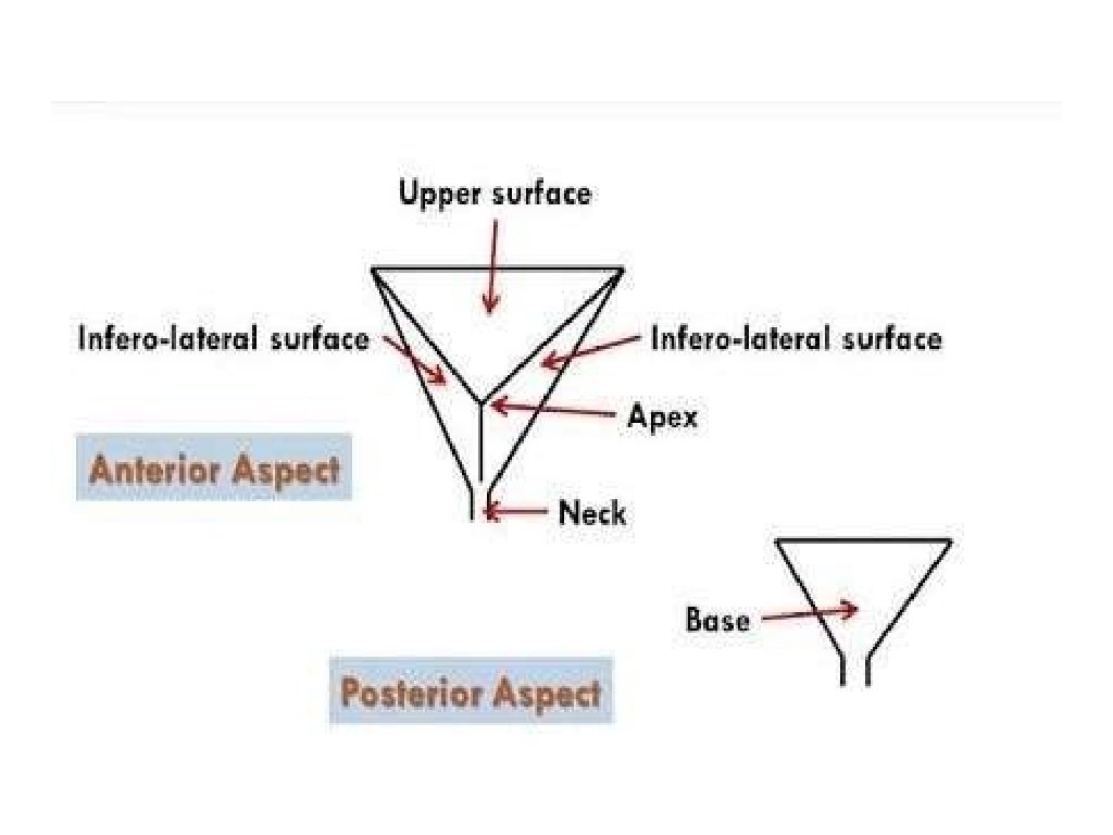
SURFACES OF THE URINARY BLADDER ▸ Superior surface. ▸ Right inferolateral surface. ▸ Left inferateral surface ▸ Posterior surface.


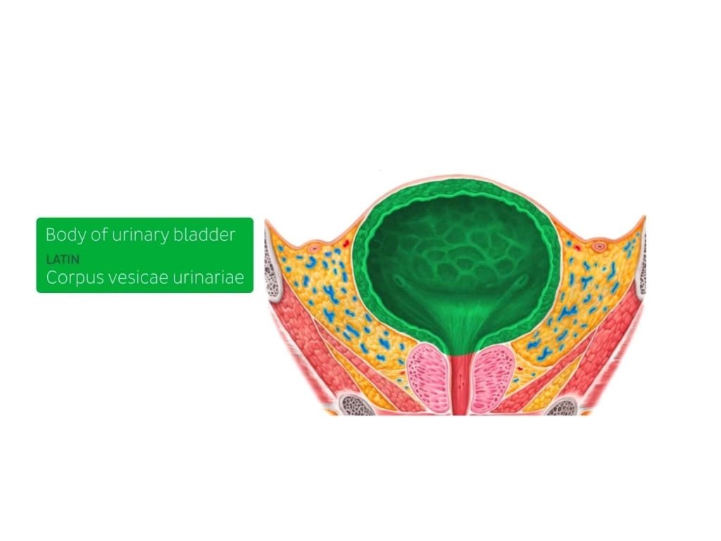
BODY OF URINARY BLADDER ▸ It is lined by Transitional epithelium, ▸ It holds the urine, before it is voided. ▸ It can hold 400 ml to 1000 ml of the urine. ▸ It is located between the apex and the fundus.

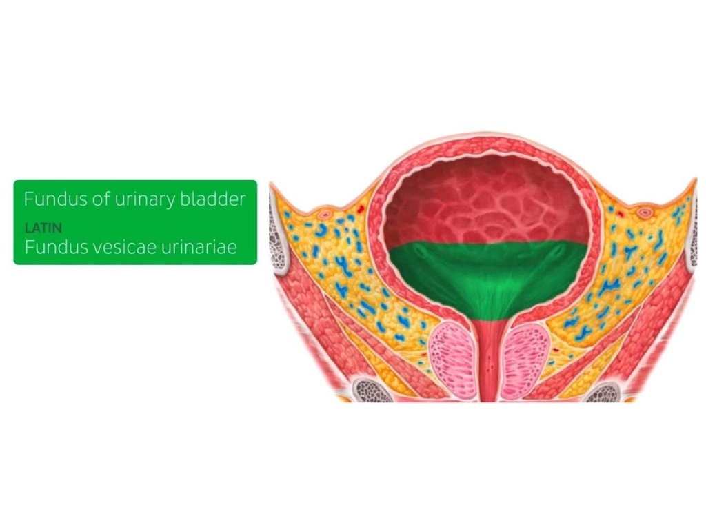
FUNDUS OF THE URINARY BLADDER ▸ It is base of the bladder. ▸ It has the shape of inverted triangle. ▸ It faces postero-inferiorly and , is formed by the posterior wall of bladder. ▸ Trigone of the urinary bladder is found on the fundus.

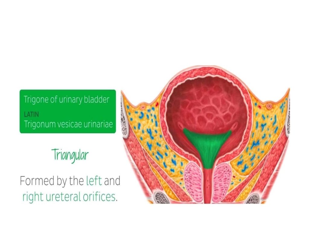
TRIGONE OF URINARY BLADDER ▸ It is smooth triangular part of urinary bladder. ▸ Mucosal lining of trigone is smooth and firmly attached to the underlying wall of the bladder. ▸ Formed by Right and left ureteral orifices. ▸ Once the trigone of urinary bladder is stretched to a certain degree, siganl is sent to the brain that bladder needs to be emptied.

URETERAL ORIFICES ▸ These are Slit like openings through which ureters enter the bladder on the posterolateral angles of the trigone of urinary bladder.

NECK OF URINARY BLADDER ▸ It is the lowest portion of the bladder through which the "Urethra" arrises.

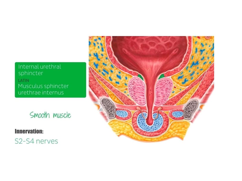
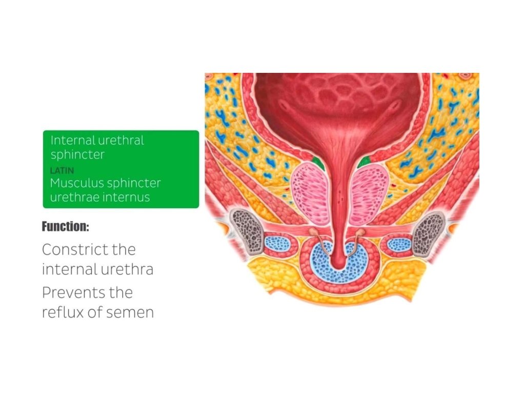
INTERNAL URETHRAL SPHINCTER ▸ It is comprised of smooth muscle that is located at the junction of urethra and the urinary bladder. ▸ It is innervated by S 2 -S 4 nerves of the pelvic plexus. ▸ It's function is to constrict the internal urethra , preventing the urine leakage and also prevents the Retrograde ejaculation ( Ejaculatory Reflex ) of semen into the bladder.

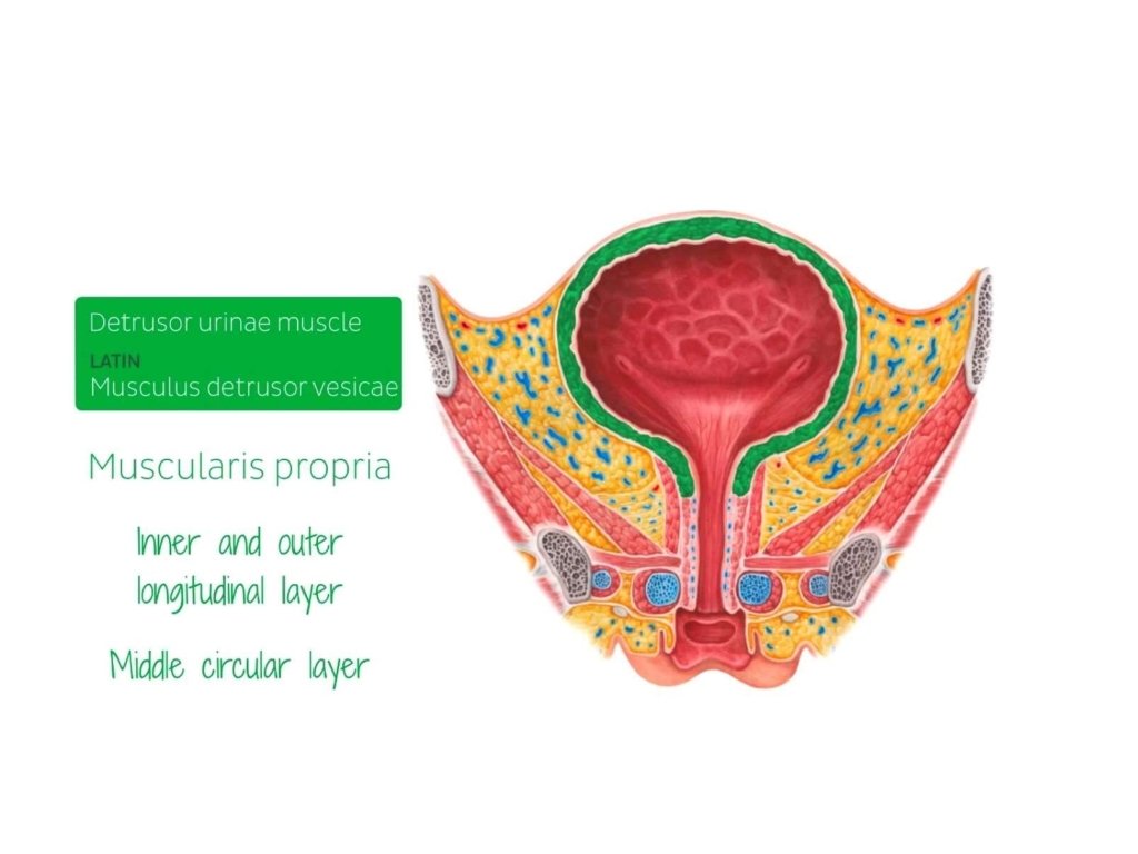
DETRUSOR MUSCLE ▸ It is also referred as " Muscularis Propria". ▸ It is smooth muscle , found around the wall of bladder. ▸ It is comprised of inner and outer longitudinal, and middle circular layer. ▸ This muscle is relaxing during accomulation of urine in the bladder, and contracts only during urination to void and empty the bladder.

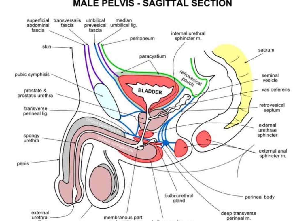
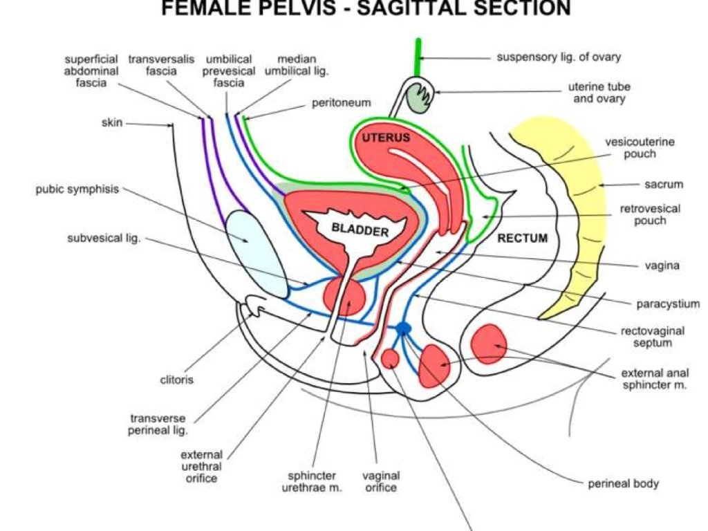
POSTERIOR RELATIONS OF URINARY BLADDER In males : ▸ Vas deferan ▸ Seminal Vesicle ▸ Rectum ▸ Retro. Vesical Fascia ▸ Peritoneum In famales : - ▸ Vagina ▸ Part of Uterus

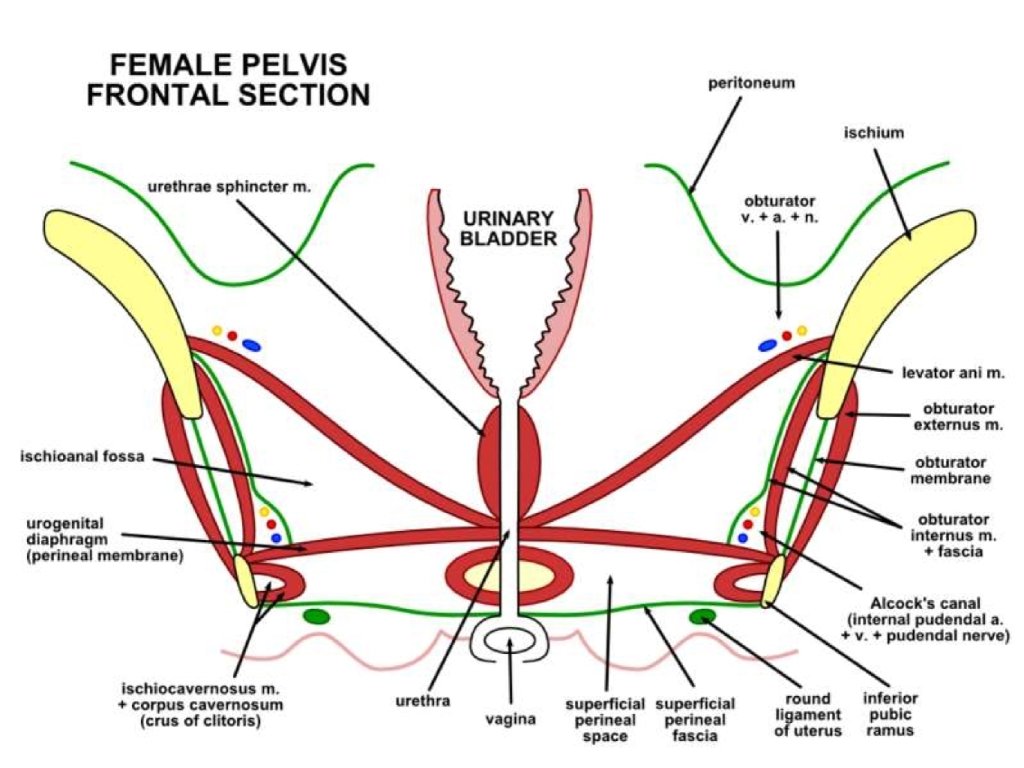
RELATIONS CONTINUED Superior Relations in male: - ▸ Peritoneum ▸ Coils of ileum ▸ Sigmoid colon Superior Relations in female: - ▸ Uterus Lateral Relations : - ▸ Obturator internus ▸ Levator ani

ARTERIAL SUPPLY ▸ Branches of internal iliac arteries. ▸ Superior vesical arteries supply anterosuperior parts of the bladder. ▸ In males, inferior vesical arteries supply the fundus and neck of the bladder. ▸ In females, vaginal arteries replace the inferior vesical arteries and send small branches to posteroinferior parts of the bladder. ▸ Obturator and inferior gluteal arteries also supply small branches to the bladder.
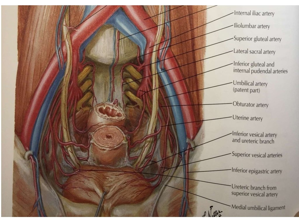

VENOUS DRAINAGE ▸ The veins draining from the bladder correspond to the arteries. ▸ Veins from the Vesical venous plexus drain into the internal iliac veins.
 comes from the sacral region")
INNERVATION OF URINARY BLADDER ▸ Pelvic Nerve (Parasympathetic nerve) comes from the sacral region of spinal cord. It is not under our control. It causes contraction of the Detrusor muscle. ▸ Pudendal nerve (Somatic nerve) causes contraction of External Sphincter. We are firing pudendal nerve when we are trying to hold our urine. ▸ Hypogastric nerve (Sympathetic nerve) causes relaxation of Detrusor muscle and contraction of Internal sphincter. ▸ Afferent Pelvic nerve that is sensory and comes from the detrusor muscle. It is stimulated when the bladder is stretched.

LYMPHATIC DRAINAGE OF BLADDER ▸ In both sexes, lymphatic vessels leave the superior surface of the bladder and pass to the "External iliac lymph nodes". ▸ Those from fundus pass to the "Internal iliac lymph nodes". ▸ Some vessels from the neck of bladder drain into the "Sacral" to "Common iliac lymph nodes".
- Slides: 29