Urinary bladder and Urethra Dr Tabrez Associate Professor

Urinary bladder and Urethra Dr. Tabrez Associate Professor Dept of Anatomy 22/6/2020 MBBS Batch 2019 -20

URINARY BLADDER • IT IS A HOLLOW MUSCULOMEMBRANOUS SAC WHICH ACTS AS A RESORVOIR FOR THE URINE. • IT IS THE MOST ANTERIOR ELEMENT OF THE PELVIC VISCERA. • IT IS A SUBPERITONEAL ORGAN AND HAS PARIETAL PERITONEUM ONLY ON ITS SUPERIOR SURFACE. • URINE ENTERS THE BLADDER VIA URETERS AND EXITS VIA THE URETHRA.

ANATOMICAL LOCATION ▸ When "Empty" , the adult urinary bladder is located in the "Lesser pelvis" lying partially superior to and partially postetior to the pubic Bones. ▸ As the bladder fills it enters the "Greater Pelvis". ▸ In some individuals, a full bladder may ascend to the level of the "Umbilicus". ▸ In infants and young children, the urinary bladder is in the abdomen even when empty. ▸ The Bladder usually enters the Greater Pelvis by 6 Years of age.

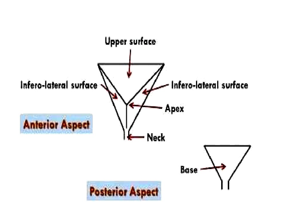
SURFACES OF THE URINARY BLADDER ▸ Superior surface. ▸ Right inferolateral surface. ▸ Left inferateral surface ▸ Posterior surface.
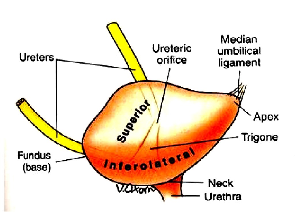

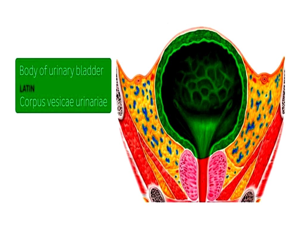
BODY OF URINARY BLADDER ▸ It is lined by Transitional epithelium, ▸ It holds the urine, before it is voided. ▸ It can hold 400 ml to 1000 ml of urine. ▸ It is located between the apex and the fundus.

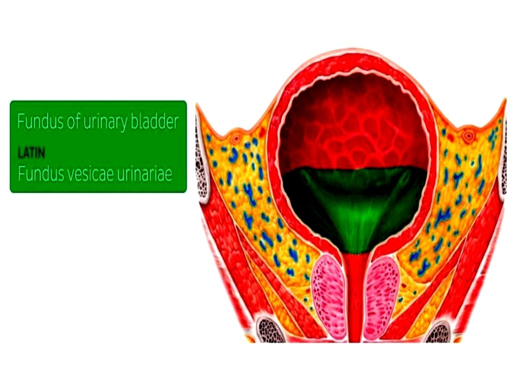
FUNDUS OF THE URINARY BLADDER ▸ It is base of the bladder. ▸ It has the shape of inverted triangle. ▸ It faces postero-inferiorly and , is formed by the posterior wall of bladder. ▸ Trigone of the urinary bladder is found on the fundus.

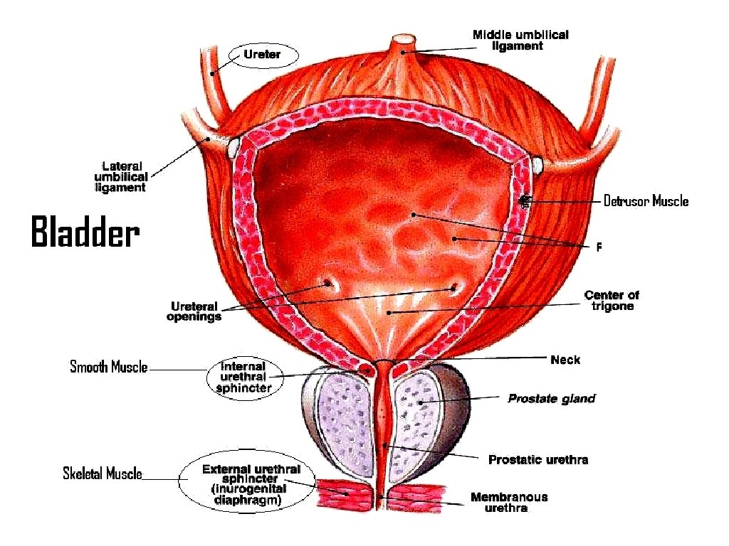
TRIGONE OF URINARY BLADDER ▸ It is smooth triangular part of urinary bladder. ▸ Mucosal lining of trigone is smooth and firmly attached to the underlying wall of the bladder. ▸ Formed by Right and left ureteral orifices. ▸ Once the trigone of urinary bladder is stretched to a certain degree, siganl is sent to the brain that bladder needs to be emptied.
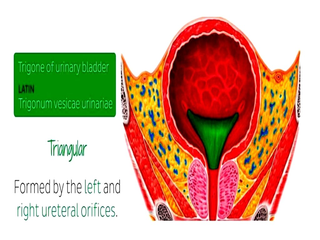

URETERAL ORIFICES ▸ These are Slit like openings through which ureters enter the bladder on the posterolateral angles of the trigone of urinary bladder.

MERCIER'S BAR ▸ It is a mucous membane present between the two ureteral orifices. ▸ It is also called "Inter. Ureteral Fold"

NECK OF URINARY BLADDER ▸ It is the lowest portion of the bladder through which the "Urethra" arrises.

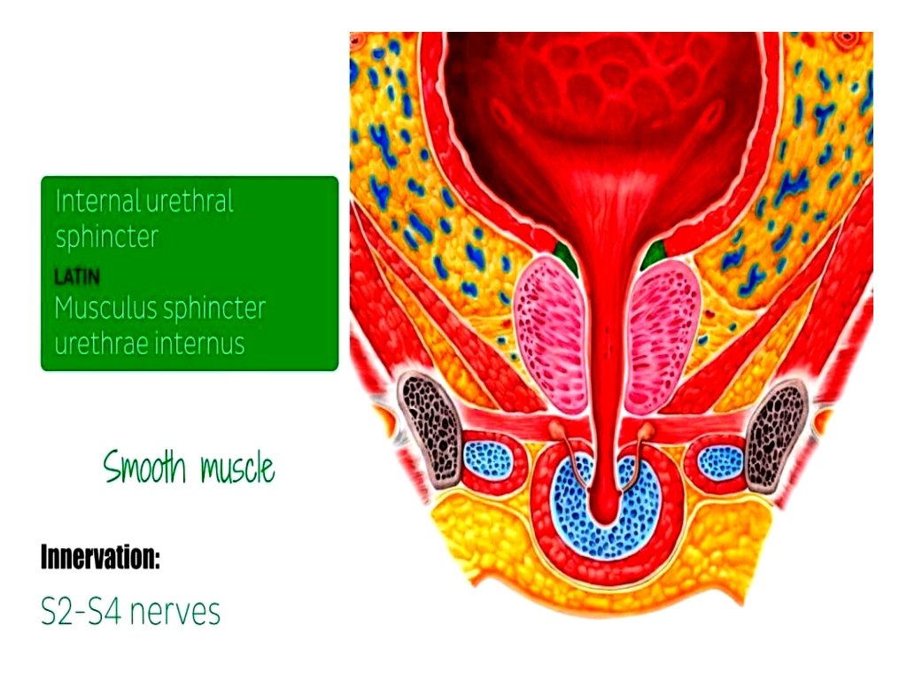
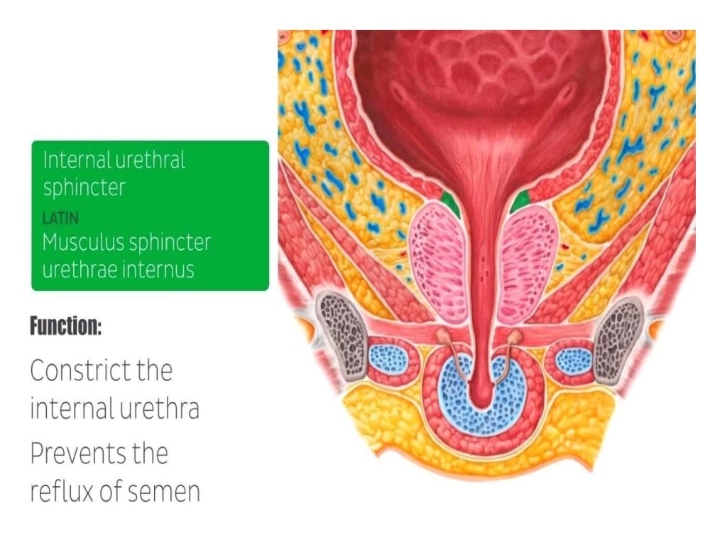
INTERNAL URETHRAL SPHINCTER ▸ It is comprised of smooth muscle that is located at the junction of urethra and the urinary bladder. ▸ It is innervated by S 2 -S 4 nerves of the pelvic plexus. ▸ It's function is to constrict the internal urethra , preventing the urine leakage and also prevents the Retrograde ejaculation ( Ejaculatory Reflex ) of semen into the bladder.

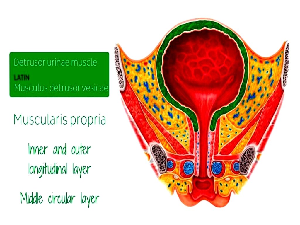
DETRUSOR MUSCLE ▸ It is also referred as " Muscularis Propria". ▸ It is smooth muscle , found around the wall of bladder. ▸ It is comprised of inner and outer longitudinal, and middle circular layer. ▸ This muscle is relaxing during accomulation of urine in the bladder, and contracts only during urination to void and empty the bladder.

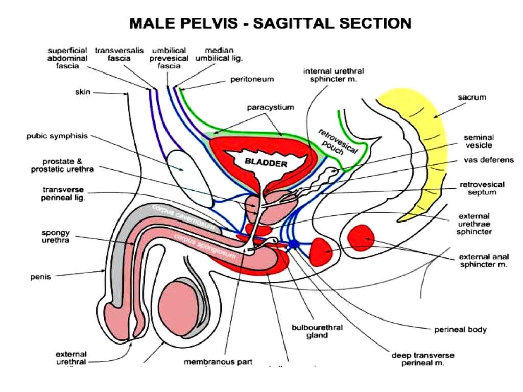
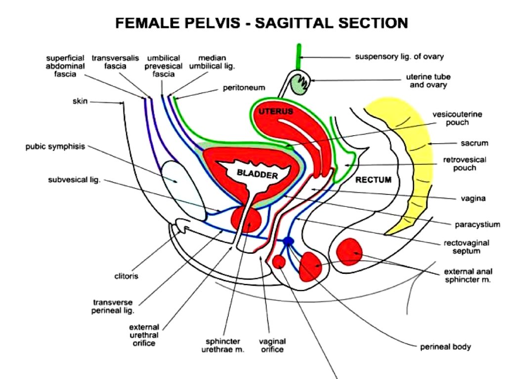
POSTERIOR RELATIONS OF URINARY BLADDER In males : ▸ Vas deferan ▸ Seminal Vesicle ▸ Rectum ▸ Retro. Vesical Fascia ▸ Peritoneum In famales : ▸ Vagina ▸ Part of Uterus

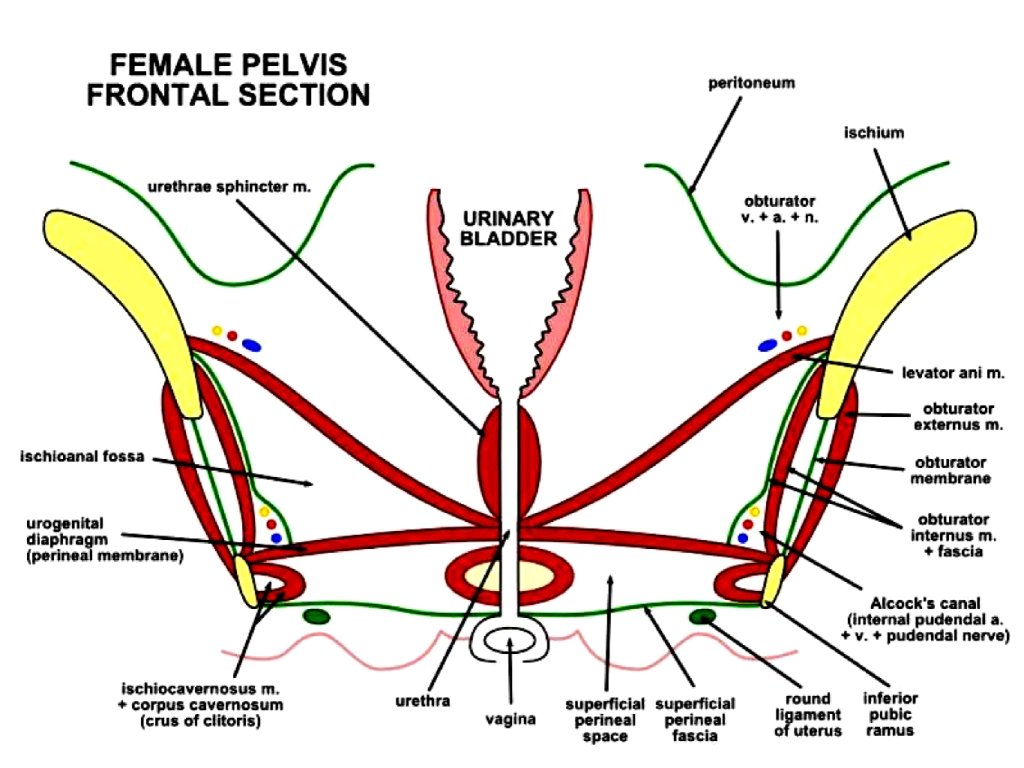
RELATIONS CONTINUED Superior Relations in male: ▸ Peritoneum ▸ Coils of ileum ▸ Sigmoid colon Superior Relations in female: ▸ Uterus Lateral Relations : ▸ Obturator internus ▸ Levator ani

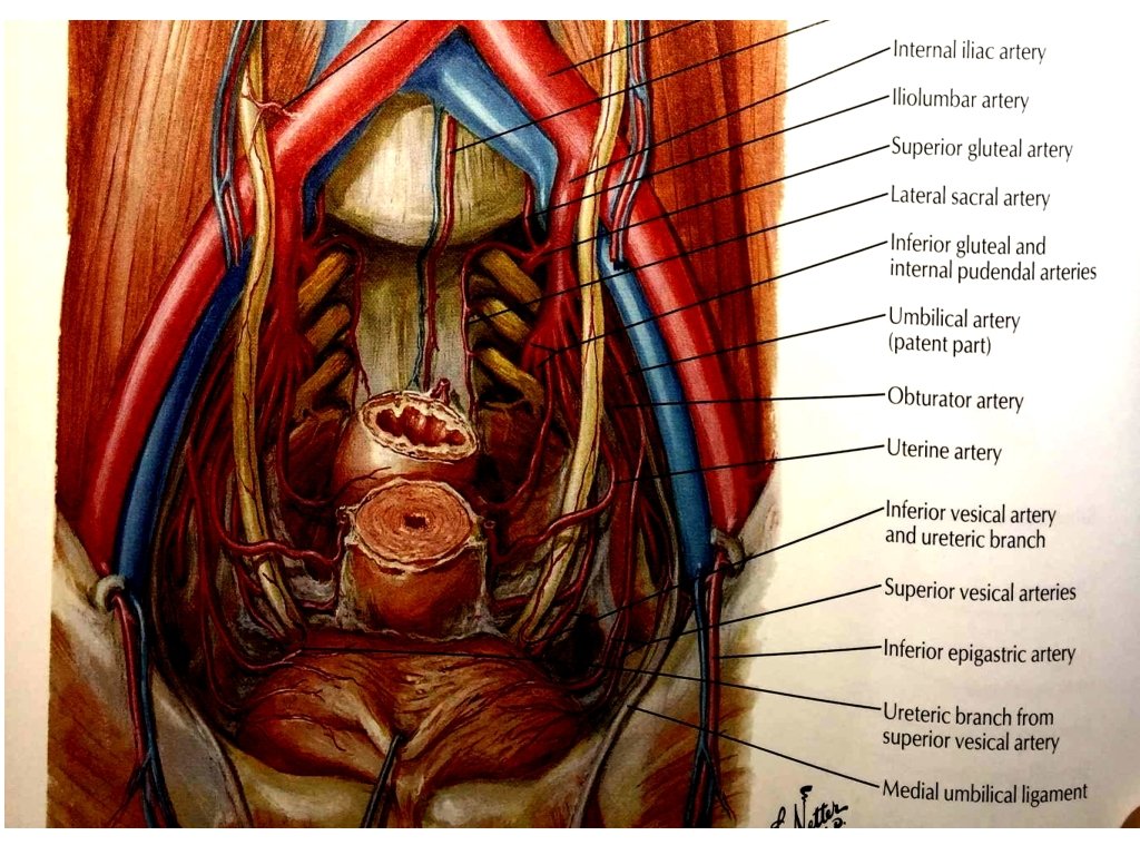
ARTERIAL SUPPLY ▸ Branches of internal iliac arteries. ▸ Superior vesical arteries supply anterosuperior parts of the bladder. ▸ In males, inferior vesical arteries supply the fundus and neck of the bladder. ▸ In females, vaginal arteries replace the inferior vesical arteries and send small branches to posteroinferior parts of the bladder. ▸ Obturator and inferior gluteal arteries also supply small branches to the bladder.

VENOUS DRAINAGE ▸ The veins draining from the bladder correspond to the arteries. ▸ Veins from the Vesical venous plexus drain into the internal iliac veins.
 comes from the sacral region")
INNERVATION OF URINARY BLADDER ▸ Pelvic Nerve (Parasympathetic nerve) comes from the sacral region of spinal cord. It is not under our control. It causes contraction of the Detrusor muscle. ▸ Pudendal nerve (Somatic nerve) causes contraction of External Sphincter. We are firing pudendal nerve when we are trying to hold our urine. ▸ Hypogastric nerve (Sympathetic nerve) causes relaxation of Detrusor muscle and contraction of Internal sphincter. ▸ Afferent Pelvic nerve that is sensory and comes from the detrusor muscle. It is stimulated when the bladder is stretched.

LYMPHATIC DRAINAGE OF BLADDER ▸ In both sexes, lymphatic vessels leave the superior surface of the bladder and pass to the "External iliac lymph nodes". ▸ Those from fundus pass to the "Internal iliac lymph nodes". ▸ Some vessels from the neck of bladder drain into the "Sacral" to "Common iliac lymph nodes".

Urethra • The urethra begins at the base of the bladder and ends with an external opening in the perineum. • The urethra differs significantly in women and men.

Urethra In women • The urethra is short, being about 4 cm long. • It travels a slightly curved course as it passes inferiorly through the pelvic floor into the perineum, where it passes through the deep perineal pouch and perineal membrane before opening in the vestibule that lies between the labia minora.

Urethra In men • The urethra is long, about 20 cm, and bends twice along its course. • Beginning at the base of the bladder and passing inferiorly through the prostate, it passes through the deep perineal pouch and perineal membrane and immediately enters the root of the penis. • The urethra exits the deep perineal pouch, it bends forward to course anteriorly in the root of the penis. • When the penis is flaccid, the urethra makes another bend, this time inferiorly, when passing from the root to the body of the penis. During erection, the bend between the root and body of the penis disappears.
")
Urethra • The urethra in men is divided into: preprostatic, membranous, and spongy (penile) parts.

Parts of the Urethra Preprostatic part Prostatic part • The preprostatic part of the urethra is about 1 cm long. • It extends from the base of the bladder to the prostate, and is associated with a circular cuff of smooth muscle fibers (the internal urethral sphincter). Contraction of this sphincter prevents retrograde movement of semen into the bladder during ejaculation. • The prostatic part of the urethra is 3 -4 cm long and is surrounded by the prostate. In this region, the lumen of the urethra is marked by a longitudinal midline fold of mucosa (the urethral crest). (Read more)

Parts of the Urethra Membranous part • The membranous part of the urethra is narrow and passes through the deep perineal pouch. • During its transit through this pouch, the urethra, in both men and women, is surrounded by skeletal muscle of the external urethral sphincter. Spongy (Penile) urethra • The spongy urethra is surrounded by erectile tissue (the corpus spongiosum) of the penis. • It is enlarged to form a bulb at the base of the penis and again at the end of the penis to form the navicular fossa). • The external urethral orifice is the sagittal slit at the end of the penis.

- Slides: 38