URETERS Pair of narrow thick walled muscular tubes

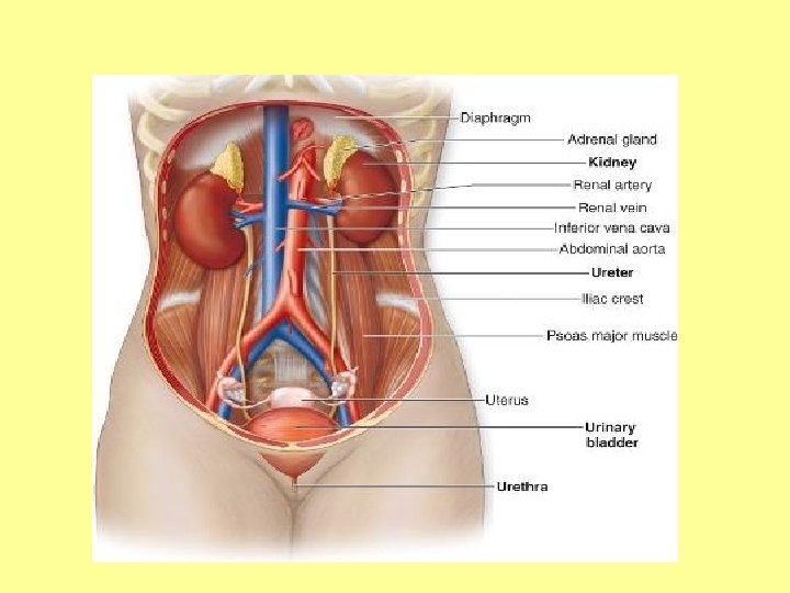

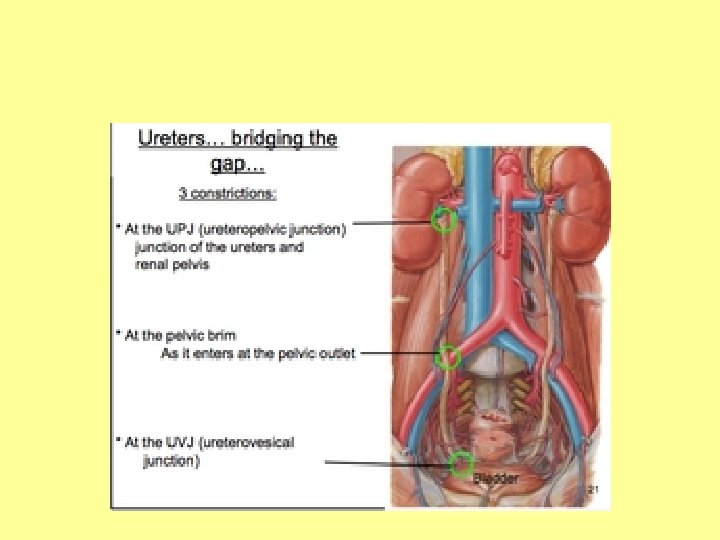
URETERS • Pair of narrow, thick walled muscular tubes which convey urine from kidneys to the urinary bladder. • 25 cms long & 3 mm in diameter. • lower half in abdomen, upper half in pelvis. • Constricted at 3 places: i. At pelvi ureteric junction. ii. At brim of lesser pelvis. iii. At its passage through bladder wall.

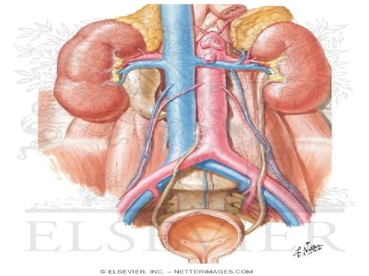
COURSE: § Begins within the renal sinus as renal pelvis. § Descends along medial margin of kidneys and narrows at the lower end of kidney. ABDOMINAL PART : Passes downwards and slightly medially on psoas major muscle to enter pelvis by crossing in front of termination of common iliac artery.

PELVIC PART OF URETER • Crosses in front of the bifurcation of common iliac artery to reach the pelvis. • Descends downward & backward, along the lower border of internal iliac artery, crossing (from above downward): 1. External iliac artery & vein 2. Obturator nerve, artery & vein curves forward & medially.

TERMINATION: • It reaches the posterosuperior angle of urinary bladder. • It runs an oblique course of about 2 cm through the wall of bladder before it opens into its lumen (intramural part of ureter). This part forms a valve-like mechanism that prevents reflux of urine into the ureter when bladder is distended.

RELATIONS : 1. RENAL PELVIS : Anteriorly- Right ureter –Right renal vessels and 2 nd part of duodenum Left ureter – Left renal vessels, pancreas, peritoneum, jejunum Posteriorly - psoas major muscle.

2. ABDOMINAL PART OF URETER : Anteriorly. Right ureter- 3 rd part of duodenum, peritoneum, right colic vessels, ileocolic vessels, gonadal vessels, root of mesentery, terminal part of ileum. Left ureter – Peritoneum, testicular artery, left colic vessels, sigmoid colon, sigmoid mesocolon.

Posteriorly – Psoas major, tips of transverse process of lumbar vertebra, genital branch of genitofemoral nerve. Medially – Right ureter- IVC, abdominal aorta. Left ureter–Left gonadal vein, abdominal aorta.

IN MALE • It is crossed anteriorly by vas deferens

IN FEMALE • It passes below the root of broad ligament, lateral to lateral fornix of vagina & is crossed superiorly by the uterine artery.

BLOOD SUPPLY Upper part – renal vessels. Middle part – branches from abdominal aorta/tributaries into the inferior vena cava. Pelvic(lower)part –branches from vesical, middle rectal, uterine vessels.

NERVE SUPPLY Sympathetic – T 10 – L 1 Parasympathetic – S 2 - S 4

THANK U
- Slides: 18