Ureteropelvic junction obstruction Intern Supervisor Identification n n

Ureteropelvic junction obstruction 報告者: Intern 黃暉程 Supervisor: 主治醫師: 邱元佑

Identification n n n Name: 黃小弟 Birth date: 05/31/03 → 19 d/o G 2 P 2, NSD, Apgar score: 9’→ 10’ GA 41 weeks, BW: 3000 g(10~25%) BL: 52. 5 cm(10~25%), HC: 35 cm(10~25%) DOIC(-), PROM(-)

Chief complaint: left abdominal mass for 2 days
;")
Present Illness GA 28 -30 wks Prenatal exam at 姚博琳’s clinic: Left hydronephrosis; Oligohydromino(-); Other abnormality() 92/05/31 One mass over left abdomen noted by his mother 92/06/18 92/06/16 GA 41 wks, NSD: Renal echo at 姚博琳’s clinic: Hydronephrosis is not identified Brought to Dr. 邱: A mass over LUQ palpable
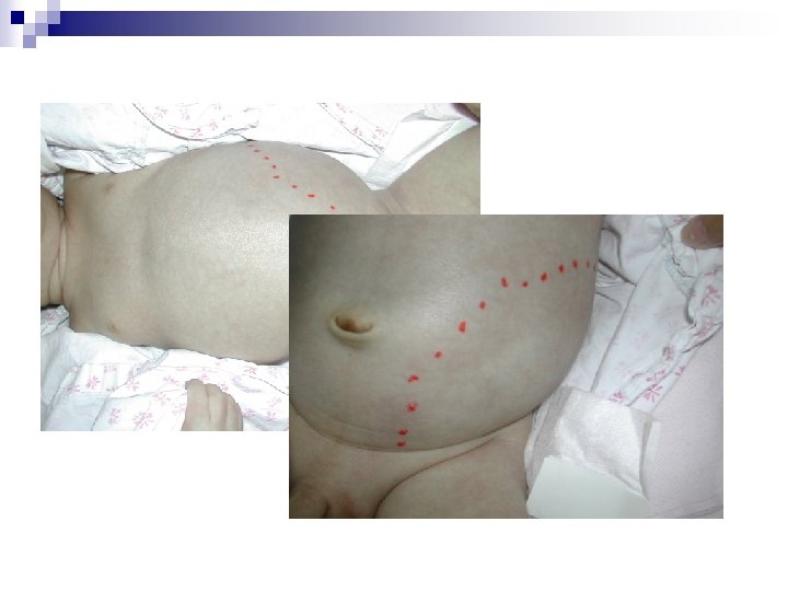

One 11 x 7 cm soft mass over left abdomen; percussion: spongy-filling

Abdominal mass ~ approach n Inspection, Percussion, Palpation

Abdominal mass by age n Age-group 1 m~1 yr Newborns After 1 yr

Differential diagnosis n Non-urologic Abdominal distention, pyloric stenosis, hepatosplenomegaly, intestinal obstruction, malignany, feces n Urologic Hydronephrosis, cystic disease, Wilms’ tumor, neuroblastoma, distended bladder
 Left severe hydronephrosis Cortex thickness: about 0. 2 cm AP")
Renal echo (Jun 18) Left severe hydronephrosis Cortex thickness: about 0. 2 cm AP diameter: 4. 48 cm (>1. 5 cm) Right moderate hydronephrosis No parenchyma involvement AP diameter: 1. 2 cm (>1 cm) Imp: suspect left ureteropelvic junction obstruction
 VCUG (2) Antegrade pyelography 92/06/18 Left PCN 92/06/19 Admission PE LAB:")
Present Illness (1) VCUG (2) Antegrade pyelography 92/06/18 Left PCN 92/06/19 Admission PE LAB: CBC/DC, Biochemistry, U/A 92/06/27 92/06/23 92/7/2: discharge (1) Left dismembered pyeloplasty (2) Pathologic Dx: Muscular hyperplasia and fibrosis, compatible with stenosis

Indication of PCN n n n n Obstruction with infection Obstruction without infection Stone disease Prelude to endoscopic/ interventional procedures Delivery of medications/ chemotherapy Urinary leaks Urinary diversion for hemorrhagic cystitis
 Imp: No evidence of vesicoureteral reflux")
VCUG (Jun 23) Imp: No evidence of vesicoureteral reflux

Antegrade pyelography: Left UPJ stenosis is considered
 VCUG (2) Antegrade pyelography Left PCN 92/06/19 92/06/27 92/06/23 Discharge !")
Present Illness (1) VCUG (2) Antegrade pyelography Left PCN 92/06/19 92/06/27 92/06/23 Discharge ! 92/7/2 (1) Left dismembered pyeloplasty : UPJ obstruction, high insertion (2) Pathologic Dx: Muscular hyperplasia and fibrosis, compatible with stenosis

Whitaker test during operation n Measure the pressure gradient between the pelvis & the bladder under fixed infusion rate Less than 12 mm. Hg: no obstruction Above 20 mm. Hg: obstruction Pressure gradient was 14~15 mm. Hg → 1. intermediate 2. good compliance of pelvis and ureter n

Diagnosis Left UPJ stenosis

Discussion UPJ obstruction

UPJ obstruction generally a congenital condition n male, left-sided lesions predominating n most frequently diagnosed cause of urinary obstruction in children n causes hydronephrosis which may damage the kidney n

Pathology Various interpretationsn Preponderance of longitudinal muscle fibers n Excessive collagen fibers in & around muscle bundles n Compromised or attenuated muscle bundles Our case: moderately lymphocytic infiltration & focal suppurative inflammation

Symptoms & signs Back or flank pain n UTI with fever old children n Hematuria n Abdominal mass → infants n }

Diagnosis & tests Prenatal Maternal pregnancy ultrasound: hydronephrosis n Postnatal Ccr, BUN, electrolytes, AP, DTPA, MAG 3, VCUG n

Etiology Intrinsic: Narrowed, dysfunctional or adynamic segments n Extrinsic: Upper ureter is angulated, kinked or compressed by bands or adhesions n

Intrinsic obstruction mechanical: narrowed → incomplete embryological ureteric bud recanalization; muscular invaginations overdevelop as flaps or valves n functional: adynamic or dysfunctional segment → inability to initiate, form or conduct peristaltic waves across the UPJ n

Extrinsic obstruction vessel or fibrous band may pass anterior to the pelvis & ureter: most common n may secondary to intrinsic disturbance which produces pelvic overdistension & rotation n high insertion of the ureter into the pelvis n

Extrinsic ~ High insertion

Whitaker test: flow across UPJ obstructions Pressure dependent Volume dependent Intrinsic obstruction Extrinsic obstruction

Treatment influenced by renal function, infection n surgical correction of the obstruction n infants: dismembered pyeloplasty n adults: percutaneous or endoscopic technique n a nephrostomy stent is placed to drain urine until the patients heals n

Surgical indication Bilateral UPJO n Palpable mass n Unilateral UPJO with hydronephrosis Grade 4 (Massive pelvic & calyceal dilatation with thinned parenchyma); DTPA < 30% or worsen > 10% in f/u n
 Poulsen et al, 1987 35")
Prognosis ~ pyeloplasty Author and Year Patients/Kidneys Success (%) Poulsen et al, 1987 35 100 O’Reilly, 1989 30 83– 93 Mac. Neily et al, 1993 75 85 32/33 (<2 mo old) 97 30/33 (>2 mo old) 93 100 98 79 90 Austin et al, 2000 135/137 91 Houben et al, 2000 186/203 93 Shaul et al, 1994 Salem et al, 1995 Mc. Aleer and Kaplan, 1999

Prognosis ~ pyeloplasty

Expectantions Rapid decompression of the kidney immediately following birth can substantially improve kidney function in an infant with UPJ obstruction diagnosed before the child is born. n Most patients do well with no long-term consequences n

Complications n Permanent loss of kidney function-renal failure n require dialysis at some point in their lives as a result of this problem

Thanks for your attention!
- Slides: 34