Upper Respiratory Tract Lectures Objectives Describe the structure

Upper Respiratory Tract

Lectures Objectives • Describe the structure of nasal cavity including nasal septum. • Describe the structure of lateral wall of nasal cavity including conchae and meatuses. • Locate the openings of the paranasal air sinuses and naso‐lacrimal duct in the meatuses. • Describe nasal innervations, blood supply, and its relation to epistaxis. • Study the structure of nasopharynx and associated openings with their clinical importance. • Describe the structure of various cartilages and membranes of the larynx. • Describe muscles of the larynx including their action, nerve and blood supply. • Describe the structure of vocal cords and the mechanism of voice production and control of air passageway.

Nose

Nose • External nose – portion visible on face • Internal nose – large cavity beyond nasal vestibule • Internal nares or choanae • Ducts from paranasal sinuses and nasolacrimal ducts open into internal nose • Nasal cavity divided by nasal septum • Nasal conchae subdivide cavity into meatuses • Increase surface area and prevents dehydration • Olfactory receptors in olfactory epithelium
 • Ala")
External Nose • Parts Root Dorsum Apex Naris (nostrils, anterior nasal apertures) • Ala • •

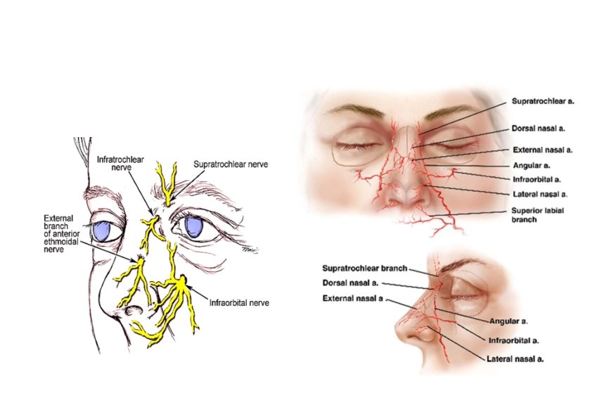
External Nose • Skeleton of the nose • Bony part • Frontal, nasal, maxillary bones • Cartilaginous part • Lateral, septal, alar cartilages • Blood supply: branches of ophthalmic, maxillary & facial aa. • Nerve supply • Infratrochlear (V 1) • External nasal (V 1) • Infraorbital (V 2)

Nasal septum • Bony part • Perpendicular plate of ethmoid • Vomer • Cartilaginous part ‐ Septal cartilage

Nasal Cavity • Parts • Nasal vestibule • Nasal septum • Choanae (posterior nasal apertures) • Walls of the nasal cavity • Floor • Roof • Lateral wall • Sphenoethmoidal recess • Nasal conchae • Superior, middle, & inferior • Meatuses • Superior, middle, & inferior

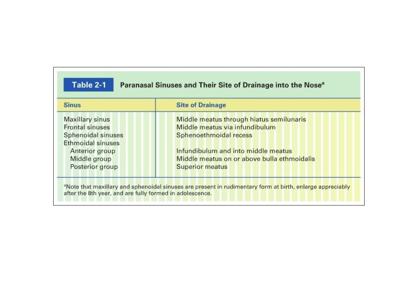
Nasal Meatuses • Sphenoethmoidal recess Sphenoid sinus • Meatuses • Superior Posterior ethmoid sinus • Middle • Bulla ethmoidalis Middle ethmoid sinus • Hiatus semilunaris Maxillary sinus • Infundibulum Frontal sinus Anterior ethmoid sinus • Inferior Nasolacrimal duct

Paranasal Sinuses • Paired cavities in ethmoid, sphenoid, frontal and maxillary bones • Lined with mucous membranes and open into nasal cavity • Resonating chambers for voice, lighten the skull • Sinusitis is inflammation of the membrane

Paranasal Sinuses Maxillary sinus • Between floor of orbit and roots of upper molars and premolars • Superior alveolar nn. (V 2) Frontal sinus • Supraorbital nn. (V 1) Sphenoid sinus • Body of sphenoid • Posterior ethmoidal nn. (V 1) Ethmoid sinus • Anterior, middle & posterior • Between nasal cavity and orbit • Anterior & posterior ethmoidal nn. From nasociliary n. (V 1)

Paranasal Sinuses: X‐ray

Nasal Cavity: Innervation • Olfactory nerve • Trigeminal nerve • Ophthalmic… • Maxillary …

Nasal Cavity: Blood Supply • Anterior & posterior ethmoidal aa. • From ophthalmic a. • Sphenopalatine a. • From maxillary a. • Septal branch from facial a. • Kiesselbach’s area & plexus • Epistaxis Lymph drainage • Deep cervical lymph nodes • Vestibule – submandibular lymph nodes

Pharynx
 hanging from skull • Skeletal muscle &")
Pharynx • Muscular tube (5 inch long) hanging from skull • Skeletal muscle & mucous membrane • Completed posteriorly & deficient anteriorly (openings into nose, mouth & larynx) • Extends from internal nares to cricoid cartilage (C 6) • Funnel shape – wide superiorly & narrow inferiorly (1. 5 cm) • Functions • Passageway for food and air • Resonating chamber for speech production • Tonsil (lymphatic tissue) in the walls protects entryway into body

Regions of the Pharynx • Distinct regions ‐‐ nasopharynx, oropharynx and laryngopharynx

Nasopharynx Above soft palate • Openings • Internal nares • Pharyngeal isthmus • Auditory (Eustachian, pharyngotympanic) tube • Structures • Pharyngeal tonsil (adenoids) • Tubal elevation • Tubal tonsils • Salpingopharyngeal fold • Salpingopharyngeus m. • Pharyngeal recess

Oropharynx From soft palate to epiglottis • Structures At floor • Posterior ⅓ of tongue • Lingual tonsils • Median glossoepiglotic fold • Lateral glossoepiglotic folds • Valleculae

Oropharynx At lateral wall • Palatoglossal fold • Palatoglossus m. • Oropharyngeal isthmus • Palatopharyngeal fold • Palatopharyngeus m. • Palatine tonsil

Laryngopharynx • Extends from epiglottis to cricoid cartilage • Posterior to laryngeal inlet • Piriform fossa • Between aryepiglotic fold and thyroid cartilage

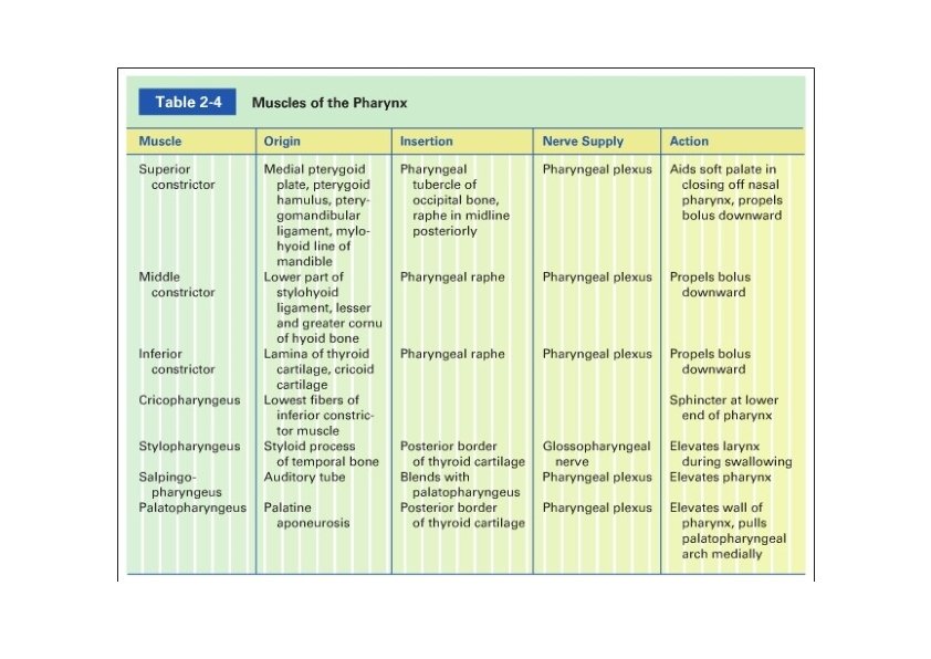
Pharyngeal Muscles • External circular muscles Inserts into the pharyngeal raphe • Superior, middle, & inferior constrictor mm. • Overlap each other (inferior is more superficial) • Cricopharyngeus m. • Lower end • Sphincter • Internal longitudinal muscles • Stylopharyngeus m. • Palatopharengeus m. • Salpingopharyngeus m. • Nerve supply: pharyngeal plexus except stylopharyngeus muscle (IX)
 • Oropharynx (IX) • Laryngopharynx (X)")
Pharynx • Sensory innervation • Nasopharynx (V 2) • Oropharynx (IX) • Laryngopharynx (X) – internal laryngeal nerve • Blood supply • Ascending pharyngeal aa. • Tonsillar branch of facial aa. • Branches of maxillary & lingual aa. • Lymph drainage • Deep cervical nodes • Retropharyngeal or paratracheal nodes → deep cervical

Larynx Cartilage & connective tissue tube Below hyoid bone Anterior to C 4 to C 6 Short passageway connecting laryngopharynx with trachea • Constructed of 3 single & 3 paired cartilages • Functions • • • Passageway for air • Voice production • Prevent entrance of food

Larynx • Relations • Infrahyoid mm. • Thyroid gland • Major blood vessels

Larynx: Cartilages

Larynx: Cartilages • Thyroid cartilage The largest Incomplete ring • Laminae • Laryngeal prominence (Adam’s apple) • Oblique line • Superior & inferior cornua • Cricothyroid joints
 •")
Larynx: Cartilages • Cricoid cartilage Complete ring Below thyroid cartilage • Lamina (posteriorly) • Cricoarytenoid joints • Arch (anteriorly) • Arytenoid cartilages • Apex, base, vocal process, & muscular process
 • Stalk – attached to")
• Epiglottis • Leaf shape cartilage (elastic cartilage) • Stalk – attached to thyroid cartilage • Aryepiglottic fold • Median and lateral glossoepiglottic folds • valleculae Larynx: Cartilages
 • Cuneiform")
Larynx: Cartilages • Corniculate cartilages • Above arytenoids (attachment of aryepiglottic fold) • Cuneiform cartilages • In the aryepiglottic fold (support)

Larynx: Membranes & Ligaments • Thyrohyoid membrane • Median thyrohyoid ligament • Cricotracheal ligament • Quadrangular membrane • Between epiglottis & arytenoid • Vestibular ligament (inferior margin) • Vestibular fold • Immovable, vascular (pinkish) • Cricothyroid ligament • Vocal ligament (superior margin) • Vocal fold • Mobile, avascular (whitish) • Rima glottidis (glottis)

Larynx: Cavity • Inlet of larynx • Orientation • Boundaries Vestibule • Between inlet & vestibular folds Middle region • Between laryngeal folds • Laryngeal sinus (ventricle) • Laryngeal saccule Lower region (infraglottic cavity) • Between vocal folds & lower border of cricoid
")
Larynx: Muscles • Extrinsic muscles • Elevators • Suprahyoids (Digastric, stylohyoid, mylohyoid, & geniohyoid) • Longitudinal pharyngeal (stylopharyngeus, salpingopharyngeus, & palatopharyngeus) • Depressors (infrahyoid) • Sternothyroid, sternohyoid, & omohyoid

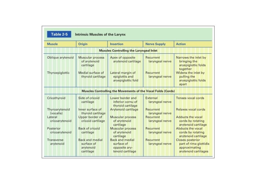
Larynx: Muscles • Intrinsic muscles • Modification of laryngeal inlet • Narrowing • Oblique arytenoid m. • Widening • Thyroepiglottic m. • Movement of vocal cords • Tensing • Cricothyroid m. • Relaxing • Thyroarytenoids (vocalis) m. • Adducting • Lateral cricoarytenoid m. • Abducting • Posterior cricoarytenoid m. • Approximating aretyneoids • Transverse arytenoid m.

Larynx: Muscles

Voice Production • Vocal folds are adducted • Muscle contraction pulls elastic ligaments which stretch vocal folds out into airway • Vibrate and produce sound with released air (frequency or pitch) • Folds can move apart or together, elongate or shorten, tighter or looser • Androgens make folds thicker and longer – slower vibration and lower pitch • Quality of voice determined by other structures (mouth, lips, tongue, pharynx, soft palate, & teeth)
 • Above vocal cords: internal laryngeal")
Larynx • Nerve supply • Sensory innervation (X) • Above vocal cords: internal laryngeal n. • Below vocal cords: recurrent laryngeal. • Motor innervation • All intrinsic muscles innervated by recurrent laryngeal except cricothyroid muscle (external laryngeal n. ) • Blood supply • Upper half: superior laryngeal branch of superior thyroid a. • Lower half: inferior laryngeal branch of the inferior thyroid a. • Lymph drainage • Deep cervical nodes
- Slides: 41