UPDATE ON UVEAL MELANOMA Presented by Dr Carlo

UPDATE ON UVEAL MELANOMA Presented by Dr. Carlo J. Pelino Assistant Professor Retina / Emergency Service The Eye Institute Philadelphia Pa 7/29/2016 October 13 th, 2016
 also called neoplasm. a new growth of tissue characterized")
Tumor Definition tumor /tu·mor/ (too´mer) also called neoplasm. a new growth of tissue characterized by progressive, uncontrolled proliferation of cells. The tumor may be localized or invasive, benign or malignant. A tumor may be named for its location, for its cellular makeup, or for the person who first identified it. Mosby's Medical Dictionary, 8 th edition © 2009, Elsevier.

Lecture Outline • Choroidal Nevus • Choroidal Melanoma • Congenital Hypertrophy of the RPE • Choroidal Metastasis • Melanocytoma • Combined Hamartoma of the Retina & RPE • Choroidal Hemangioma • Choroidal Osteoma • Retinal Astrocytic Hamartoma • Retinoblastoma

Conjunctival melanoma Cutaneous Melanoma Updated ways to diagnose uveal melanoma New treatment plans for uveal melanoma Gene expression profiling Adjuvant treatments - liver metastasis

Retinal Anatomy Retinal and Choroidal Anatomy
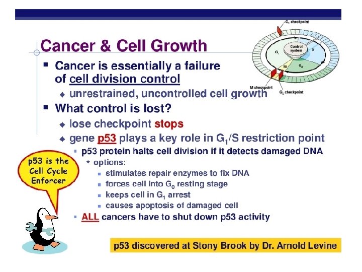

Protect p 53 = Natural food sources include: Glutathione found in sulfur foods such as onions, garlic, milk thistle Curcumin (23) found in turmeric Anthocyanins (24) found in berries, red cabbage & red onions among others Resveratrol found in berries and grape skins Carotenoids (27) found in a number of fruits, vegetables and pasture-raised animal foods

Longevity: • Insulin is very low • IGF is low • m. TORC 1 is low • AMP kinase is high • Ras is low 1/8/2016

Uveal melanoma is the second-most common form of melanoma and the most common primary intraocular malignancy. Up to one-half of patients are at risk for fatal metastatic disease. 7/29/2016

Population by Race for the US: 2012 RACE Total population. . . 313, 914, 040 100. 0% Caucasian. . . . 244, 852, 951 78. % Source: quickfacts. census. gov

Gene Mutations in Cutaneous Melanoma

Uveal Melanoma Stats – SEER Database Incidence 4. 3 cases per / million Gender: Race: Age: Male / Female 52: 48 W / B 150: 1 Overall 60. 4 mean Incidence of uveal melanoma has remained the same over the last 25 years 1/8/2016

Where does uveal melanoma come from Choroidal Nevus • Most common intraocular tumor • Proliferation of choroidal melanocytes • Present in ~ 7. 9% of Caucasians • Growth is rare after puberty ? ?
 and melanin producing cells (melanocytes)")
Nevus of Ota have increased amounts of melanin (pigment) and melanin producing cells (melanocytes) in and around their eyes. This includes the intraocular blood vessel layer called the uvea (choroid, ciliary body and iris), on the sclera, and in the eyelids. 7/29/2016 Risk Factors of Melanoma

• Baseline fundus photography • Consider OCT if location permits • Consider B-Scan if suspicious • Yearly dilated fundus examination Choroidal Nevus: Treatment & Management
 q Fair skin q Propensity to")
q Caucasian ethnicity q Light colored eyes (blue) q Fair skin q Propensity to burn when exposed to UV light q Cutaneous nevi or freckles q Iris nevi q Welders Risk Factors of Melanoma 7/29/2016

Nevoma 7/29/2016 Make sure to photodocument and measure

To Find Small Ocular Melanoma • • • T= thickness F= subretinal fluid S= symptoms O= orange pigment M= tumor margin touches disk • • No risk factors (<4%) 1 risk factor (36%) 3 risk factors (50%) 5 risk factors (70%) DOCUMENTED GROWTH - MEANS EVERYTHING Using Helpful Hints = Ultrasound hollow, Halo absent 7/29/2016

Special Testing OCT • Can detect sub-retinal fluid • Has been shown to detect early seeding • Helpful in monitoring response to treatment • Enhanced depth imaging B Scan • Acoustically hollow • Choroidal excavation with orbital shadowing • Classic mushroom appearance (less common) • Can identify extraocular extension FA • No pathognomonic pattern • Typically, mottled fluorescence during arteriovenous phasefollowed by leakage and staining CAN J OPHTHALMOL—VOL. 42, NO. 2, 2007

Collaborative Ocular Melanoma Study • Organized and funded in 1985 to address issues related to the management of choroidal melanoma. > 4000 patients. 65% pts eligible • Primarily to study the overall survival of patient following treatment Small melanomas < 2. 5 mm in height Medium melanomas 2. 5 – 10. 0 mm in height Large melanomas > 10. 0 mm in height • Secondary outcomes = metastasis-free survival, years of useful vision 7/29/2016

Treatment d e pends on • Enucleation • Radioactive plaques • Proton beam radiotherapy Most widely accepted … 1. state of ot her eye 2. lo cation 3. extent of t u m or 3. vision 4. • s iz Transpupillary e 5. health 6. age of p thermotherapy t • Local resection Less common Treatment & Management

Treatment Options for Uveal Melanoma It is left in place for 4 to 7 days to provide 8, 000 centigray of radiation to the entire tumor. The remainder of the body receives a small amount of radiation, about the equivalent of 1 chest x-ray.

• Using fluorescence in situ hybridization and molecular assay techniques, several genetic abnormalities in uveal melanoma were found on chromosomes 1, 3, 6, and 8 • Monosomy 3 • Found in up to 50% of uveal melanomas • Imparts a worse prognosis. • In small melanoma it provokes the argument for earlier treatment than observation. 7/29/2016 Role of Cytogenetics
 divides uveal melanoma into 2 molecular subgroups: Class 1 A")
Gene expression profiling (GEP) divides uveal melanoma into 2 molecular subgroups: Class 1 A and B (low risk) and Class 2 (high risk) GEP allows oncologists to accurately predict which patients with uveal melanoma will get metastatic disease. Technology is now available as a routine clinical test (Decision. DX-UM, Castle Biosciences) Improving the prognosis for uveal melanoma 7/29/2016

Mutations in the Gq alpha subunits GNAQ and GNA 11 are mutually exclusive and represent early or initiating events that constitutively activate the MAPK pathway. Mutations in BRCA 1 -associated protein-1 (BAP 1) and splicing factor 3 B subunit 1 (SF 3 B 1) also appear to be largely mutually exclusive, and they occur later in tumor progression. BAP 1 mutations are strongly associated with metastasis (liver) SF 3 B 1 mutations are associated with a more favorable outcome. BAP 1 mutations can arise in the germ line, leading to a newly described BAP 1 familial cancer syndrome (mesothelioma, cutaneous melanoma, basal cell carcinoma and renal cell carcinoma) 7/29/2016 Cell-Signaling Advances in Uveal Melanoma

Types of Choroidal Melanoma Class 1 A tumors account for about 45% of all uveal melanomas Class 1 B tumors about 15% Class 2 about 40%. The five-year risk of metastasis is less than 5% for Class 1 A, about 15% for Class 1 B, and 70 -80% for Class 2. The goal is to be able to offer adjuvant therapy to all Class 2 patients and to selected Class 1 B patients in the near future. 7/29/2016

Adjuvant Therapy Adjuvant: A substance that helps and enhances the effect of a drug, treatment, or biologic system. From the late 16 th century: from Latin adjuvant- 'helping toward', from the verb adjuvare, from ad- 'toward' The first class of compounds consists of various types of agents that activate the patient’s immune system to kill tumor cells. Such agents include interferon and ipilimumab or Yervoy. 7/29/2016

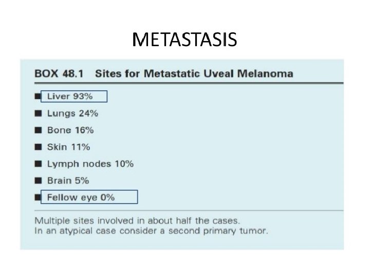
Choroidal Melanoma • 10 year mortality for uveal melanoma – Large 50% – Medium 25% – Small 12% • Most common sites of metastasis for uveal melanoma – – Liver 89% Lung 29% Bone 17% Skin / Subcutaneous 12% • Pattern of metastasis • Uveal: hematogenous • Cutaneous: lymphatic • Median survival after dx of metastasis - 6 months

Dr. Takami Sato Chemoembolization Immunoembolization – GM-CSF Radioembolization 7/29/2016

Adjuvant Therapy The second class of compounds consists of inhibitors of proteins that promote cell growth, such as mitogen activated protein kinases (MAP K pathway) The goal of treatment with such compounds is primarily to shrink large, proliferating tumors.

Adjuvant Therapy The third class of compounds could be referred to as “epigenetic modifiers” because they alter the expression of many proteins. Such compounds include histone deacetylase (HDAC) inhibitors, which appear to work by delaying or preventing tiny, undetectable metastatic deposits from growing by causing them to go into a dormant state.

Class 2 uveal melanoma = Micrometastasis to the liver

Courtesy: Dr. Lauren Richards and Dr. Britt Parvis
 x 11 mm (V) x 4 mm")
Choroidal nevus vs Melanoma 11 mm (H) x 11 mm (V) x 4 mm (Height)

Melanoma Summary • Most often seen in caucasians • Usually in adults • 80 % choroid, 2 % iris, 18 % ciliary body • 2, 500 cases per year compared to 32, 000 cases of cutaneous melanomas. • ~ 5 cases per million people annually • Classification = small < 3 mm medium 3 -8 mm large > 8 mm

1/8/2016

The most common intraocular malignancy in children is retinoblastoma 1/8/2016

The End! Any Questions ? ? Special Thanks to Dr. Lauren Richards
- Slides: 41