UNIVERSITY OF MEDICINE AND PHARMACY Victor Babe TIMISOARA

UNIVERSITY OF MEDICINE AND PHARMACY “Victor Babeş” TIMISOARA MEDICAL INFORMATICS DEPARTMENT www. medinfo. umft. ro/dim

COURSE 9 MEDICAL IMAGING
 image improvement for easier human interpretation")
1. WHY IMAGE PROCESSING? • Aplications: – (a) image improvement for easier human interpretation – (b) image data processing area for autonomous machine perception - automated recognition • Milestones: – · ~ 1920 – cable image transmision between London and New. York – · 1964 – imagines transmitted from Moon by Ranger 7
 medicine, geography, meteorology, physics, astronomy, defense, industrial applications")
• Application domains: • (a) medicine, geography, meteorology, physics, astronomy, defense, industrial applications • (b) optical character recognition (OCR), industrial artificial vision systems, digital fingerprint processing, meteorologic prediction, automated blood analysers • The human visual system – highest performance

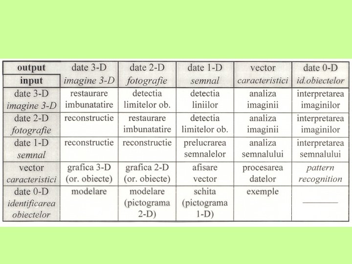
2. BASIC NOTIONS AN IMAGE MODEL Definition: image – Function of light intensity, f(x, y), which represents the “image” intensity (luminosity) in the point (x, y) – Function f(x, y) nature can be characterized by two components: – (1) luminance i(x, y) – (2) reflectance r(x, y)
 coordinates")
• Definition: • A monochrom image intensity f for the (x, y) coordinates = image grey level al in that point, - l • Lmin l Lmax • Lmin=imin rmin si Lmax=imax rmax • [Lmin , Lmax] - grey scale • in practice: [0, L] • · l=0 corresponds to BLACK • · l=L corresponds to WHITE

MEDICAL IMAGING • X-RAY IMAGES • 1895 – Julius Roentgen • X Rays – wavelengths between 0. 1 and 1Å used in medicine • • Is it useful to process X-Ray 2 -D images by computer?

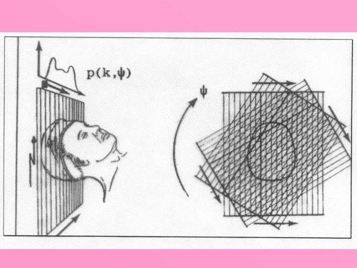
COMPUTER TOMOGRAPHY • Images – reconstructed by a large number of “attenuation profiles", taken at constant angular intervals for a given incidence Back-projection principle
 – spin small magnet")
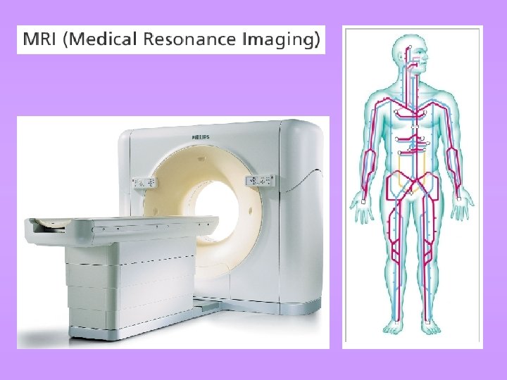
Nuclear magnetic resonance Hydrogen nucleus (proton) – spin small magnet

Comparision between CT and NMR images
")
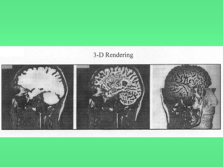
3 D images reconstruction (Rendering)

Scintigraphy images Radioopaque images – gamma camera

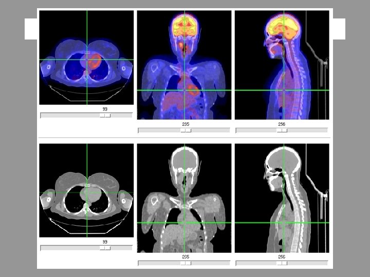
• · Heart imaging – the acquisition timing is controlled by ECG signal • · 3 -D reconstruction – SPECT (Single Photon Emission Computed Tomography) • · PET (Positron Emission Tomography) – the dynamic properties of biochemical processes

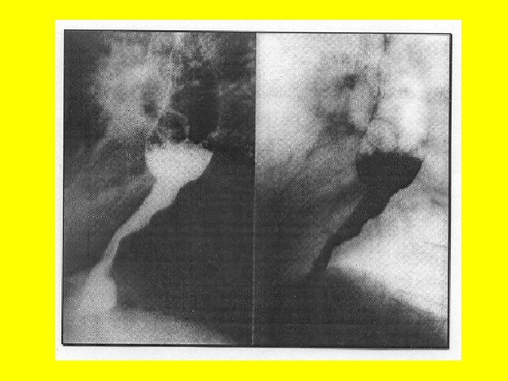
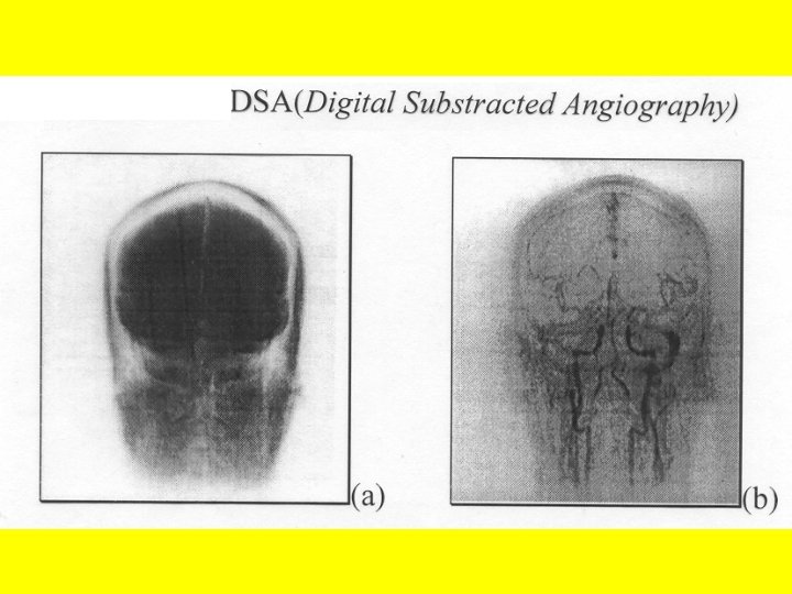
X-Ray images – contrast enhancement

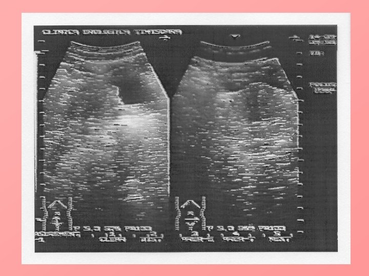
ULTRASOUND IMAGING • • · Ultrasounds – soundwaves with frequency over 20 k. Hz · Properties: speed, specific impedance and attenuation coefficient which differ by the material through which the ultrasounds propagate · Limitations – almost completely reflected by gas containing structures Image composition - transducers

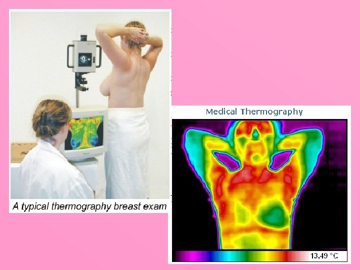
MEDICAL THERMOGRAPHY • · complementary to radiology methods • · applications: diagnosing deep vein thrombosis and other circulation problems, breast cancer, inflammatory joint disease, localization of placenta, identification and location of intraocular and orbital tumors

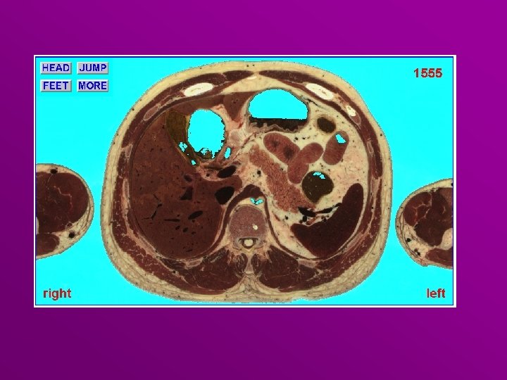
THE VISIBLE HUMAN PROJECT • · sponsored by the U. S. National Library of Medicine • · goal- to provide image data sets of the human body for use in: anatomy, research, applications for education, diagnostic and treatment planning, simulations, virtual reality • · 1 st phase : CT, MRI and cryosection: • Ø male – 1 mm resolution(15 Gb) • Ø female – 0. 33 mm resolution (40 Gb)
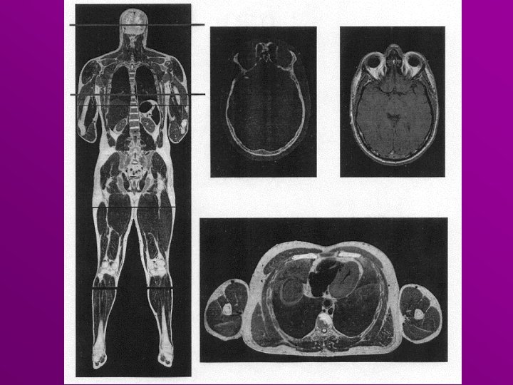

www. nlm. nih. gov/research/visible_human. html

BREAK
- Slides: 31