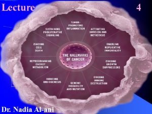
UNIVERSITTSKLINIKUM SchleswigHolstein Hallmarks of cancer Sustained proliferation evading


- Slides: 50

UNIVERSITÄTSKLINIKUM Schleswig-Holstein Hallmarks of cancer: Sustained proliferation; evading growth suppressors; replicative immortality Prof. Dr. rer. nat. Norbert Arnold Hanahan DW, Weinberg RA (2011). Hallmarks of Cancer the next generation. Cell 144: 646 -674 Dept. Gynecology and Obstetrics Oncology Laboratory

UNIVERSITÄTSKLINIKUM Schleswig-Holstein Educational objectives § Understanding factors leading to cellular dysfunction and cancer growth § Risk factors § Breast cancer as an example § Disturbed cell growth signaling § such as epidermal growth factor signaling disturbance § Disturbance of cell proliferation and DNA repair efficiency § leading to genomic instability § Understanding genetic alteration § leading to new therapeutic options Dept. Gynecology and Obstetrics Oncology Laboratory

UNIVERSITÄTSKLINIKUM Schleswig-Holstein What is cancer § Cancer arise by an evolutionary process § driven by mutation and selection analogous to Darwian evolution § Cancer is characterized by unregulated cell growth § Spread of cells from the origin to other sides of the body § Transformation § in epithelial cells § classified as carcinomas (approximately 85% of cancers) § in mesoderm cells (e. g. bone, muscle) § Classified as sarcomas § In glandular tissue (e. g. breast) § Classified as adenocarcinomas Dept. Gynecology and Obstetrics Oncology Laboratory

UNIVERSITÄTSKLINIKUM Schleswig-Holstein Influentional factors in human carcinogenesis § Cancer is a disease of the genome at the cellular level influenced by § Environment § Example: Unprotected exposure to the sun § Reproductive life § Observation: Nuns are more likely to develop breast cancer than other women § Diet § Observation: The risk of stomach cancer in Japanese people who have migrated to the USA decreases only if they adopt the American diet, but not if they retain a Japanese diet These lifestyle factors can, in principle, be altered to prevent most cancers Dept. Gynecology and Obstetrics Oncology Laboratory

UNIVERSITÄTSKLINIKUM Schleswig-Holstein Dept. Gynecology and Obstetrics Oncology Laboratory

UNIVERSITÄTSKLINIKUM Schleswig-Holstein Breast cancer - Risk factors § early first and late last menstruation (i. e. number of cycles) § no children (i. e. number of cycles) § increased age at first birth § hormone replacement with estrogens § obesity § physical inactivity § alcohol, smoking § age § history of breast or ovarian cancer increases the risk for subsequent breast cancer Dept. Gynecology and Obstetrics Oncology Laboratory

UNIVERSITÄTSKLINIKUM Schleswig-Holstein Breast cancer - Obesity as risk factor § elevated plasma level of insuline § has several effects in favoring tumor development § anti-apoptotic § enhances cellular growth and angiogenesis § stimulate synthesis of insuline-growth-factor 1 associated with tumor growth and metastasis § adipocyte has direct and indirect affects on growth regulating factors like m. TOR-signaling and AMP-activating protein kinases Dept. Gynecology and Obstetrics Oncology Laboratory

UNIVERSITÄTSKLINIKUM Schleswig-Holstein Cancer Distribution in Germany (RKI 2012) Krebs in Deutschland für 2013 -2014; RKI 2017 Dept. Gynecology and Obstetrics Oncology Laboratory

UNIVERSITÄTSKLINIKUM Schleswig-Holstein Survival rate Germany (RKI 2013 -14) Krebs in Deutschland für 2013 -2014; RKI 2017 Dept. Gynecology and Obstetrics Oncology Laboratory

UNIVERSITÄTSKLINIKUM Schleswig-Holstein Breast cancer - Epidemiology § Most frequent cause of cancer in women § 1 in 8 women develops breast cancer § 25% are younger than 55 years at diagnosis § disease incidence increases (better screening? ) § survival significantly improved over the past 20 years Dept. Gynecology and Obstetrics Oncology Laboratory

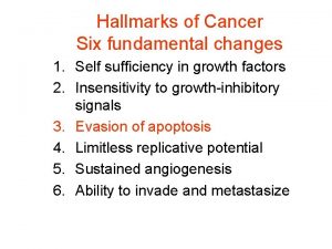
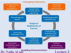
Figure 1 UNIVERSITÄTSKLINIKUM Schleswig-Holstein First description of Cancer Hallmarks (2000) by Hanahan and Weinberg Source: Cell , Volume 144, Issue 5, Pages 646 -674 (DOI: 10. 1016/j. cell. 2011. 02. 013) Dept. Gynecology and Obstetrics Oncology Laboratory

UNIVERSITÄTSKLINIKUM Schleswig-Holstein Sustaining proliferative signaling Growth signal autonomy § Normal cells need external signals from growth factors to divide § Cancer cells are not dependent on normal growth factor signaling § Acquired mutations short-circuit growth factor pathsways § Leading to unregulated growth Dept. Gynecology and Obstetrics Oncology Laboratory

UNIVERSITÄTSKLINIKUM Schleswig-Holstein Evading growth suppressors Evasion of growth inhibitory signals § Normal cells respond to inhibitory signals to maintain homeostasis § Most cells of the body are not actively dividing § Cancer cells do not respond to growth inhibitory signals § Acquired mutations or gene silencing interfere with the inhibitory pathways Recent high-throughput sequencing studies suggests that this two hallmarks can be consolidated into one! Dept. Gynecology and Obstetrics Oncology Laboratory

UNIVERSITÄTSKLINIKUM Schleswig-Holstein aus http: //www. hartnell. edu/tutorials/biology/signaltransduction. html Dept. Gynecology and Obstetrics Oncology Laboratory

UNIVERSITÄTSKLINIKUM Schleswig-Holstein Receptor signaling depends on § Hormone concentration § Receptor activity and modification Dept. Gynecology and Obstetrics Oncology Laboratory

UNIVERSITÄTSKLINIKUM Schleswig-Holstein Schematic view of the 3 steps aus http: //www. hartnell. edu/tutorials/biology/signaltransduction. html Dept. Gynecology and Obstetrics Oncology Laboratory

UNIVERSITÄTSKLINIKUM Schleswig-Holstein Localisation of receptors § Transmission of information from extracellular environment to the inside of the cell through § Intracellular receptors § Membrane receptors Dept. Gynecology and Obstetrics Oncology Laboratory

UNIVERSITÄTSKLINIKUM Schleswig-Holstein Are found inside the cell (cytoplasm or nucleus Chemical messengers capable passing the cell are hydrophobic or very small (p. e. steroid hormones) Once bound activated by the signal molecule, activated receptor can initiate cellular response, such as change in gene expression aus http: //www. hartnell. edu/tutorials/biology/signaltransduction. html Dept. Gynecology and Obstetrics Oncology Laboratory

UNIVERSITÄTSKLINIKUM Schleswig-Holstein Functioning of membrane receptors? § Binding of the signal molecule (ligand) § cause the production of a second signal (second messenger) § cause cellular response § Transmission of information by § changing shape § Joining with another protein once a specific ligand binds to it § Examples § G-Protein-coupled Receptors § Receptor Tyrosine Kinases Dept. Gynecology and Obstetrics Oncology Laboratory

UNIVERSITÄTSKLINIKUM Schleswig-Holstein Schematic view of membrane receptor functioning aus http: //www. hartnell. edu/tutorials/biology/signaltransduction. html Dept. Gynecology and Obstetrics Oncology Laboratory

UNIVERSITÄTSKLINIKUM Schleswig-Holstein Mechanism of physiological and oncogenic RTK activation Du Z and Lovly CM Molecular Cancer 17: 58 (2018) Dept. Gynecology and Obstetrics Oncology Laboratory

UNIVERSITÄTSKLINIKUM Schleswig-Holstein HER-Signaling (Heterodimer formation and downstream signaling) Leads to • • Proliferation Motility Invasiveness Resistance to apoptosis • Angiogenesis Dept. Gynecology and Obstetrics Oncology Laboratory

UNIVERSITÄTSKLINIKUM Schleswig-Holstein EGFR Family of receptor tyrosine kinases Taken as an example because of its therapeutic relevance in breast cancer § HER 1/EGFR; HER 2/Erb. B 2; HER 3/Erb. B 3; HER 4/Erb. B 4 § important regulators of normal growth and cell differentiation § HER 2 overexpression counts for approx. 20% of all breast cancer § First targeted drug (humanized monoclonal antibody trastuzumab/Herceptin) § HER 3 dimerization partner of HER 2 § HER 3 possesses no signal domain; but binding domain for HRG § promoting malignant transformation mainly via PI 3 K pathway § Heterodimer stabilized by heregulin (HRG/Type I NRG 1 neuregulin 1) § Different family members of neuregulin are produced by alternative splicing or by use of several cell type specific transcription initiation sites § loss of HER 3 reduces cell proliferation and decreases PI 3 K activity Dept. Gynecology and Obstetrics Oncology Laboratory

UNIVERSITÄTSKLINIKUM Schleswig-Holstein Structural composition of EGFR-Family § Composite extracellular domain with well defined ligand-binding site (at least HER 2, HER 3 and HER 4) § Single pass transmembran domain § Intracellular domain with distinguishable § tyrosine kinase domain § C-terminal non-catalytic signaling tail § Signaling mediated through § Homo- or heterodimerization with other family members Dept. Gynecology and Obstetrics Oncology Laboratory

UNIVERSITÄTSKLINIKUM Schleswig-Holstein Method of action for particular drugs Name Drug Type Action Trastuzumab/ Herceptin® Anti-HER 2 Humanized Mol. Ab Binds to HER 2 blocking its downstream signaling pathway; promotes HER 2 endocytotic degradation Trastuzumab (DM 1)/ Kadcyla® Anti-HER 2 Immunoconjugate Direct delivery of chemotherapeutic agent (mertansine) to cancerous tissue. DM 1 disrupts tubulin polymerization and micrutubule assembly Pertuzumab/Perjeta® Anti-HER 2 Humanized Mol. Ab Bind to HER 2 and prevents heterodimerization Lapatinib/Tyverb® Anti. HER 2/EGFR Small molecule TKE Inhibits. TK autophosphorylation, Blocks intracellular signal transduction of EGFR and HER 2 Gefitinib/Iressa™ Anti. HER 2/EGFR TKI Inhibits. TK autophosphorylation, Blocks intracellular signal transduction of EGFR and HER 2 Dept. Gynecology and Obstetrics Oncology Laboratory

UNIVERSITÄTSKLINIKUM Schleswig-Holstein Possible personalized therapy based on molecular profiling of breast cancer Patient with HER 2 positive Br. Ca Pathology results, including tumor genotyping PI 3 K mutation Trastuzumab + pertuzumab ± PI 3 K inhibitor HER 3 amplification Trastuzumab + pertuzumab ± HER 3 inhibitor FGFR amplification Trastuzumab + pertuzumab ± FGFR inhibitor No actionable mutation, HR positive Trastuzumab + pertuzumab ± endocrine therapy No actionable mutation, HR nebative Trastuzumab + pertuzumab ± chemotherapy Dept. Gynecology and Obstetrics Oncology Laboratory

UNIVERSITÄTSKLINIKUM Schleswig-Holstein Enabling replicative immortality Unlimited replicative potential § Normal cells have an autonomous counting device to define a finite number of cell doublings after which they become senescent § shortening of chromosomal ends (telomeres), occurs during every round of DNA replication § Cancer cells maintain the length of there telomeres § Altered regulation of telomere maintenance results in unlimited replicative potential Dept. Gynecology and Obstetrics Oncology Laboratory

UNIVERSITÄTSKLINIKUM Schleswig-Holstein Cell Cycle § Critical regulator § Process of cell proliferation and growth § Cell division after DNA damage § Governs § Transition from quiescence (G 0) to cell proliferation § Checkpoints § Ensure fidelity of the genetic transcript Mechanism by which cells reproduce Dept. Gynecology and Obstetrics Oncology Laboratory

UNIVERSITÄTSKLINIKUM Schleswig-Holstein Dept. Gynecology and Obstetrics Oncology Laboratory

UNIVERSITÄTSKLINIKUM Schleswig-Holstein Dept. Gynecology and Obstetrics Oncology Laboratory

UNIVERSITÄTSKLINIKUM Schleswig-Holstein Cell cycle § Promoted by a number of cyclin-dependent kinases (CDKs) § Positively regulated by cyclins § for driving the cells through the cell cycle § Negatively regulated by CDK inhibitors (CDKIs) § stop the cell from proceeding to the next phase of cell cycle – INK 4 (inhibitor of cdk 4) class of CDKIs inhibit CDK 2, -4, -6) – KIP (kinase inhibitor protein) class of CDKIs inhibit cyclin E/CDK 2 and cyclin A/CDK 2 complexes § Specific cyclin expression pattern § defines the relative position of the cell within the cell cycle Dept. Gynecology and Obstetrics Oncology Laboratory

UNIVERSITÄTSKLINIKUM Schleswig-Holstein Fluctuation of cyclin levels during the cell cycle Figure 8. 10 The Biology of Cancer (© Garland Science 2007) Dept. Gynecology and Obstetrics Oncology Laboratory

UNIVERSITÄTSKLINIKUM Schleswig-Holstein G 0 -G 1 Transition § Healthy cells need to decide when to divide and when to stay in G 0 § G 0 is an active phase in which cellular functions and cellular growth occur § Entry into the cell cycle (G 1) is governed by the restriction point § Beyond the R-Point cell progression through the cell cycle is independent of external stimuli – nutrients or mitogen activation § R-Point divide the early and late G 1 -phase § Mitogenic activation of a variety growth signals is mediated by RAS/RAF/MAPK pathway § Endpoint is stimulation of D-type cyclin production Dept. Gynecology and Obstetrics Oncology Laboratory

UNIVERSITÄTSKLINIKUM Schleswig-Holstein G 1/S-Transition § Retinoblastoma tumor suppressor gene product (Rb) governs the G 1/STransition § Active state hypophosphorylated and forms inhibitory complex with a group of transcription factors known as E 2 F-DP controlling the G 1/S transition § E 2 F is still able to transcribe some genes such as Cyclin E § Activity of Rb is modulated by phosphorylation by CDK 4/6 -Cyclin D and CDK 2 -Cyclin E § Completely hyperphosphorylated Rb releases the E 2 F-DP complex resulting in activation of numerous S-phase proteins § Cyclin D and E are targeted by ubiquitination for proteasome degradation in early S-phase Dept. Gynecology and Obstetrics Oncology Laboratory

UNIVERSITÄTSKLINIKUM Schleswig-Holstein J Clin Oncol 23: 9408 -9421 (2005) Dept. Gynecology and Obstetrics Oncology Laboratory

UNIVERSITÄTSKLINIKUM Schleswig-Holstein Late S and throughout G 2 § Orderly S-phase progression requires timely inactivation of E 2 F § done by Cyclin A-CDK 2 phosphorylation of E 2 F/DP heterodimer, neutralizing DNA binding capacity § Cells prepare for mitosis by increasing levels of cyclins A and B § Cyclin B forms a complex with cdc 2 (CDK 1) in cytoplasm and by starting mitosis it shuttles into the nucleus Dept. Gynecology and Obstetrics Oncology Laboratory

UNIVERSITÄTSKLINIKUM Schleswig-Holstein Genomic instability and mutation § Acquiring the core hallmarks of cancer usually depends on genomic alterations § Faulty DNA repair pathways can contribute to genomic instability Dept. Gynecology and Obstetrics Oncology Laboratory

UNIVERSITÄTSKLINIKUM Schleswig-Holstein Genetic Component of Breast Cancer Euhus D Ann Surg Oncol 21(10): 3209 -15 (2014) ) Dept. Gynecology and Obstetrics Oncology Laboratory

UNIVERSITÄTSKLINIKUM Schleswig-Holstein Breast Cancer Lifetime Risk of various genetic alterations Euhus D Ann Surg Oncol 21(10): 3209 -15 (2014) ) Dept. Gynecology and Obstetrics Oncology Laboratory

UNIVERSITÄTSKLINIKUM Schleswig-Holstein Difference between hereditary and sporadic cancers in regard of the defined hallmarks Dept. Gynecology and Obstetrics Oncology Laboratory

UNIVERSITÄTSKLINIKUM Schleswig-Holstein Main pathways implicated in genomic instability I § Base and nucleotide excision repair § Excise and repair abnormal bases or nucleotides such as deaminated cytosines or ultraviolet radiation induced pyrimidine dimers § Germline mutations in components of these pathways predispose p. e. to skin cancers § Mismatch repair § Act during DNA replication to correct base mismatches, insertions, deletions at repetitive sequences § Loss of function of MSH 2 and MLH 1 results in hypermutation and microsatellite instability § Telomere maintenance § Telomere erosion or uncapping results in catastrophic chromosomal instability due to „breakage-fusion-bridge“ cycles § Reaktivation of telomerase expression can alleviate this instability Dept. Gynecology and Obstetrics Oncology Laboratory

UNIVERSITÄTSKLINIKUM Schleswig-Holstein Main pathways implicated in genomic instability II § Double-strand break repair § Homologous recombination repair of double-strand breaks uses the sister DNA molecule as a template to repair the break § Defects result in chromosomal instability with elevated rate of structural chromosomal rearrangements and point mutations § DNA replication § Deregulated DNA replication can result in replication fork stalling, reversal and collapse § Deregulation can occur through oncogene activation, loss of certain tumor suppressors, DNA polymerase inhibition etc and trigger DNA DSB formation, unscheduled recombination events, chromosomal rearrangements § Chromosome segregation § Arise directly through defects in the mitotic checkpoint, sister chromatid cohesion, spindle geometry, spindle dynamics § Deregulation generates aneuploid daughter cells Dept. Gynecology and Obstetrics Oncology Laboratory

UNIVERSITÄTSKLINIKUM Schleswig-Holstein Number of somatic mutations in solid tumors Ton N. Schumacher, and Robert D. Schreiber Science 2015; 348: 69 -74 Dept. Gynecology and Obstetrics Oncology Laboratory

UNIVERSITÄTSKLINIKUM Schleswig-Holstein Distribution of somatic variants in solid tumors González et al. Seminars in Cancer Biology https: //doi. org/10. 1016/j. semcancer. 2017. 12. 001 Dept. Gynecology and Obstetrics Oncology Laboratory

UNIVERSITÄTSKLINIKUM Schleswig-Holstein Alteration classes in breast cancer 2014 Vasan N et al. Oncologist 19(5): 453 -458 Dept. Gynecology and Obstetrics Oncology Laboratory

UNIVERSITÄTSKLINIKUM Schleswig-Holstein Induction of cell death in BRCA deficient cancer cells 2019 Gourley C et al. J Clin Oncol doi 10. 1200/JCO. 18. 02050 Dept. Gynecology and Obstetrics Oncology Laboratory

UNIVERSITÄTSKLINIKUM Schleswig-Holstein Mode of action by PARPi Dept. Gynecology and Obstetrics Oncology Laboratory

UNIVERSITÄTSKLINIKUM Schleswig-Holstein Characteristics of tumor cells § High mutation rate § Genetic instability § Epigenetic alterations § Dysfunction of cell-cycle checkpoints and/or repair enzymes § Growth advantage § Independence of mitogenic signals § Loss of contact inhibition § Survival in foreign tissues Dept. Gynecology and Obstetrics Oncology Laboratory

UNIVERSITÄTSKLINIKUM Schleswig-Holstein Exam questions examples: multiple choice Q 1: a) b) c) d) e) In most cases, protein kinases? Hydrolyze proteins Stimulate adenylyl cyclase Add phosphate groups to proteins Bind DNA are components of the extracellular space Answer: c Recommendation for the exam: use lecture material only / no further literature needed Dept. Gynecology and Obstetrics Oncology Laboratory

UNIVERSITÄTSKLINIKUM Schleswig-Holstein Exam questions examples: free text Q 1: Describe the four steps which are necessary to activate a receptor tyrosine kinase protein? Answer: Ligand binding of inactive monomers (1 P); leading to mono- or dimerization (1 P); phosphorylation of the functional side/tyrosine kinase domain (1 P); activated receptor phosphorylates inactive relay protein and turns it active (1 P) Recommendation for the exam: use lecture material only / no further literature needed; Please give the answer in a few sentences or with key words Dept. Gynecology and Obstetrics Oncology Laboratory
 Benedictine hallmarks
 Contoh keterbatasan metode ilmiah
 Folliculocentric basaloid proliferation
 Reed sternberg cells
 Proliferation of interest groups
 Helmer strik
 Intimal proliferation
 Uncontrolled clonal proliferation
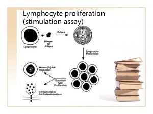
 Lymphocyte proliferation
 Intimal proliferation
 Industrial proliferation
 Proliferation of advertising example
 Sustained attention
 What is controlled release
 How hpw contributes to employee engagement
 Sustained attention example
 Sustained silent reading rules
 Firm resources and sustainable competitive advantage
 Sustained silent reading
 Principles of assessment flexibility
 Ocusert definition
 How are local cultures sustained
 What is extended metaphor
 Convention synoynm
 Extended release vs sustained release
 Sustained release
 Vocabulary of dance
 Small amplitude rhythmic oscillations
 Cancer osseo
 Ca 125
 The new prostate cancer infolink
 Simbolos de lucha contra el cancer
 Cancer personnalité
 Conclusion of breast cancer
 The biology of cancer
 Sindromes paraneoplasicos cancer de pulmon
 Endometrial cancer
 The genetic basis of cancer
 Conclusion of breast self examination
 Follicular adenoma
 Tnm breast cancer
 Cejas ralas hipotiroidismo
 Symptoms of metal fume fever
 Jod basedow phenomenon
 Nat rev cancer
 Abcd skin cancer
 What empties into the left subclavian vein
 Paraganglioma
 Ikea cancer
 Inflammatory breast cancer pictures
 Equator line

