Unit R 023 Body Systems Disease coursework unit
 Week A Thur sday")
Unit R 023 - Body Systems & Disease (coursework unit) Week A Thur sday Lesso n 7
 Student Task- Complete and")
Unit R 023 - Body Systems & Disease (coursework unit) Student Task- Complete and submit the third section of the Mr Walsh coursework. This is, • The fact file about the Digestive system. The deadline is Friday 5 th June 2020. When complete, please email: kswales@sjfchs. org. uk

Section 3: The Digestive System Include details about: 1. The organs in the digestive system 2. Mechanical digestion 3. Chemical digestion (including enzymes)
 •")
Make sure you include • Lots of diagrams and images. (use google images) • Add the web site addresses from any sources you use. DON’T WORRY about hand drawn diagramsthese can be added when we are back at school. Feel free to use the notes and images provided in this power point.

Parts of the digestive system to research-

STOMACH PANCREAS LARGE INT SMALL INT RECTUM

Our digestive system uses both: chemical digestion physical The shape of the food is physically changed so that it can fit through the small diameter of the digestive system. • Teeth & Tongue • Churning of Stomach chemical Bonds between Food Molecules are broken. • Enzymes • HCl • Bile

The Mouth- The Buccal Cavity MECHANICAL DIGESTION We chew food with our teeth, tongue and jaw so that it breaks down into small pieces to swallow. This action creates a larger Surface area of food.
 Softens and lubricates")
In the Mouth- Chemical Digestion Salivary glands produce saliva which: 1) Softens and lubricates food making it easier to swallow 2) Allows food to be tasted when moist food comes into contact with taste buds 3) Contains an enzyme called SALIVARY AMYLASE which begins to break down Starch.

Epiglottis –Valve at the back of the Buccal cavity that closes over your trachea during swallowing, prevents food entering trachea and causing choking. Peristalsis- Muscles in the oesophagus push the food towards the stomach.

The stomach is a muscular bag, filled with hydrochloric acid and gastric juices cross section of stomach digested food leaves through a sphincter into the small intestine food enters from the oesophagus muscle tissue contracts to churn the contents- known as Chyme Lining tissue makes hydrochloric acid, mucus and the enzyme Pepsin (protein breakdown)

Mechanical digestion: • Muscles of stomach churn food • Breaks food into smaller pieces/chyme Chemical digestion: • Produces enzyme Pepsin. • Action triggered by hydrochloric acid • Pepsin breaks down large proteins into smaller ones (peptides).
 2. Jejunum")
The liquid then travels into the small intestine 1. Duodenum (30 cm) 2. Jejunum (2 m) 3. Ileum (4 m) • • Almost all digestion takes place in the duodenum. Almost all absorption takes place in the Jejunum an Ileum.
 The Role of the Pancreas The PANCREAS connects")
Digestion in the Small Intestine (Duodenum) The Role of the Pancreas The PANCREAS connects to the duodenum via the Pancreatic duct It releases; • Amylase enzyme (starch digestion into sugars) • Lipase enzyme (fat digestion into fatty acids and glycerol) • Protease enzyme (protein digestion into amino acids) • An Alkaline solution to neutralise acid/raise p. H

Role of the Liver and Gall Bladder • The Liver cells make Bile Salts • Bile salts are stored in the Gall Bladder • Bile salts enter the small intestine via the Bile duct.

Fat Digestion Bile Salts are made in the Liver and stored in the Gall bladder. BILE SALTS emulsify large fat molecules into tiny fat droplets Smaller fat droplets have a larger Surface Area Lipase enzyme is secreted by the Pancreas into the small intestine. Fats are digested to Fatty acids and glycerol which are easily absorbed into the blood stream.
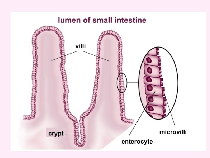
 is where digested")
Absorption in the Small Intestine The small intestine (Jejunum and Ileum) is where digested food molecules (glucose, amino acids, fatty acids) are absorbed into the blood. • It has a Large surface area due to Microvilli on Villi • Villi contain blood capillaries to absorb sugars, amino acids and water soluble vitamins • Contains lymph vessels/lacteals to absorb fats/fatty acids VILLI
 • Absorbs water from the waste food and it passes into")
LARGE INTESTINE (Colon) • Absorbs water from the waste food and it passes into the blood. • Contains digestive bacteria • Synthesises Vitamin K Rectum: Stores the faeces until it is ready to be expelled Anus: A ring of muscle (sphincter) which relaxes to let faeces exit.
- Slides: 19