UNIT B Chapter 10 Circulatory System and Lymphatic

UNIT B Chapter 10: Circulatory System and Lymphatic System Section 10. 2 Blood is a liquid connective tissue that has many different functions. • Transports nutrients, wastes, and hormones • Regulates body temperature by dispersing body heat • Regulates blood pressure (plasma proteins contribute to osmotic pressure of blood) • Protects the body against invasion by disease-causing pathogens • Clotting mechanisms protect the body against loss of blood. TO PREVIOUS SLIDE

UNIT B Chapter 10: Circulatory System and Lymphatic System Section 10. 2 Composition of Blood separates into three layers when centrifuged. • Upper layer: plasma (liquid portion of blood) • Lower layers: formed elements (white blood cells, platelets, red blood cells) TO PREVIOUS SLIDE Figure 10. 3 Composition of blood. When blood is collected into a test tube containing an anticoagulant to prevent clotting and then centrifuged, it consists of three layers. The transparent straw-coloured or yellow top layer is the plasma, the liquid portion of the blood. The thin, middle buffy coat layer consists of leukocytes and platelets. The bottom layer contains the erythrocytes.

UNIT B Chapter 10: Circulatory System and Lymphatic System Plasma also contains plasma proteins, which have many functions: • Transport: albumin transports bilirubin; lipoproteins transport cholesterol • Blood clotting: fibrinogen • Fighting infection: antibodies • Maintaining blood volume: plasma proteins are too large to leave the capillaries − Blood in capillaries has a higher solute concentrate than tissue fluid, causing water to diffuse in TO PREVIOUS SLIDE Section 10. 2

UNIT B Chapter 10: Circulatory System and Lymphatic System Section 10. 2 The Red Blood Cells Red blood cells (erythrocytes) are manufactured in the red bone marrow of the skull, ribs, vertebrae, and ends of the long bones. They transport oxygen and carbon dioxide in the blood. TO PREVIOUS SLIDE

UNIT B Chapter 10: Circulatory System and Lymphatic System Mature red blood cells have no nuclei and are biconcave. • Only live about 120 days (possibly due to lack of nuclei) and are destroyed in the liver and spleen • Biconcave shape increases flexibility (for moving through capillary beds) and surface area (for diffusion of gases) • Contain hemoglobin, a respiratory pigment that carries oxygen o Iron in hemoglobin acquires oxygen in the lungs and gives it up in the tissues TO PREVIOUS SLIDE Section 10. 2

UNIT B Chapter 10: Circulatory System and Lymphatic System Section 10. 2 Figure 10. 4 Physiology of red blood cells. a. Red blood cells move in single file through the capillaries. b. Each red blood cell is a biconcave disk containing many molecules of hemoglobin, the respiratory pigment. c. Hemoglobin contains four polypeptide chains (purple). There is an iron-containing heme group in the centre of each chain. Oxygen combines loosely with iron when hemoglobin is oxygenated. Oxyhemoglobin is bright red, and deoxyhemoglobin is a dark maroon colour. TO PREVIOUS SLIDE

UNIT B Chapter 10: Circulatory System and Lymphatic System Section 10. 2 Anemia is a common blood disorder that results in a tired, rundown feeling. Anemia has three basic causes: • Decreased production of red blood cells • Loss of red blood cells from the body • Destruction of red blood cells within the body Most common type of anemia: iron-deficiency anemia • Caused by decreased production of red blood cells due to a diet that lacks adequate iron TO PREVIOUS SLIDE

UNIT B Chapter 10: Circulatory System and Lymphatic System The White Blood Cells White blood cells (leukocytes) are usually larger than red blood cells, have a nucleus, lack hemoglobin, and appear translucent without staining. They fight infection and play a role in developing immunity. TO PREVIOUS SLIDE Section 10. 2

UNIT B Chapter 10: Circulatory System and Lymphatic System Section 10. 2 Figure 10. 5 Micrograph of the Formed Elements. The formed elements include red blood cells, different types of white blood cells, and platelets. TO PREVIOUS SLIDE
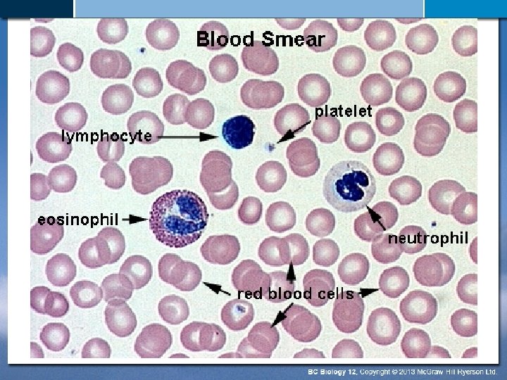

UNIT B Chapter 10: Circulatory System and Lymphatic System Section 10. 2 White blood cells can be divided on the basis of structure into granular leukocytes and agranular leukocytes: Granular leukocytes • Neutrophils • Eosinophils • Basophils Agranular leukocytes • Monocytes • Lymphocytes TO PREVIOUS SLIDE

UNIT B Chapter 10: Circulatory System and Lymphatic System Granular leukocytes are filled with spheres that contain enzymes and proteins that help white blood cells defend against microbes. • Neutrophils: phagocytize pathogens • Eosinophils: phagocytize antigenantibody complexes and allergens • Basophils: release histamine to promote blood flow to injured tissues TO PREVIOUS SLIDE Section 10. 2

UNIT B Chapter 10: Circulatory System and Lymphatic System Agranular leukocytes include cells that can phagocytize pathogens and cells that are involved in specific immunity. • Monocytes: largest white blood cells; differentiate into phagocytic dendritic cells and macrophages • Lymphocytes: o B lymphocytes (B cells): produce antibodies o T lymphocytes (T cells): helper T cells regulate the responses of other cells; cytotoxic T cells kill other cells TO PREVIOUS SLIDE Section 10. 2

UNIT B Chapter 10: Circulatory System and Lymphatic System Section 10. 2 White Blood Cells and Disease The number of white blood cells increases or decreases beyond normal if disease is present. • Neutrophils: increase in response to bacterial infections • B cells: increase in response to infectious mononucleosis • T cells: a low number of T cells indicates if an HIVinfected person has AIDS • A large number of abnormal white blood cells is a characteristic of leukemia, a form of cancer TO PREVIOUS SLIDE

UNIT B Chapter 10: Circulatory System and Lymphatic System Section 10. 2 Figure 10. 6 Macrophage (red) engulfing bacteria. Monocyte-derived macrophages are the body’s scavengers. They engulf microbes and debris in the body’s fluids and tissues, as illustrated in this colourized scanning electron micrograph. TO PREVIOUS SLIDE

UNIT B Chapter 10: Circulatory System and Lymphatic System Section 10. 7 Adaptive Immunity Adaptive immunity is activated when innate defences fail to prevent an infection. The adaptive immune system recognizes, responds to, and usually eliminates antigens from the body. • Antigens: any molecules that stimulate an adaptive immune response • Adaptive defences take 5 to 7 days to become fully activated and last for many years Watch: http: //kidshealth. org/kid/htbw/_bfs_ISmoviesource. html TO PREVIOUS SLIDE

UNIT B Chapter 10: Circulatory System and Lymphatic System Section 10. 7 The adaptive immune system depends on the activity of B cells and T cells. • Both cells recognize antigens because they have specific antigen receptors • Each lymphocyte has only one type of receptor • Large diversity of antigen receptors on B and T cells o There are specific B cells and/or T cells for almost any possible antigen TO PREVIOUS SLIDE

UNIT B Chapter 10: Circulatory System and Lymphatic System Section 10. 7 B Cells and Antibody-Mediated Immunity Defence by B cells (antibody-mediated immunity) • B cells are activated in a lymph node or the spleen, when their receptors bind to specific antigens • Cytokines secreted by T cells stimulate B cells to divide o Most cells become plasma cells, which secrete antibodies − Antibodies are the secreted form of the receptor of the B cell that was activated o Some cells become memory B cells, which allow for longterm immunity − If the same antigen enters again, memory B cells divide and give rise to more plasma cells that can produce the antibody against the antigen TO PREVIOUS SLIDE

UNIT B Chapter 10: Circulatory System and Lymphatic System Section 10. 7 B Cells and Antibody-Mediated Immunity Antibody Structure • Antibodies are also called immunoglobulins (Ig), which are Y-shaped molecules with two arms made of polypeptides o Heavy (long) polypeptide chain o Light (short) polypeptide chain o C (constant) region: set sequence of amino acids o V (variable) region: amino acid sequence varies between antibodies; forms the antigen-binding site TO PREVIOUS SLIDE

UNIT B Chapter 10: Circulatory System and Lymphatic System Figure 10. 24 Structure of an antibody. a. An antibody contains two heavy (long) polypeptide chains and two light (short) chains arranged so that there are two variable regions, where a particular antigen is capable of binding with an antibody (V = variable region, C = constant region). The shape of the antigen fits the shape of the binding site. b. Computer model of an antibody molecule. The antigen combines with the two side branches. TO PREVIOUS SLIDE Section 10. 7

UNIT B Chapter 10: Circulatory System and Lymphatic System Section 10. 7 B Cells and Antibody-Mediated Immunity • Antigens combine with an antibody at the antigen-binding site in a lock-and-key manner • Antigen-antibody reactions can result in immune complexes (antigens combined with antibodies) o Immune complexes may mark the antigens for destruction by a neutrophil or macrophage o Antibodies can “neutralize” toxins by preventing them from binding to cells TO PREVIOUS SLIDE

UNIT B Chapter 10: Circulatory System and Lymphatic System Section 10. 7 T Cells and Cell-Mediated Immunity Defence by T cells (cell-mediated immunity) • T cells have a unique T-cell receptor but cannot recognize antigens on their own o Require antigens be “presented” to the receptors by an MHC (major histocompatibility complex) protein on the surface of another cell • There are two types of T cells: o Helper T cells (TH cells) o Cytotoxic T cells (TC cells or CTLs) TO PREVIOUS SLIDE

UNIT B Chapter 10: Circulatory System and Lymphatic System Section 10. 7 T Cells and Cell-Mediated Immunity Helper T cells (TH cells) • Only recognize antigens presented by specialized antigenpresenting cells (APCs) with MHC class II proteins on their surface Cytotoxic T cells (TC cells or CTLs) • Only recognize antigens presented by various cells with MHC class I proteins on their surface • Some T cells become memory T cells o Live for many years and can quickly initiate immune response to an antigen previously present in the body TO PREVIOUS SLIDE

UNIT B Chapter 10: Circulatory System and Lymphatic System T Cells and Cell-Mediated Immunity Cell-mediated immunity by TC cells • Activated TC cells bound to a virus-infected or cancer cell release perforin, which forms pores in the plasma membranes of the abnormal cell o This allows enzymes called granzymes to enter the target cell and induce apoptosis Figure 10. 25 Cell-mediated immunity. a. How a TC cell destroys a virusinfected cell or cancer cell. TO PREVIOUS SLIDE Section 10. 7

UNIT B Chapter 10: Circulatory System and Lymphatic System T Cells and Cell-Mediated Immunity • TC cells are capable of moving on to kill other target cells Figure 10. 25 Cell-mediated immunity. b. The scanning electron micrograph shows cytotoxic T cells attacking and destroying a cancer cell (target cell). TO PREVIOUS SLIDE Section 10. 7

UNIT B Chapter 10: Circulatory System and Lymphatic System Section 10. 2 The Platelets and Blood Clotting Platelets (also called thrombocytes) result from fragmentation of large cells called megakaryocytes in the red bone marrow. Platelets are involved in blood clotting (coagulation). • There at least 12 clotting factors in the blood that help platelets in the formation of a blood clot (examples: prothrombin, fibrinogen). TO PREVIOUS SLIDE

UNIT B Chapter 10: Circulatory System and Lymphatic System Section 10. 2 Blood Clotting • The process of clotting begins when a blood vessel is damaged. • Platelets clump at the site of damage and form a plug to partially seal the leak. TO PREVIOUS SLIDE

UNIT B Chapter 10: Circulatory System and Lymphatic System Section 10. 2 Blood Clotting • Platelets and damaged tissue release prothombin activator, which converts prothrombin (a clotting factor) to thrombin in the presence of calcium ions. • Thrombin acts as an enzyme on fibrinogen (a clotting factor) to form fibrin threads. TO PREVIOUS SLIDE

UNIT B Chapter 10: Circulatory System and Lymphatic System Section 10. 2 Blood Clotting • Fibrin threads wind around the platelet plug and trap red blood cells to form the framework of the clot. • When blood vessel repair is initiated, plasmin (an enzyme) destroys the fibrin framework and restores the fluidity of the plasma. TO PREVIOUS SLIDE

UNIT B Chapter 10: Circulatory System and Lymphatic System Section 10. 2 Blood Clotting • If blood is allowed to clot in a test tube, a yellowish material, called serum, develops above the clotted material TO PREVIOUS SLIDE

UNIT B Chapter 10: Circulatory System and Lymphatic System Figure 10. 7 Blood clotting. a. A scanning electron micrograph of a blood clot shows red blood cells caught in the fibrin threads. b. Platelets and damaged tissue cells release prothrombin activator, which acts on prothrombin in the presence of Ca 2+ (calcium ions) to produce thrombin. Thrombin acts on fibrinogen in the presence of Ca 2+ to form fibrin threads. TO PREVIOUS SLIDE Section 10. 2

UNIT B Chapter 10: Circulatory System and Lymphatic System Section 10. 2 Hemophilia: A Blood Clotting Disorder Hemophilia is a group of inherited clotting disorders caused by a deficiency in a clotting factor. Hemophilia A: 90% of all hemophilia cases • Occurs frequently in males since the faulty gene is on the X chromosome • Individuals with hemophilia are more prone to bleeding • Bleeding in the muscles can lead to nerve damage • Bleeding into the brain can lead to neurological damage or death • Individuals require frequent blood transfusions or injections of the deficient clotting factor TO PREVIOUS SLIDE

UNIT B Chapter 10: Circulatory System and Lymphatic System Bone Marrow Stem Cells A stem cell is a cell that is ever capable of dividing and producing new cells that go on to differentiate into particular types of cells. TO PREVIOUS SLIDE Section 10. 2

UNIT B Chapter 10: Circulatory System and Lymphatic System Section 10. 2 Figure 10. 8 Blood cell formation in red bone marrow. Multipotent stem cells give rise to two specialized types of stem cells. The myeloid stem cells give rise to still other cells, which become red blood cells, platelets, and all the white blood cells except lymphocytes. The lymphoid stem cells give rise to lymphoblasts, which become lymphocytes. TO PREVIOUS SLIDE

UNIT B Chapter 10: Circulatory System and Lymphatic System Section 10. 2 Multipotent stem cells in red bone marrow have the potential to give rise to other stem cells for the various formed elements. • Can also differentiate into other cells (liver, bone, fat, cartilage, heart, neurons) • A patient’s own bone marrow stem cells (adult stem cells) could be used to treat diabetes, heart disease, liver disease, or brain disorders • Some researchers prefer using embryonic stem cells since they may be more likely to become any type of cell o Can be collected from unused embryos in fertility clinics, or umbilical cord blood TO PREVIOUS SLIDE

UNIT B Chapter 10: Circulatory System and Lymphatic System Plasma contains a variety of inorganic and organic substances dissolved or suspended in water. TO PREVIOUS SLIDE Section 10. 2
- Slides: 36