UNIT 7 THE NERVOUS SYSTEM NEURAL TISSUE Contains


 Peripheral nervous")

 Consists of the spinal cord and brain Contain neural")


 that surround the brain and spinal cord Pia")


 Thalamus: relays and processes sensory information Hypothalamus: hormone production")

 Mesencephalon Also called midbrain Processes sight, sound,")
 Medulla Oblongata Connects brain to spinal cord")

 Interface between")


 Gyri of neural cortex: increase neurons) surface")
 Longitudinal separates fissure: cerebral hemispheres Lobes: divisions")
 Central sulcus divides: anterior Lateral sulcus divides:")


 Receives")
: is associated with general interpretive area coordinates")

 controls: reading, writing, and")
")

 long 1/2 inch (14")

 Includes all neural tissue outside the CNS")


 Autonomic nervous system (ANS)")
 Includes all somatic motor neurons that innervate skeletal muscles")
 Visceral motor neurons innervate all other peripheral effectors: Controls")


")

 short, branched dendrites long,")

 to target Axon structure is")


")

")
")
")

: powered by ATP +")


")


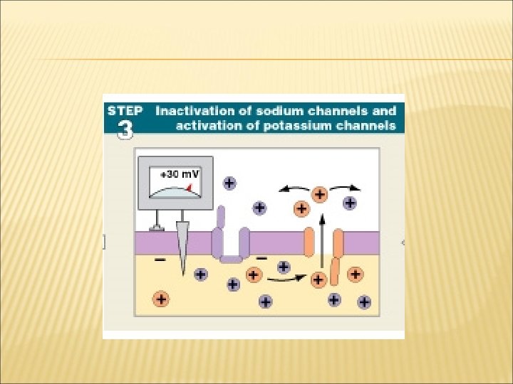


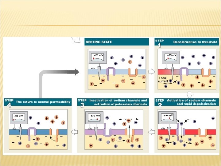


 Figure 12– 15 (Steps 1, 2)")
 Figure 12– 15 (Steps 3, 4)")


 dopamine serotonin gamma aminobutyric acid (GABA)")
 Released by adrenergic synapses Excitatory and depolarizing effect Found in brain and")


 Inhibitory effect Functions in CNS Not well understood")
- Slides: 132
UNIT 7 – THE NERVOUS SYSTEM
NEURAL TISSUE Contains 2 kinds of cells: neurons: cells that send and receive signals neuroglia cells (glial cells): that support and protect neurons
ORGANS OF THE NERVOUS SYSTEM Brain and spinal cord Sensory receptors of sense organs (eyes, ears, etc. ) Nerves connect nervous system with other systems
ANATOMICAL DIVISIONS OF THE NERVOUS SYSTEM 1. 2. Central nervous system (CNS) Peripheral nervous system (PNS)
THE CENTRAL NERVOUS SYSTEM
THE CENTRAL NERVOUS SYSTEM (CNS) Consists of the spinal cord and brain Contain neural tissue, connective tissues, and blood vessels
FUNCTIONS OF THE CNS Are to process and coordinate: sensory data: motor commands: higher functions of brain: intelligence, memory, learning, emotion
THE BRAIN
THE HUMAN BRAIN Ranges from 750 cc to 2100 cc Contains almost 98% of the body’s neural tissue Average weight about 1. 4 kg (3 lb)
MENINGES There are 3 layers (meninges) that surround the brain and spinal cord Pia mater – covers the brain Arachnoid – middle layer Dura mater – outermost layer that is attached to the interior of the skull
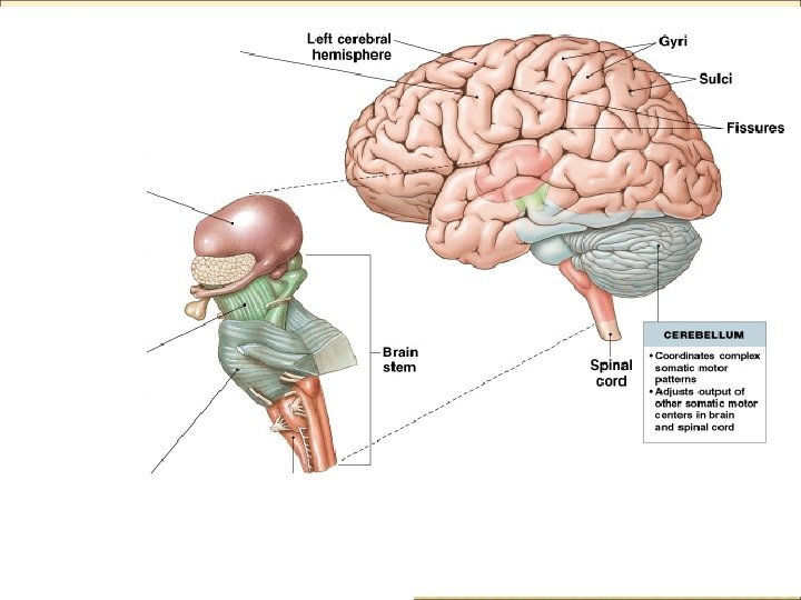
6 REGIONS OF THE BRAIN Cerebrum Cerebellum Diencephalon Mesencephalon Pons Medulla oblongata
REGIONS OF BRAIN Cerebrum Largest part of brain Controls higher mental functions Memory storage Skeletal Muscle Control Divided into left and right cerebral hemispheres Surface layer of gray matter (neural cortex)
REGIONS OF BRAIN Cerebrum Also Neural Cortex called cerebral cortex Folded surface increases surface area Elevated ridges (gyri) Shallow depressions (sulci) Deep grooves (fissures)
REGIONS OF BRAIN
REGIONS OF BRAIN Cerebellum Second largest part of brain Coordinates repetitive body movements 2 hemispheres Covered with cerebellar cortex
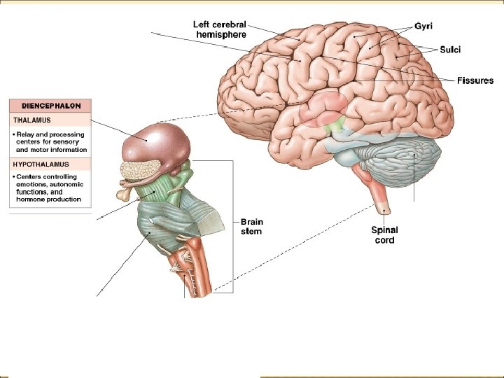
REGIONS OF BRAIN Diencephalon Located under cerebrum and cerebellum Links cerebrum with brain stem Contains thalamus and hypothalamus
REGIONS OF BRAIN Diencephalon (Con’t) Thalamus: relays and processes sensory information Hypothalamus: hormone production emotion thirst/hunger body temp. controls circadian rhythms (day–night cycles)
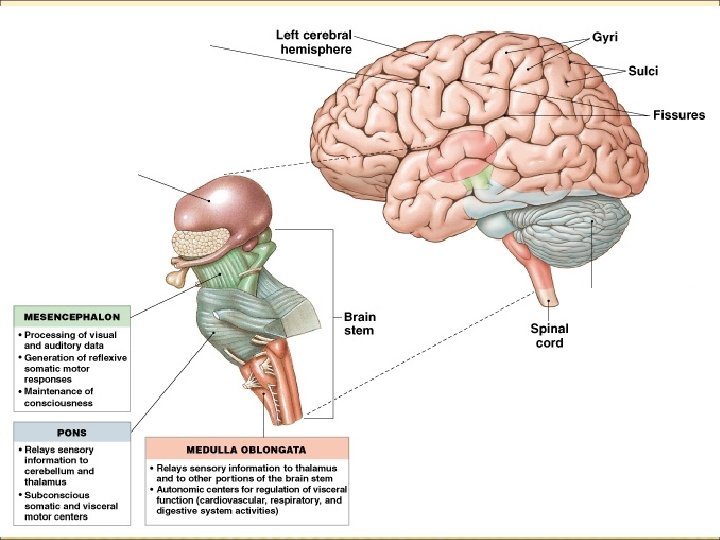
REGIONS OF BRAIN The Brain Stem Processes spinal infomation between: cord and cerebrum or cerebellum Includes: mesencephalon pons medulla oblongata
REGIONS OF BRAIN The Brain Stem (Con’t) Mesencephalon Also called midbrain Processes sight, sound, and associated reflexes Maintains consciousness Pons Connects cerebellum to brain stem Is involved in somatic and visceral motor control
REGIONS OF BRAIN The Brain Stem (Con’t) Medulla Oblongata Connects brain to spinal cord Relays information Regulates autonomic functions: Cardiovascular, respiratory, and digestive systems
LIMBIC SYSTEM Includes tracts between the cerebrum and diencephalon. Functions are: Establishing emotional states Linking the conscious, intellectual functions of the cerebral cortex with the unconscious functions of the brain stem. Facilitating memory storage and retrieval. The cortices of the brain enable you to do tasks, the limbic system makes you want to do them.
THE LIMBIC SYSTEM Major parts to the limbic system: Amygdaloid body (amygdala) Interface between limbic system, cerebrum, and various systems. Role in: regulating heart rate control of the fight or flight response Linking emotions with specific memories Hippocampus Important in learning especially in the storage and retrieval of new long term memories. Studies have shown that the hypothalamus portion of the limbic system is responsible for emotions of rage, fear, pain, sexual arousal, and pleasure.
THE LIMBIC SYSTEM
Divisions of the cerebrum
THE CEREBRUM Is the largest part of the brain Controls all conscious thoughts and intellectual functions Processes somatic sensory and motor information
THE CEREBRAL CORTEX Figure 14– 12 b
STRUCTURES OF THE CEREBRUM (1 OF 3) Gyri of neural cortex: increase neurons) surface area (number of cortical
STRUCTURES OF THE CEREBRUM (2 OF 3) Longitudinal separates fissure: cerebral hemispheres Lobes: divisions of hemispheres
STRUCTURES OF THE CEREBRUM (3 OF 3) Central sulcus divides: anterior Lateral sulcus divides: frontal Corpus frontal lobe from posterior parietal lobe from temporal lobe Callosum Connects left and right hemispheres
STRUCTURES OF THE CEREBRUM
3 FUNCTIONAL PRINCIPLES OF THE CEREBRUM 1. 2. 3. Each cerebral hemisphere receives sensory information from, and sends motor commands to, the opposite side of body The 2 hemispheres have different functions although their structures are alike Correspondence between a specific function and a specific region of cerebral cortex is not precise
Sensory and Motor Areas of the Cerebrum
MOTOR AND SENSORY AREAS OF THE CORTEX Central sulcus separates motor and sensory areas Figure 14– 15 a
MOTOR AREAS Precentral directs gyrus of frontal lobe: voluntary movements
SENSORY AREAS Postcentral receives gyrus of parietal lobe: somatic sensory information (touch, pressure, pain, vibration, taste, and temperature)
SPECIAL SENSORY CORTEXES Visual cortex: information Auditory from sight receptors cortex: information Olfactory from sound receptors cortex: information Gustatory from odor receptors cortex: information from taste receptors
SPECIAL SENSORY CORTEXES
ASSOCIATION AREAS Sensory association areas: monitor and interpret arriving information at sensory areas of cortex Somatic motor association area (premotor cortex): coordinates motor responses (learned movements)
ASSOCIATION AREAS
SENSORY ASSOCIATION AREAS Somatic sensory association area: interprets input to primary sensory cortex (e. g. , recognizes and responds to touch) Visual association area: interprets Auditory activity in visual cortex association area: monitors auditory cortex
SPECIAL SENSORY CORTEXES
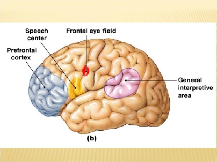
GENERAL INTERPRETIVE AREA Also called Wernicke’s area Present in only 1 hemisphere (left) Receives information from all sensory association areas Coordinates access to complex visual and auditory memories
OTHER INTEGRATIVE AREAS Speech center (Broca’s Area): is associated with general interpretive area coordinates all vocalization functions Prefrontal cortex of frontal lobe: integrates information from sensory association areas performs abstract intellectual activities (e. g. , predicting consequences of actions) Hippocampus sorts and integrates emotions and memories deep portion on the temporal lobe
HEMISPHERIC LATERALIZATION Functional differences between left and right hemispheres Figure 14– 16
THE LEFT HEMISPHERE In most people, left brain (dominant hemisphere) controls: reading, writing, and math decision-making speech and language
THE RIGHT HEMISPHERE Right cerebral hemisphere relates to: senses (touch, smell, sight, taste, feel) recognition (faces, voice inflections)
THE SPINAL CORD
SPINAL CORD Adult Spinal Cord About 18 inches (45 cm) long 1/2 inch (14 mm) wide Ends between vertebrae L 1 and L 2 Carries signals between brain and PNS Responsible for reflexes
THE PERIPHERAL NERVOUS SYSTEM
THE PERIPHERAL NERVOUS SYSTEM (PNS) Includes all neural tissue outside the CNS
FUNCTIONS OF THE PNS 1. 2. Deliver sensory information to the CNS Carry motor commands to peripheral tissues and systems
NERVES Also called peripheral nerves: bundles of axons with connective tissues and blood vessels carry sensory information and motor commands in PNS: cranial nerves—connect to brain spinal nerves—attach to spinal cord
RECEPTORS AND EFFECTORS Receptors: detect changes or respond to stimuli neurons and specialized cells complex sensory organs (e. g. , eyes, ears) Effectors: respond to efferent signals cells and organs
FUNCTIONAL DIVISIONS OF THE PNS Afferent division: carries sensory information from PNS sensory receptors to CNS Efferent carries division: motor commands from CNS to PNS muscles and glands
THE EFFERENT DIVISIONS OF THE PNS Somatic nervous system (SNS) Autonomic nervous system (ANS)
THE SOMATIC NERVOUS SYSTEM (SNS) Includes all somatic motor neurons that innervate skeletal muscles Controls skeletal muscle contractions: voluntary muscle contractions involuntary muscle contractions (reflexes)
THE AUTONOMIC NERVOUS SYSTEM (ANS) Visceral motor neurons innervate all other peripheral effectors: Controls subconscious actions: contractions of smooth muscle and cardiac muscle glandular secretions
DIVISIONS OF THE ANS Sympathetic has division: a stimulating effect Fight or flight Parasympathetic has division: a relaxing effect
NEURONS The basic functional units of the nervous system
FUNCTIONAL CLASSIFICATIONS OF NEURONS Sensory neurons: afferent Motor neurons of PNS neurons: efferent neurons of PNS
FUNCTIONS OF SENSORY NEURONS Monitor internal environment Monitor effects of external environment
MOTOR NEURONS Carry instructions from CNS to peripheral effectors Via efferent fibers (axons)
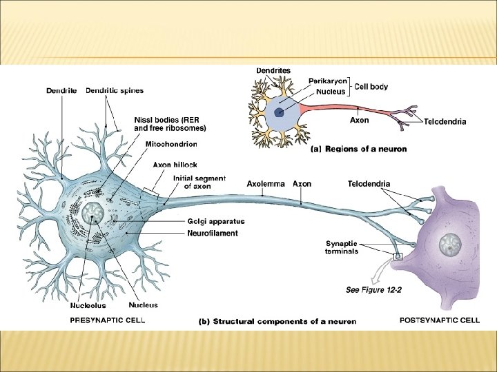
THE STRUCTURE OF NEURONS Figure 12– 1
THE MULTIPOLAR NEURON Common cell in the CNS: body (soma) short, branched dendrites long, single axon
DENDRITES Highly branched Dendritic spines: many fine processes receive information from other neurons 80– 90% of neuron surface area
THE AXON Are long Carries electrical signal (action potential) to target Axon structure is critical to function
THE SYNAPSE Area where a neuron communicates with another cell Synaptic Cleft The small gap that separates the presynaptic membrane and the postsynaptic membrane Synaptic Is Knob expanded area of axon Contains synaptic vesicles of neurotransmitters
THE SYNAPSE Figure 12– 2
NEUROTRANSMITTERS Are chemical messengers Are released at presynaptic membrane Affect receptors of postsynaptic membrane Are broken down by enzymes Are reassembled at synaptic knob
MYELINATION Increases speed of action potentials Myelin insulates myelinated axons Makes nerves appear white
WHITE MATTER AND GRAY MATTER White matter: regions Gray of CNS with many myelinated nerves matter: unmyelinated areas of CNS
SCHWANN CELLS Form myelin sheath around peripheral axons 1 Schwann cell sheaths 1 segment of axon: many Schwann cells sheath entire axon
SCHWANN CELLS Figure 12– 5 a
ELECTRICAL IMPULSES IN NERVES
ION MOVEMENTS AND ELECTRICAL SIGNALS All cell membranes produce electrical signals by ion movements Transmembrane potential is particularly important to neurons
MEMBRANE PROCESSES IN NEURAL ACTIVITIES Figure 12– 7 (Navigator)
MEMBRANE PROCESSES IN NEURAL ACTIVITIES Resting the potential: transmembrane potential of resting cell Graded potential: temporary, localized change in resting potential caused by stimulus Action is potential: an electrical impulse produced by graded potential propagates along surface of axon to synapse
RESTING POTENTIAL Figure 12– 8 (Navigator)
3 REQUIREMENTS FOR TRANSMEMBRANE POTENTIAL 1. 2. 3. Concentration gradient of ions (Na+, K+) Selectively permeable through channels Maintains charge difference across membrane (resting potential — 70 m. V)
PASSIVE FORCES ACROSS THE MEMBRANE Chemical gradients: concentration Electrical gradients of ions (Na+, K+) gradients: separated charges of positive and negative ions result in potential difference
EQUILIBRIUM POTENTIAL The transmembrane potential at which there is no net movement of a particular ion across the cell membrane
ACTIVE FORCES ACROSS THE MEMBRANE Sodium–potassium are ATPase (Na-K pump): powered by ATP + + carries 3 Na out and 2 K in balances passive forces of diffusion maintains resting potential (— 70 m. V)
CHANGES IN TRANSMEMBRANE POTENTIAL Transmembrane in potential rises or falls: response to temporary changes in membrane permeability resulting from opening or closing specific membrane channels
SODIUM AND POTASSIUM CHANNELS permeability to Na+ and K+ determines transmembrane potential Sodium and potassium channels are either passive or active Membrane
RESTING MEMBRANE POTENTIAL Resting membrane potential is -70 m. V Inside of cell is slightly negative compared to outside of cell Figure 12– 11 (Navigator)
GRADED POTENTIALS Also called local potentials Changes in transmembrane potential: that Any can’t spread far from site of stimulation stimulus that opens a gated channel: produces a graded potential
GRADED POTENTIALS Opening sodium channel produces graded potential Figure 12– 11 (Navigator)
GRADED POTENTIALS: STEP 1 Resting membrane exposed to chemical Sodium channel opens Sodium ions enter the cell Transmembrane potential rises Depolarization occurs
DEPOLARIZATION A shift in transmembrane potential toward 0 m. V
GRADED POTENTIALS: STEP 2 of Na+ through channel Produces local current Depolarizes nearby cell membrane (graded potential) Change in potential is proportional to stimulus Movement
REPOLARIZATION When the stimulus is removed, transmembrane potential returns to normal
EFFECTS OF GRADED POTENTIALS Also called local potentials At cell dendrites or cell bodies: trigger specific cell functions e. g. , exocytosis of glandular secretions At motor end plate: releases ACh into synaptic cleft
ACTION POTENTIALS
ACTION POTENTIALS Propagated changes in transmembrane potential Affect an entire excitable membrane Link graded potentials at cell body with motor end plate actions
INITIATING ACTION POTENTIAL Initial a stimulus: graded depolarization large enough (10 to 15 m. V) to change resting potential (— 70 m. V) to threshold level of voltage-regulated sodium channels (— 60 to — 55 m. V)
ALL-OR-NONE PRINCIPLE If a stimulus exceeds threshold amount: the action potential is the same no matter how large the stimulus Action potential is either triggered, or not
REMEMBER RESTING POTENTIAL
4 STEPS IN THE GENERATION OF ACTION POTENTIALS 1. 2. Depolarization to threshold Activation of Na+ channels: rapid depolarization Na+ ions rush into cytoplasm inner membrane changes from negative to positive
4 STEPS IN THE GENERATION OF ACTION POTENTIALS
4 STEPS IN THE GENERATION OF ACTION POTENTIALS 3. Inactivation of Na+ channels, activation of K+ channels: at +30 m. V Na+ channels close K+ channels open repolarization begins
4 STEPS IN THE GENERATION OF ACTION POTENTIALS 4. Return to normal permeability: K+ channels begin to close: K+ channels finish closing: when membrane reaches normal resting potential ( — 70 m. V) membrane is hyperpolarized to — 90 m. V Na+/K+ Pump returns ions to correct side of membrane, restoring resting membrane potential action potential is over
http: //www. youtube. com/watch? v=jc. ZLt. H-Uv 8 M&feature=player_embedded http: //www. youtube. com/watch? v=7 Eyhs. Oewn. H 4&feature=related
SUMMARY: GENERATION OF ACTION POTENTIALS Table 12 -3
THE REFRACTORY PERIOD The time period: from beginning of action potential to return to resting state during which membrane will not respond normally to additional stimuli
2 DIVISIONS OF THE REFRACTORY PERIOD 1. Absolute refractory period: sodium channels open or inactivated no action potential possible
2 DIVISIONS OF THE REFRACTORY PERIOD 2. Relative refractory period: membrane potential almost normal very large stimulus can initiate action potential
POWERING THE SODIUM-POTASSIUM EXCHANGE PUMP maintain concentration gradients of Na+ and K+ over time: To energy (1 ATP for each 2 K+/3 Na+ exchange) requires Without ATP: neurons stop functioning
PROPAGATION OF ACTION POTENTIALS Propagation: moves action potentials generated in axon hillock along entire length of axon a series of repeated actions, not passive flow
2 METHODS OF PROPAGATING ACTION POTENTIALS 1. Continuous propagation: 2. unmyelinated axons Saltatory propagation: myelinated axons
CONTINUOUS PROPAGATION Of action potentials along an unmyelinated axon Affects 1 segment of axon at a time Figure 12– 14
CONTINUOUS PROPAGATION: STEP 1 Action potential in segment 1 Depolarizes membrane to +30 m. V Figure 12– 14 (Step 1)
CONTINUOUS PROPAGATION: STEP 2 Local current Depolarizes second segment to threshold Figure 12– 14 (Step 2)
CONTINUOUS PROPAGATION: STEP 3 Second segment develops action potential First segment enters refractory period Figure 12– 14 (Step 3)
CONTINUOUS PROPAGATION: STEP 4 Local current depolarizes next segment Cycle repeats Action potential travels in 1 direction (1 m/sec) Figure 12– 14 (Step 4)
SALTATORY PROPAGATION Of action potential along myelinated axon Figure 12– 15
SALTATORY PROPAGATION (2 OF 3) Figure 12– 15 (Steps 1, 2)
SALTATORY PROPAGATION (3 OF 3) Figure 12– 15 (Steps 3, 4)
SALTATORY PROPAGATION Faster and uses less energy than continuous propagation Speed: 15 -150 m/s Myelin insulates axon, prevents continuous propagation Local current “jumps” from node to node Depolarization occurs only at nodes http: //www. youtube. com/watch? v=m. Og. HC 5 G 8 Lu. I&feature=fvwrel
WHAT FACTORS AFFECT THE PROPAGATION SPEED OF ACTION POTENTIALS?
AXON DIAMETER AND PROPAGATION SPEED Ion movement is related to cytoplasm concentration Axon diameter affects action potential speed The larger diameter, the lower the resistance
IMPORTANT NEUROTRANSMITTERS Other than acetylcholine: norepinephrine (NE) dopamine serotonin gamma aminobutyric acid (GABA)
NOREPINEPHRINE (NE) Released by adrenergic synapses Excitatory and depolarizing effect Found in brain and portions of ANS
DOPAMINE A CNS neurotransmitter May be excitatory or inhibitory Involved in Parkinson’s disease, cocaine use
SEROTONIN A CNS neurotransmitter Affects attention and emotional states
GAMMA AMINOBUTYRIC ACID (GABA) Inhibitory effect Functions in CNS Not well understood