Unit 2 Support and Movement in Animals Topic
 Secondary")


- Slides: 21
Unit 2: Support and Movement in Animals Topic: Human Skeleton B. Ed (Hons) Secondary Semester IV Subject: Advance Biology II Course Title: Animal Form and Function-I Represented By: Ms Sidra Younis Department of Education (Planning and Development) Lahore College for Women University , Lahore
skeleton “Skeleton is the supporting structure of an organism” There are three types of skeletons: 1. Hydrostatic skeleton 2. Exoskeleton 3. Endoskeleton
HUMAN SKELETON SYSTEM v. Human skeleton is the internal framework of the human body. It is composed of bones , cartilage and joints. v. It provide support and protection to human body.
COMPONENTS OF ENDOSKELETON Bone It is solid, hard and strong connective tissue made up of fibers and matrix. Cartilage It is specialized connective tissue which is less compact and slightly elastic.
Divisions of human skeleton
Axial skeleton is the part of skeleton that consists of the bones of head and trunk of a vertebrate. In the human skeleton, it consists of 80 bones and is composed of six parts. It serves to protect the brain, spinal cord and lungs. It also serves as the attachment sites for muscles that move the head, neck and back.
BONES OF AXIAL SKELETON Axial skeleton consists of 80 bones: Skull, which contains 22 bones, from which 8 are cranial and 14 are facial, 6 middle ear ossicles (3 in each ear), 1 hyoid bone in the neck, 26 1 bones of vertebral column, chest bone (sternum), 24 ribs (12 pairs).
skull § 29 bones exists in human skull of which 8 bones provide safety to the human mind are connected through sutures. §Rest of the bones forms the human face in which 14 bones are remarkably respondent. §Skull consist of cranium and facial bones.
cranium Cranium consists of eight flat bones joined together by sutures. There is a large opening at the base of the skull called the foramen magnum. On the either side is a projection which articulates with the first vertebra.
Facial bones There are 15 facial bones including cheek bones, nasal bone, temples, upper jaw and lower jaw. Vertebral column It is composed of 33 vertebrae and is developed from notochord. The entire vertebral can be divided into five regions, labeled in the diagram.
RIB CAGE AND STERNUM Rib cage is bony and cartilaginous structure. A typical ribcage consist of 24 ribs(12 pairs), the sternum, coastal cartilages and 12 thoracic vertebrae. First seven ribs connect directly to the sternum and are called true ribs. The remaining 5 ribs do not connect directly to the sternum and are called false ribs, with the help of diaphragm and the intercostals muscles, they increase and decrease the volume of thoracic cavity thereby allowing inhalation and exhalation to take place.
Appendicular skeleton consists of the pectoral girdle with arms and pelvic girdle with legs. It includes all bones of the upper and lower limbs plus the bones that attach each limb to the axial skeleton. There are 126 bones in the appendicular skeleton of an adult.
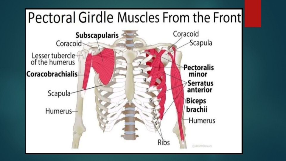
PECTORAL GIRDLE AND ARMS Pectoral girdle consists of the clavicle and the scapula, which serve to attach the upper limb to the sternum of the axial skeleton. Scapula (shoulder blade) lies on the posterior aspect of the shoulder. It is supported by the clavicle, which also articulates with the humerus (arm bone) to form the shoulder joint. Scapula is a flat, triangular-shaped bone with a prominent ridge running across its posterior surface.
FUNCTIONS OF PECTORAL GIRDLE q Pectoral girdles are responsible for providing structural support to your shoulder region on the left and right side of your body. q They also allow for a large range of motion. q Connecting muscles necessary for shoulder and arm movement.
PELVIC GIRDLE AND LEGS §Pelvic girdle consists of hip bones joined at the front by cartilage called the pubic symphysis and they are attached to sacrum at the back. §Each hip bone consists of three fused bones (ilium, ischium and pubis) portion of all three bones contribute to the formation of the acetabulum, a deep socket into which the head of the femur (thigh bone) joins to form the hip joint.
LOWER LIMBS §Femur in the leg is the largest bone. The upper end forms ball and socket joint while the lower limb articulates with the tibia. §There are two bones in the lower leg : Tibia and fibula.
Cont… The structure of foot is similar to that of hand. However the foot supports the weight of the body, so it is strongest and less mobile than hand. There are seven tarsals or ankle bones. There are metatarsal bones which form ball and arch of foot. The 14 phalanges of toes are the counter parts of those in fingers.
Functions of human skeleton Support: Skeleton provides the framework which supports the body and maintains its shape. Movement: The joints between bones allow movement, some allowing a wider range of movement than others. Protection: Skeleton helps to protect our many vital internal organs from being damaged. §Skull protects the brain §Vertebrae protect the spinal cord. §Rib cage, spine, and sternum protect the lungs, heart and major blood vessels.
CONT… Blood Cell Production: § Skeleton is the site of haematopoisis, the development of blood cells that takes place in the bone marrow. § In children, haematopoiesis occurs primarily in the marrow of the long bones such as the femur and tibia. § In adults, it occurs mainly in the pelvis, cranium, vertebrae, and sternum. Endocrine Regulation: §Bone cells release a hormone called osteocalcin, which contributes to the regulation of blood sugar (glucose) and fat deposition. §Osteocalcin increases both the insulin secretion and sensitivity, in addition to boosting the number of insulin-producing cells and reducing stores of fat.
summary There are three types of skeleton. 1. Hydrostatic skeleton 2. Exoskeleton 3. Endoskeleton Human Axial skeleton is an endoskeleton. and appendicular skeletons are the divisions of human skeleton. Human skeleton provides support, protection and movement to the body.