Unit 16 Reproductive System Overview Female Reproductive System


 Fallopian tubes Paired tubular structures Function: Passageway for sperm")


 – simple squamous & areolar")

 Functions:")
 between vaginal wall & cervix protrusion Allows for")


 Located postero-lateral to the vaginal opening -")


- Slides: 30
Unit 16 Reproductive System Overview
Female Reproductive System Use this PPT and your textbook to complete your charts.
Female Reproductive System · Ligaments · Ovaries · Duct System · Uterine tubes (fallopian tubes) · Uterus · Vagina · External genitalia Copyright © 2003 Pearson Education, Inc. publishing as Benjamin Cummings Slide
Female Reproductive System Figure 16. 8 a Copyright © 2003 Pearson Education, Inc. publishing as Benjamin Cummings Slide
Ligaments: all ligaments are primarily dense regular CT in structure Ovarian ligament Cord like portion of broad ligament Attaches upper portion of uterus to medial/inferior surface of ovary Broad ligament: Fold of the peritoneum Encloses & anchors ovaries, uterine tubes, and uterus to sides & floor of pelvic cavity Suspensory ligament Small fold of peritoneum Attaches lateral/superior surface of ovary to pelvic wall Contains ovarian artery & vein
Know the ligament locations on a diagram
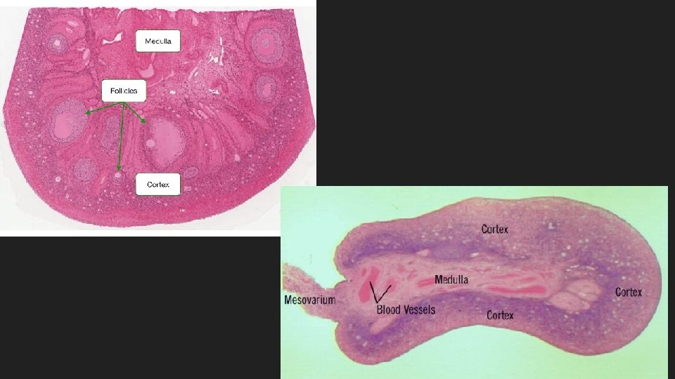
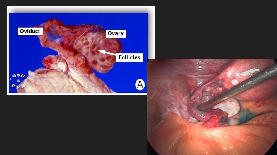
Ovaries: female gonads Paired glands Size & shape of almonds Function: Produce ova through process of oogenesis Produce & secrete female hormones
Ovaries: female gonads Tunica albuginea Dense CT layer on surface of ovary Provides structure & support Stroma Interior tissues of ovary composed of cortex & medulla Functional tissue of ovary Cortex Superficial layer Contains ovarian follicles Medulla Deep layer Contains larger BV, LV, & NF
Uterine tubes (Fallopian tubes, oviducts) Fallopian tubes Paired tubular structures Function: Passageway for sperm to reach egg Site of fertilization Passageway for fertilized egg to reach uterus
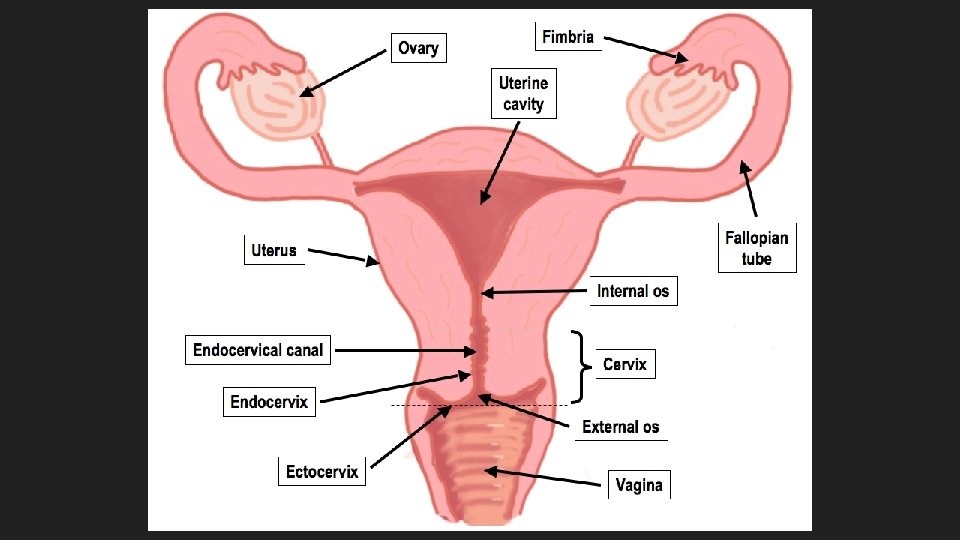
Parts of Fallopian tube Infundibulum: funnel shaped portion located closest to ovary Funnels/receives egg into Fallopian tube Fimbria: finger like projections at end of infundibulum No physical connection to uterus Function: lined with cilia: movement creates current to draw egg into infundibulum
Uterus Structure: 3” x 2” and approx. 1. 75 -3. 5 oz Hollow, muscular pear shaped Function: receives fertilized embryo protect, nourish & remove wastes for the developing child
Uterine structures Fundus: dome shaped top portion Entry point of uterine tubes Body: upper 2/3 of uterus Contains uterine cavity Hollow interior of body – contain developing child Cervix: Lower 1/3 of uterus – tubular “neck” Muscular opening into vagina Closes to hold baby in uterus
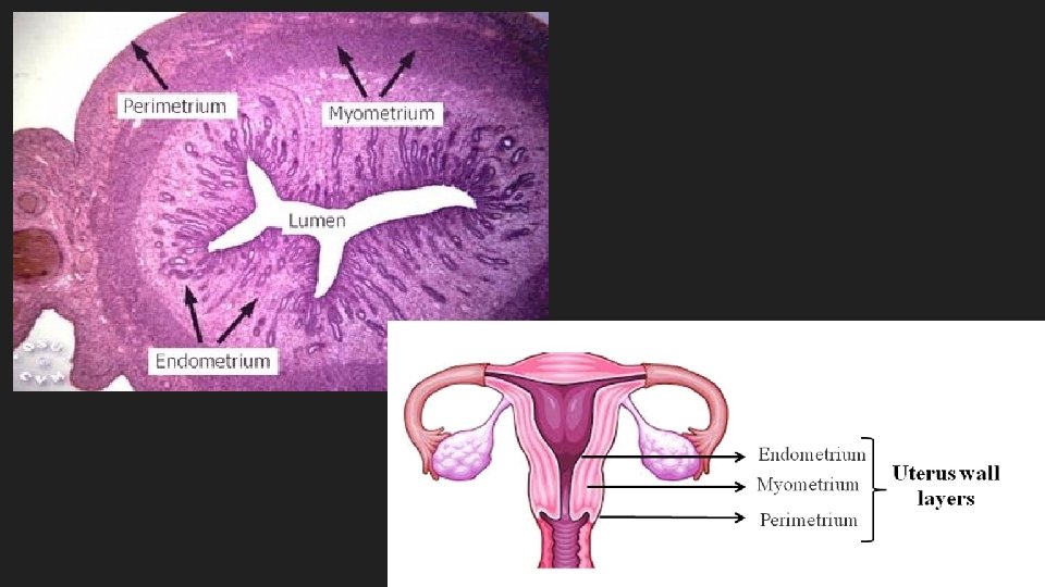
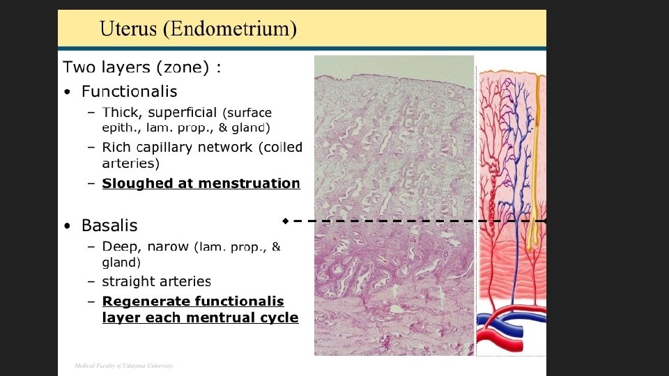
Uterine wall - layers Perimetrium: outer serosa (serous membrane) – simple squamous & areolar CT Reduce friction from surrounding organs Myometrium: middle layer – 3 layers of smooth muscle Contract during childbirth to push child out of body Contract slightly to shed functional layer of endometrium during menses
Uterine wall - layers Endometrium: Highly vascularized inner layer Two layers: functional layer : discharged during menses basal layer: permanent layer
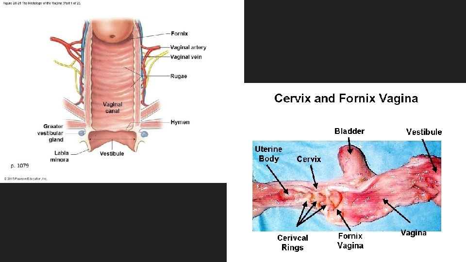
Vaginal structure Vagina: fibromuscular tube 3 -3. 5 inches long Highly distensible (stretchy) Functions: Conveys uterine secretions, such as menstrual fluids. Receives erect penis Open channel for birth
Vaginal structure Fornix: shallow recess (pockets) between vaginal wall & cervix protrusion Allows for expansion of cervix & vaginal canal during birth Allow physician to palpate AP organs/provide surgical access Hymen: elastic epithelial membrane fold Partially blocks opening to vagina
External Genitalia
Vulva – aka Pudendum Structures surrounding the openings of the urethra and vaginal canal
Structures of the vulva Labia minora: longitudinal folds of CT Encloses vestibule Protects openings to vagina and urethra Labia majora: homologous to scrotum: skin, adipose, smooth muscle Encloses and protects labia minora & other vulvar structures
Structures of the vulva Mons pubis: rounded elevation of adipose tissue over pubic symphysis Protective structure
Structures of the vulva Clitoris: homologous to penis: Produces feelings of pleasure Composed of erectile tissue - Does not have a duct through it Prepuce: skin fold Covers & protects clitoris
Female Glands Vestibular glands (aka. Bartholin’s glands) Located postero-lateral to the vaginal opening - homologous to bulbourethral glands Secretion: Mucus Function: vaginal lubrication & protection Mammary glands Modified sweat glands in the breast tissue Secretion: milk Functions: lactation: synthesis, secretion, and ejection of milk
Trace the pathway from origin of sperm until it exits the male system Figure 16. 2 Copyright © 2003 Pearson Education, Inc. publishing as Benjamin Cummings Slide
Continue the sperm pathway until it meets the egg & implants in the uterus