Unit 11 Blood Lymphatic System Immunity Human Anatomy

Unit 11: Blood, Lymphatic System & Immunity Human Anatomy & Physiology J. Kalinowski Spring 2019
")
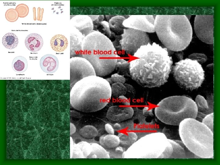
Blood • Only Fluid Tissue • Two components – Formed elements • Leukocytes (WBC) and platelets (buffy coat) equal less than 1% of blood • Erythrocytes (RBC) equal 45% of blood – Aka. Hematocrit = measure of RBC present – Plasma (fluid portion) = 55% of blood


Blood Information • p. H = 7. 35 – 7. 45 • Temperature = 38 o. C or 100. 4 o. F – Slightly higher than body temp. • Approx. 8% of body weight – Males have 5 -6 L while females have 4 -5 L although amount depends on size • Color – Oxygen rich blood is scarlet red – Oxygen poor blood is dull or rusty red – Blood is heavier & more viscous than water

Functions of Blood • Transport: – Oxygen – Waste products incl. Carbon dioxide – Hormones – Heat • Maintenance – Normal p. H – Adequate fluid volume

Blood flow thru capillaries

Functions of Blood • Prevention: – Blood loss through clotting mechanisms – Infection through white blood cells – - example: engulfing TB bacteria

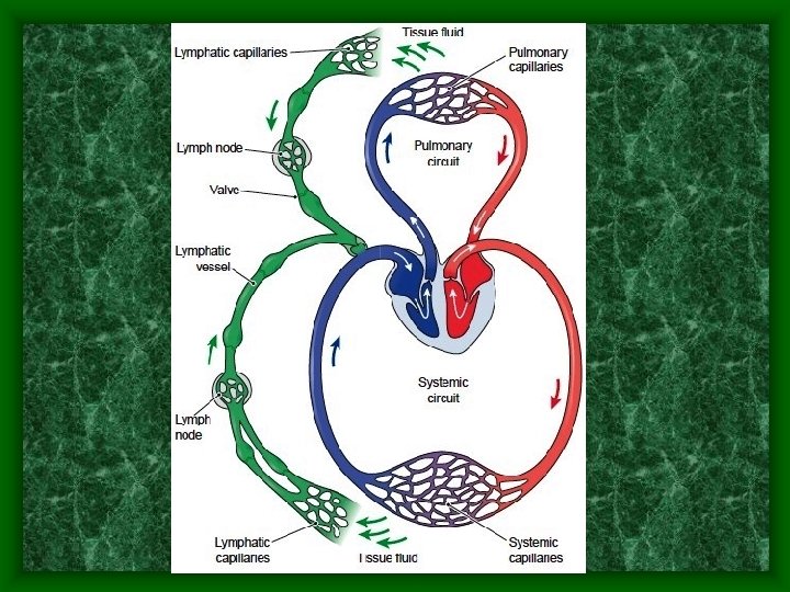
System Interactions • Circulatory and lymphatic work together to: • Return interstitial fluid to the circulatory system to maintain fluid balance

System Interactions • Respiratory and Excretory systems work together to: • Maintain a stable blood p. H • Maintain water and electrolyte balance in the blood

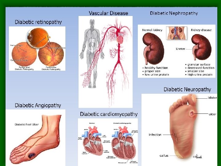
Failure to maintain Homeostasis • Blood glucose levels: –Hyper & hypoglycemia can result in • Diabetic retinopathy • Neuropathy • Nephropathy

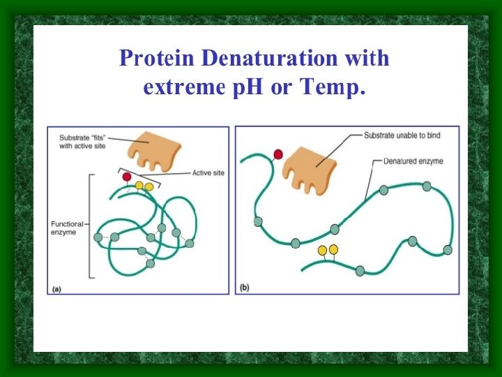
Failure to maintain Homeostasis • p. H level in blood –acidosis & alkalosis can result in enzymatic destruction through protein denaturation affecting body processes

Erythrocytes • Structure: – Few organelles – Lack mitochondria – Don’t do aerobic respiration so don’t use up the oxygen that they are carrying • Anucleate – Only survive ~120 days

RBC proteins

RBC Proteins • Hemoglobin – 33% of cell weight – Carries oxygen on iron atoms

• Spectrin RBC Proteins – to maintain membrane integrity and change cell shape so cell can travel without rupturing as it squeezes through capillaries • Misc. other proteins help with facilitating gas exchange and other functions

• Biconcave shape increases surface area to volume ratio for gas exchange

Numbers of RBCs • Outnumber WBC 1000 to 1 • # of blood cells compared to amount of plasma is major factor in blood viscosity –If blood is too viscous – heart must work too hard to pump it

Anemia vs. Polycythemia

Functions of RBCs • Major function is to carry oxygen • Single RBC contains ~250 million hemoglobin molecules each capable of carrying 4 oxygen atoms

Hemoglobin • Protection of hemoglobin – Enclosed in RBC to prevent fragmentation which would increase blood viscosity • Reminder: – If blood is too viscous – heart must work too hard to pump it

Sickle Cell Anemia
 which")
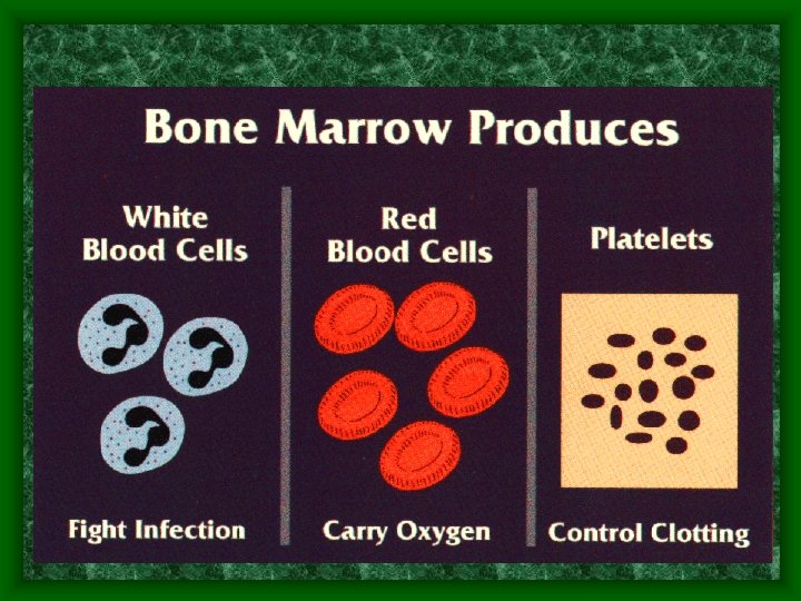
• Hematopoiesis: Blood cell formation – Occurs in red bone marrow (myeloid) which is found in the ends of long bones and in flat bones • Stem Cell is called Megakaryocyte • Hemocytoblasts convert to hemocytes – Cycle takes 3 -5 days

Stem Cells

Erythropoiesis • Erythropoiesis: Red blood cell formation – Based on oxygen demands by body • Hypoxia: too few RBCs = oxygen deprivation • Too many (polycythemia) = blood viscosity increases – Average production rate = 2 million/sec – Controlled hormonally • Based on level of available oxygen • triggers erythropoietin production in kidney

Erythropoiesis

Erythropoiesis • Production depends on: – Fe, vitamin B 12, and folic acid • Necessary for DNA synthesis and hemoglobin synthesis

Life Cycle of RBCs • After 120 days, the RBC is degraded and recycled by Macrophages (cleanup crew) • Hemoglobin is broken down to bilirubin – Goes to liver to be excreted – Liver damage can cause jaundice affecting many body organs

Life Cycle of RBCs

End of Quiz #1 Material
 – body defense system • 4000 – 11, 000 per mm 3")
Leukocytes (WBCs) – body defense system • 4000 – 11, 000 per mm 3 • Complete cells with nuclei and various organelles

Leukocyte Special Characteristics • Diapedesis – Reach infection site by slipping into and out of blood vessels • Ameboid motion – Move through tissue spaces to reach location • Chemotaxis – Respond to chemicals released by damaged cells in order to locate damaged area

Diapedesis

Leukocytes-Granulocytes Contain specialized granules and lobed nuclei

Neutrophils Active phagocytes – attracted to inflammation through chemotaxis • Numbers increase during bacterial & fungal infections • Produce white/yellow pus and snot & mucus

Basophils • Basophils – Located in certain tissues – aka. Mast cells – Increase in # during allergy attacks – Produce heparin & histamine to cause vasodilation and attract other WBCx to area of attack – Produce clear watery snot

Eosinophils • Located in intestinal & pulmonary mucosa and in dermis • Increase in number during – Parasitic infestations and produce chemicals to counteract allergic reactions – Produce greenish snot

Leukocytes-Agranulocytes

Leukocytes • Lack granules • Formed in bone marrow and then migrate to lymphatic tissues – rarely circulate in blood unless needed • 2 types

Leukocytes-Agranulocytes • Lymphocytes • Monocytes

Leukocytes • Lymphocytes: 3 types that play an immune system role – (B-cells, T-cell, and Natural Killer cells) • T-cells (several types) – Attack virus infected & tumor cells • B-cells (several types) – Produce antibodies (immunoglobulins) for long term immunity

Natural Killer Cells

Leukocytes • Monocytes – Very mobile, aggressive macrophages – Increase in number during chronic infections (such as tuberculosis) and act against viruses and bacteria in long term infections – Activate lymphocytes to start immune response

Leukopoiesis • Activated by specific chemicals in response to infections, toxins, tumor cells, etc. • Granulocytes produced and stored in bone marrow as needed • Granulocytes have short life span -. 5 to 9 days – die fighting invaders • Agranulocytes may live days to years depending on type

Plasma • Straw colored, sticky fluid matrix • 90% water - 10% dissolved proteins, gases, wastes, etc. • Plasma proteins produced by liver: know functions: – – Albumin – water balance Fibrinogen - clotting Alpha & beta globulins - transport Gamma globulins - immunity • Homeostatic levels maintained by various organs
 • Formed by megakaryocytes (stem cells) • Fragments of cells that clump")
Platelets (Thrombocytes) • Formed by megakaryocytes (stem cells) • Fragments of cells that clump together to form a seal at damaged BV locations • Not a complete cell – lack nuclei and organelles so short life span

End of Quiz #2 Material

Clot formation • Know the chemicals involved in each step. • Know the sequence of events

Steps of Hemostasis • Platelet plug formation – Normally, platelets and endothelium are both positively charged so they repel each other and the endothelial wall of BV – When endothelium ruptured, +platelets contact negative collagen fibers – Chemical changes cause platelets to swell and stick together and to the wall – Chemicals are released to attract more platelets to seal cuts – Platelet plug is formed – effective in sealing small vascular nicks

Aspirin • Aspirin inhibits platelet plug formation and prolonged bleeding may occur – In small doses, it inhibits unnecessary clotting thus preventing heart attacks & strokes • Aspirin is an anticoagulant

Steps to Hemostasis • Vascular Spasms –. initiated by serotonin released from anchored platelets and stimulation of local pain receptors cause BV to spasm – vasoconstriction narrows BV decreasing blood loss

Steps to Hemostasis • Coagulation – blood clotting • Critical events that occur: – Thromboplastin released by injured tissue – interacts with prothrombin activator (PF 3) – Which converts prothrombin to thrombin – Which joins fibrinogen molecules into a fibrin mesh – Which traps RBCs and pulls edges closer together
 • Blood Clotting Animation")
Hemostasis (blood stopping) • Blood Clotting Animation

Hemostasis • More than 30 substances involved – Procoagulant – promotes clotting – Anticoagulant – inhibits clotting

Homeostasis • When body is in homeostasis – there are several mechanisms that prevent clotting when it should NOT occur and enhance clotting when it SHOULD occur.
")
Fibrinolysis (clot busting)
")
Fibrinolysis • When normal cell regeneration begins, clot becomes unnecessary • Plasmin (clot buster) is released until clot is dissolved totally. • release typically begins within 2 days and continues until clot dissolved

HUMAN BLOOD TYPING
 for cell recognition – common antigens -")
Blood Groups • RBCs contain antigens (glycoproteins) for cell recognition – common antigens - ABO and Rh cause vigorous transfusion reactions • ABO blood groups – based on presence or absence of A or B antigens on RBCs – plasma antibodies act against foreign antigens not present on that individual's RBCs

Blood Types

Rh factor • Rh+ 85% of Americans - carry Rh antigen on RBC • Rh- don't have antigen on RBC • Rh- can go into Rh+ BUT Rh+ cannot go into Rh • less severe transfusion reaction (hemolysis of donor RBCs) doesn't usually occur until 2 nd transfusion due to body's reaction time
 – if Rh-")
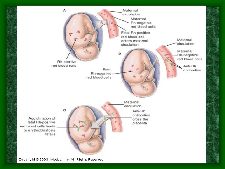
In Pregnant Women • Erythroblastosis fetalis (hemolytic disease of the newborn) – if Rh- woman – carries Rh+ baby – Has maternal blood to fetal blood contact due to: • Birth • Abortion • Miscarriage • Placental problems during pregnancy
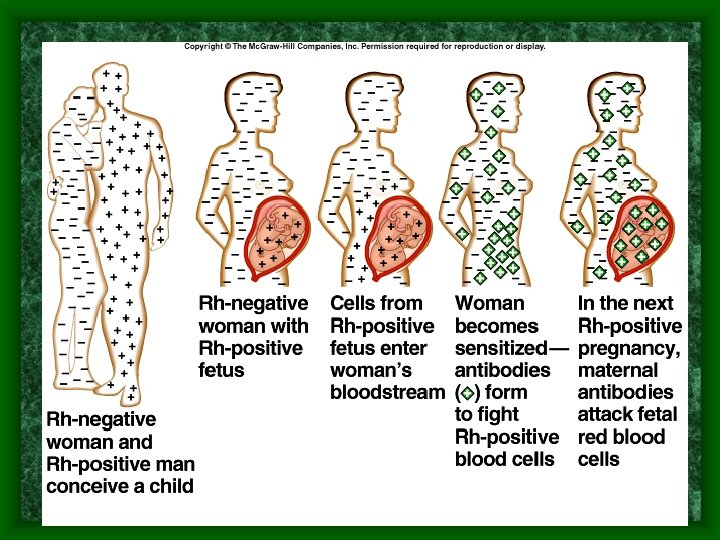

Erythroblastosis Fetalis • 1 st baby is usually okay due to reaction time unless there was a bleeding problem during the pregnancy or a previous miscarriage or abortion. • 2 nd baby will have its blood cells attacked by mother’s antibodies • Rhogam shot can prevent this if injected at 28 weeks of pregnancy and again right after birth.

Transfusions • In case of blood loss, body tries to: • • • – 1. reduce BV volume to maintain circulation to vital organs – 2. step up production of RBCs for replacement 15 -30% loss - pallor & weakness over 30% - severe shock may be fatal substantial blood loss - whole blood transfusion Plasma, electrolyte solutions ( Ringer's solution) etc. can be used to increase blood volume while body steps up production of RBCs Whole Blood transfusions can be given but blood must be typed and matched to prevent transfusion reaction

Know the information contained in this chart

Transfusion Reaction • Mismatched RBCs antigens attacked by plasma antibodies • agglutination of foreign RBCs can: – clog small BV - reduce blood flow – lysed RBCs release hemoglobin into bloodreduced oxygen capacity - blocks kidney tubules and causes renal shutdown • Reactions: fever, chills, vomiting • Treatment: alkaline fluids to dilute hemoglobin, diuretics to increase urine flow to flush kidneys

Agglutination

Inheritance of blood type
- Slides: 76