Undesirable Clotting Thrombus A clot in an unbroken

Undesirable Clotting • Thrombus • A clot in an unbroken blood vessel • Can be deadly in areas such as the heart • Embolus • A thrombus that breaks away and floats freely in the bloodstream • Can later clog vessels in critical areas such as the brain

Bleeding Disorders • Thrombocytopenia • Platelet deficiency • Even normal movements can cause bleeding from small blood vessels that require platelets for clotting • Evidenced by petechiae (small purplish blotches on the skin) • Hemophilia • Hereditary bleeding disorder • Normal clotting factors are missing

Blood Groups and Transfusions • Large losses of blood have serious consequences • Loss of 15 to 30 percent causes weakness • Loss of over 30 percent causes shock, which can be fatal • Blood transfusions are given for substantial blood loss, to treat severe anemia, or for thrombocytopenia

Human Blood Groups • Blood contains genetically determined proteins • Antigens are substances that the body recognizes as foreign and that the immune system may attack • Antibodies are the “recognizers” • Blood is “typed” by using antibodies that will cause blood with certain proteins to clump (agglutination) and lyse

Human Blood Groups • There are over 30 common red blood cell antigens • The most vigorous transfusion reactions are caused by ABO and Rh blood group antigens https: //www. youtube. com/watch? v=Bh 4 j. KXsqu. WA

ABO Blood Groups • Based on the presence or absence of two antigens: 1. Type A 2. Type B • The lack of these antigens is called type O • The presence of both antigens A and B is called type AB • The presence of antigen A is called type A • The presence of antigen B is called type B • The lack of both antigens A and B is called type O

• Blood type AB can receive A, B, AB, and O blood • Universal recipient • Blood type B can receive B and O blood • Blood type A can receive A and O blood • Blood type O can receive O blood • Universal donor

Rh Blood Groups • Named because of the presence or absence of one of eight Rh antigens (agglutinogen D) that was originally defined in Rhesus monkeys • Most Americans are Rh+ (Rh positive) • Problems can occur in mixing Rh+ blood into a body with Rh– (Rh negative) blood • Hemolysis does not occur with first transfusion, because it takes time to make antibodies • Second, and subsequent, transfusions involve antibodies attacking donor’s Rh+ RBCs

Rh Dangers During Pregnancy • Danger occurs only when the mother is Rh– and the father is Rh+, and the child inherits the Rh+ factor • Rho. GAM shot can prevent buildup of anti-Rh+ antibodies in mother’s blood • The mismatch of an Rh– mother carrying an Rh+ baby can cause problems for the unborn child • The first pregnancy usually proceeds without problems • The immune system is sensitized after the first pregnancy • In a second pregnancy, the mother’s immune system produces antibodies to attack the Rh+ blood (hemolytic disease of the newborn)

Blood Typing • Blood samples are mixed with anti-A and anti-B serum • Agglutination or the lack of agglutination leads to identification of blood type • Typing for ABO and Rh factors is done in the same manner

Blood being tested Anti-A Serum Anti-B Type AB (contains antigens A and B; agglutinates with both sera) Agglutinated RBCs Type B (contains antigen B; agglutinates with anti-B serum) Type A (contains antigen A; agglutinates with anti-A serum) Type O (contains no antigens; does not agglutinate with either serum)

The Cardiovascular System • A closed system of the heart and blood vessels • The heart pumps blood • Blood vessels allow blood to circulate to all parts of the body • Functions of the cardiovascular system: • Deliver oxygen and nutrients to cells and tissues • Remove carbon dioxide and other waste products from cells and tissues

The Heart • Location • Thorax, between the lungs in the inferior mediastinum • Orientation • Pointed apex directed toward left hip • Base points toward right shoulder • About the size of a human fist
 Pulmonary trunk Left lung Pericardium (cut) Diaphragm")
Superior vena cava Aorta Parietal pleura (cut) Pulmonary trunk Left lung Pericardium (cut) Diaphragm (a) Apex of heart
 Point of maximal intensity (PMI)")
Midsternal line 2 nd rib Sternum Diaphragm (b) Point of maximal intensity (PMI)
 Posterior")
Mediastinum Heart Left lung (c) Posterior

Coverings and Walls of the Heart • Pericardium—a double-walled sac • Fibrous pericardium is loose and superficial • Serous membrane is deep to the fibrous pericardium and composed of two layers: 1. Parietal pericardium: outside layer that lines the inner surface of the fibrous pericardium 2. Visceral pericardium: next to heart; also known as the epicardium • Serous fluid fills the space between the layers of pericardium (decreases friction!)

Problem • Pericarditis • Inflammation of pericardium • Roughens membrane surfaces, causing pericardial friction rub (creaking sound) heard with stethoscope • Cardiac tamponade • Excess fluid that leaks into pericardial space • Can compress heart’s pumping ability • Treatment: fluid is drawn out of cavity (usually with syringe)

Pulmonary trunk Pericardium Myocardium Fibrous pericardium Parietal layer of serous pericardium Pericardial cavity Epicardium (visceral layer of serous pericardium) Heart wall Myocardium Endocardium Heart chamber

Coverings and Walls of the Heart • Three layers of the heart wall: 1. Epicardium • Outside layer • This layer is the visceral pericardium • Connective tissue layer 2. Myocardium • Middle layer • Mostly cardiac muscle 3. Endocardium • Inner layer known as endothelium

Pulmonary trunk Pericardium Myocardium Fibrous pericardium Parietal layer of serous pericardium Pericardial cavity Epicardium (visceral layer of serous pericardium) Heart wall Myocardium Endocardium Heart chamber

The heart wall is composed of three layers. The deepest layer is called the • epicardium. • myocardium. • endocardium. • pericardium.

The heart wall is composed of three layers. The deepest layer is called the • epicardium. • myocardium. • endocardium. • pericardium.

Chambers and Associated Great Vessels • Right and left side act as separate pumps • Four chambers: • Atria (right and left) • Receiving chambers • Ventricles (right and left) • Discharging chambers

Atria: the receiving chambers Small, thin-walled chambers; contribute little to propulsion of blood Auricles: appendages that increase atrial volume Right atrium: receives deoxygenated blood from body Three veins empty into right atrium: Superior vena cava: returns blood from body regions above the diaphragm Inferior vena cava: returns blood from body regions below the diaphragm Coronary sinus: returns blood from coronary veins Left atrium: receives oxygenated blood from lungs Pectinate muscles found only in auricles Four pulmonary veins return blood from lungs

• Ventricles: the discharging chambers • • • Make up most of the volume of heart Right ventricle: most of anterior surface Left ventricle: posteroinferior surface Thicker walls than atria Actual pumps of heart Right ventricle • Pumps blood into pulmonary trunk • Left ventricle • Pumps blood into aorta (largest artery in body)

Superior vena cava Aorta Left pulmonary artery Right pulmonary artery Left atrium Right atrium Left pulmonary veins Right pulmonary veins Fossa ovalis Right atrioventricular valve (tricuspid valve) Right ventricle Chordae tendineae Pulmonary semilunar valve Left atrioventricular valve (bicuspid valve) Aortic semilunar valve Left ventricle Interventricular septum Inferior vena cava Myocardium (b) Frontal section showing interior chambers and valves. Visceral pericardium (epicardium)

Left ventricle Right ventricle Muscular interventricular septum

Chambers and Associated Great Vessels • Interventricular septum • Separates the two ventricles • Interatrial septum • Separates the two atria

Superior vena cava Aorta Left pulmonary artery Right pulmonary artery Left atrium Right atrium Left pulmonary veins Right pulmonary veins Fossa ovalis Right atrioventricular valve (tricuspid valve) Right ventricle Chordae tendineae Pulmonary semilunar valve Left atrioventricular valve (bicuspid valve) Aortic semilunar valve Left ventricle Interventricular septum Inferior vena cava Myocardium (b) Frontal section showing interior chambers and valves. Visceral pericardium (epicardium)

The Pulmonary and Systemic Circuits • Heart is a transport system consisting of two side-by-side pumps • Right side receives oxygen-poor blood from tissues • Pumps blood to lungs to get rid of CO 2, pick up O 2, via pulmonary circuit • Left side receives oxygenated blood from lungs • Pumps blood to body tissues via systemic circuit

• Receiving chambers of heart • Right atrium • Receives blood returning from systemic circuit • Left atrium • Receives blood returning from pulmonary circuit • Pumping chambers of heart • Right ventricle • Pumps blood through pulmonary circuit • Left ventricle • Pumps blood through systemic circuit

Pulmonary circulation • Blood flows from the right side of the heart to the lungs and back to the left side of the heart • Blood is pumped out of right side through the pulmonary trunk, which splits into pulmonary arteries and takes oxygen-poor blood to lungs • Oxygen-rich blood returns to the heart from the lungs via pulmonary veins

Systemic circulation • Blood flows from the left side of the heart through body tissues, and back to the right side of the heart • Blood returned to the left side of the heart is pumped out into the aorta • Oxygen-poor blood circulates to systemic tissues, and returns to the right atrium via systemic veins, which empty blood into the superior and inferior venae cavae

Capillary beds of lungs where gas exchange occurs Pulmonary Circuit Pulmonary arteries Pulmonary veins Aorta and branches Venae cavae Left atrium Right atrium Left ventricle Heart Right ventricle Systemic Circuit KEY: Oxygen-rich, CO 2 -poor blood Oxygen-poor, CO 2 -rich blood Capillary beds of all body tissues where gas exchange occurs

Oxygen-poor blood is pumped through the venae cavae to the right side of the heart, and then through the pulmonary arteries to the lungs and back to the left side of the heart. • True • False

Oxygen-poor blood is pumped through the venae cavae to the right side of the heart, and then through the pulmonary arteries to the lungs and back to the left side of the heart. • True • False

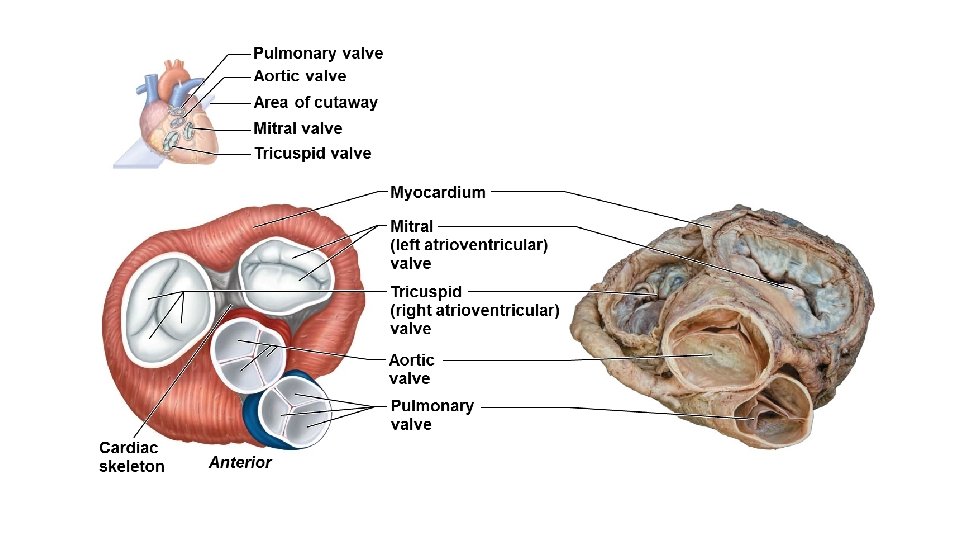
Heart Valves • Allow blood to flow in only one direction, to prevent backflow • Four valves • Atrioventricular (AV) valves—between atria and ventricles • Bicuspid (mitral) valve (left side of heart) • Tricuspid valve (right side of heart) • Semilunar valves—between ventricle and artery • Pulmonary semilunar valve • Aortic semilunar valve No valves are found between major veins and atria; not a problem because: Inertia of incoming blood prevents backflow Heart contractions compress venous openings

The right AV valve is the • tricuspid valve. • bicuspid or mitral valve. • semilunar valve. • pulmonary valve.

The right AV valve is the • tricuspid valve. • bicuspid or mitral valve. • semilunar valve. • pulmonary valve.
")
Heart Valves • AV valves • Anchored in place by chordae tendineae (“heart strings”) • Open during heart relaxation and closed during ventricular contraction • Semilunar valves • Closed during heart relaxation but open during ventricular contraction • These valves open and close in response to pressure changes in the heart
 Operation of the AV valves 1 Blood returning to the atria")
Slide 2 (a) Operation of the AV valves 1 Blood returning to the atria puts pressure against AV valves; the AV valves are forced open. Ventricles
 Operation of the AV valves 1 Blood returning to the atria")
Slide 3 (a) Operation of the AV valves 1 Blood returning to the atria puts pressure against AV valves; the AV valves are forced open. 2 As the ventricles fill, AV valve flaps hang limply into ventricles. Ventricles
 Operation of the AV valves 1 Blood returning to the atria")
Slide 4 (a) Operation of the AV valves 1 Blood returning to the atria puts pressure against AV valves; the AV valves are forced open. 2 As the ventricles fill, AV valve flaps hang limply into ventricles. 3 Atria contract, forcing additional blood into ventricles. AV valves open; atrial pressure greater than ventricular pressure Ventricles
 Operation of the AV valves 1 Blood returning to the atria")
Slide 5 (a) Operation of the AV valves 1 Blood returning to the atria puts pressure against AV valves; the AV valves are forced open. 4 Ventricles contract, forcing blood against AV valve flaps. 2 As the ventricles fill, AV valve flaps hang limply into ventricles. 3 Atria contract, forcing additional blood into ventricles. AV valves open; atrial pressure greater than ventricular pressure Ventricles
 Operation of the AV valves 1 Blood returning to the atria")
Slide 6 (a) Operation of the AV valves 1 Blood returning to the atria puts pressure against AV valves; the AV valves are forced open. 4 Ventricles contract, forcing blood against AV valve flaps. 5 AV valves close. 2 As the ventricles fill, AV valve flaps hang limply into ventricles. 3 Atria contract, forcing additional blood into ventricles. AV valves open; atrial pressure greater than ventricular pressure Ventricles
 Operation of the AV valves 1 Blood returning to the atria")
Slide 7 (a) Operation of the AV valves 1 Blood returning to the atria puts pressure against AV valves; the AV valves are forced open. 4 Ventricles contract, forcing blood against AV valve flaps. 2 As the ventricles fill, AV valve flaps hang limply into ventricles. 6 Chordae tendineae tighten, preventing valve flaps from everting into atria. 3 Atria contract, forcing additional blood into ventricles. AV valves open; atrial pressure greater than ventricular pressure 5 AV valves close. Ventricles AV valves closed; atrial pressure less than ventricular pressure
 Operation of the semilunar valves Pulmonary trunk 1 As ventricles contract")
Slide 2 (b) Operation of the semilunar valves Pulmonary trunk 1 As ventricles contract and intraventricular pressure rises, blood is pushed up against semilunar valves, forcing them open. Semilunar valves open Aorta
 Operation of the semilunar valves Pulmonary trunk 1 As ventricles contract")
Slide 3 (b) Operation of the semilunar valves Pulmonary trunk 1 As ventricles contract and intraventricular pressure rises, blood is pushed up against semilunar valves, forcing them open. Semilunar valves open Aorta 2 As ventricles relax and intraventricular pressure falls, blood flows back from arteries, filling the leaflets of semilunar valves and forcing them to close. Semilunar valves closed

Problems • Two conditions severely weaken heart: • Incompetent valve • Blood backflows so heart repumps same blood over and over • Valvular stenosis • Stiff flaps that constrict opening • Heart needs to exert more force to pump blood • Defective valve can be replaced with mechanical, animal, or cadaver valve

Cardiac Circulation • Blood in the heart chambers does not nourish the myocardium • The heart has its own nourishing circulatory system consisting of: • Coronary arteries—branch from the aorta to supply the heart muscle with oxygenated blood • Cardiac veins—drain the myocardium of blood • Coronary sinus—a large vein on the posterior of the heart, receives blood from cardiac veins • Blood empties into the right atrium via the coronary sinus

Brachiocephalic trunk Left common carotid artery Superior vena cava Left subclavian artery Right pulmonary artery Aortic arch Ascending aorta Pulmonary trunk Right pulmonary veins Right atrium Right coronary artery in coronary sulcus (right atrioventricular groove) Anterior cardiac vein Right ventricle Marginal artery Small cardiac vein Inferior vena cava (a) Ligamentum arteriosum Left pulmonary artery Left pulmonary veins Left atrium Auricle of left atrium Circumflex artery Left coronary artery in coronary sulcus (left atrioventricular groove) Left ventricle Great cardiac vein Anterior interventricular artery (in anterior interventricular sulcus) Apex

These blood vessels branch from the ascending aorta and deliver blood supply to the heart. • carotid arteries • pulmonary arteries • coronary arteries • systemic arteries

These blood vessels branch from the ascending aorta and deliver blood supply to the heart. • carotid arteries • pulmonary arteries • coronary arteries • systemic arteries

Blood Flow Through the Heart • Superior and inferior venae cavae dump blood into the right atrium • From right atrium, through the tricuspid valve, blood travels to the right ventricle • From the right ventricle, blood leaves the heart as it passes through the pulmonary semilunar valve into the pulmonary trunk • Pulmonary trunk splits into right and left pulmonary arteries, which carry blood to the lungs

Blood Flow Through the Heart • In the lungs, blood picks up oxygen and drops off carbon dioxide • Oxygen-rich blood returns to the heart through the four pulmonary veins • Blood enters the left atrium and travels through the bicuspid valve into the left ventricle • From the left ventricle, blood leaves the heart via the aortic semilunar valve and aorta

Capillary beds of lungs where gas exchange occurs Pulmonary Circuit Pulmonary arteries Pulmonary veins Aorta and branches Venae cavae Left atrium Right atrium Left ventricle Heart Right ventricle Systemic Circuit KEY: Oxygen-rich, CO 2 -poor blood Oxygen-poor, CO 2 -rich blood Capillary beds of all body tissues where gas exchange occurs

Pathway of Blood Through Heart • Right side of the heart • • Superior vena cava (SVC), inferior vena cava (IVC), and coronary sinus Right atrium Tricuspid valve Right ventricle Pulmonary semilunar valve Pulmonary trunk Pulmonary arteries Lungs

Pathway of Blood Through Heart • Left side of the heart • • Four pulmonary veins Left atrium Mitral valve Left ventricle Aortic semilunar valve Aorta Systemic circulation
 Inferior vena cava (IVC) Coronary sinus Right atrium Tricuspid valve")
Superior vena cava (SVC) Inferior vena cava (IVC) Coronary sinus Right atrium Tricuspid valve Right ventricle Pulmonary semilunar valve Oxygen-poor blood Pulmonary trunk Pulmonary arteries SVC Coronary sinus Right atrium IVC Pulmonary trunk Tricuspid valve Right ventricle Pulmonary semilunar valve Oxygen-poor blood is carried in two pulmonary arteries to the lungs (pulmonary circuit) to be oxygenated. To lungs Pulmonary capillaries Oxygen-rich blood returns to the heart via the four pulmonary veins. To heart Oxygen-rich blood

To heart Oxygen-poor blood returns from the body tissues back to the heart. Oxygen-poor blood Oxygen-rich blood Systemic capillaries To body Oxygen-rich blood is delivered to the body tissues (systemic circuit). Aorta Pulmonary veins Aortic semilunar valve Left atrium Mitral valve Left ventricle Aorta Aortic semilunar valve Left ventricle Mitral valve Left atrium Four pulmonary veins
 Inferior vena cava (IVC) Coronary sinus Right atrium Tricuspid valve")
Superior vena cava (SVC) Inferior vena cava (IVC) Coronary sinus Right atrium Tricuspid valve Right ventricle Oxygen-poor blood Pulmonary semilunar valve Pulmonary trunk Pulmonary arteries SVC Coronary sinus Pulmonary trunk Right atrium Tricuspid valve Pulmonary semilunar valve Right ventricle IVC To heart Oxygen-poor blood is carried in two pulmonary arteries to the lungs (pulmonary circuit) to be oxygenated. Oxygen-poor blood returns from the body tissues back to the heart. Systemic capillaries To body To lungs Pulmonary capillaries Oxygen-rich blood returns to the heart via the four pulmonary veins. Oxygen-rich blood is delivered to the body tissues (systemic circuit). Aorta To heart Pulmonary veins Aortic semilunar valve Left atrium Mitral valve Left ventricle Aorta Aortic semilunar valve Left ventricle Mitral valve Left atrium Four pulmonary veins Oxygen-rich blood

• Equal volumes of blood are pumped to pulmonary and systemic circuits • Pulmonary circuit is short, low-pressure circulation • Systemic circuit is long, high-friction circulation • Anatomy of ventricles reflects differences • Left ventricle walls are 3 times thicker than right • Pumps with greater pressure

Coronary Circulation • Coronary circulation • • Functional blood supply to heart muscle itself Shortest circulation in body Delivered when heart is relaxed Left ventricle receives most of coronary blood supply

• Coronary arteries • Both left and right coronary arteries arise from base of aorta and supply arterial blood to heart • Both encircle heart in coronary sulcus • Branching of coronary arteries varies among individuals • Arteries contain many anastomoses (junctions) • Provide additional routes for blood delivery • Cannot compensate for coronary artery occlusion • Heart receives 1/20 th of body’s blood supply

• Left coronary artery supplies interventricular septum, anterior ventricular walls, left atrium, and posterior wall of left ventricle; has two branches: • Anterior interventricular artery • Circumflex artery • Right coronary artery supplies right atrium and most of right ventricle; has two branches: • Right marginal artery • Posterior interventricular artery
 Left coronary")
Aorta Pulmonary trunk Superior vena cava Left atrium Anastomosis (junction of vessels) Left coronary artery Right atrium Circumflex artery Right coronary artery Right ventricle Right marginal artery Left ventricle Anterior interventricular artery Posterior interventricular artery

• Coronary veins • Cardiac veins collect blood from capillary beds • Coronary sinus empties into right atrium; formed by merging cardiac veins • Great cardiac vein of anterior interventricular sulcus • Middle cardiac vein in posterior interventricular sulcus • Small cardiac vein from inferior margin • Several anterior cardiac veins empty directly into right atrium anteriorly

Superior vena cava Great cardiac vein Anterior cardiac veins Coronary sinus Small cardiac vein Middle cardiac vein

Problems • Angina pectoris • Thoracic pain caused by fleeting deficiency in blood delivery to myocardium • Cells are weakened • Myocardial infarction (heart attack) • Prolonged coronary blockage • Areas of cell death are repaired with noncontractile scar tissue

Aorta Left pulmonary artery Superior vena cava Right pulmonary artery Right pulmonary veins Left pulmonary veins Auricle of left atrium Left atrium Right atrium Inferior vena cava Great cardiac vein Coronary sinus Posterior vein of left ventricle Right coronary artery (in coronary sulcus) Left ventricle Posterior interventricular artery (in posterior interventricular sulcus) Middle cardiac vein Apex Posterior surface view Right ventricle

A “heart attack” is also known as • angina. • pericarditis. • congestive heart failure. • myocardial infarction.

A “heart attack” is also known as • angina. • pericarditis. • congestive heart failure. • myocardial infarction.
- Slides: 74