Undescended Testis Ectopic testis Dr Manish Kumar Gupta


, causing regression of")


 is more common than left-sided undescent(42– 47%). •")


 recommend")


, the")


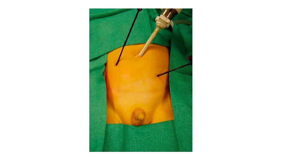
 After intra-abdominal mobilization of the testis, the gubernaculum has been grasped with forceps")
- Slides: 69
Undescended Testis/ Ectopic testis Dr. Manish Kumar Gupta Assistant Professor Department of Paediatric Surgery AIIMS, Rishikesh
INTRODUCTION • Numerous factors interact to affect normal testicular descent. • Any abnormality in this process can result in an undescended testis (UDT). • Also known as cryptorchidism, an UDT carries fertility and malignancy implications.
• Undescended testis is best defined as a testis that cannot be manipulated to the bottom of the scrotum without undue tension on the spermatic cord. • A normally descended testis resides spontaneously in the lower scrotum even if it was retracted when the patient was first examined.
Embryology • Early gonadal differentiation occurs in the urogenital ridge and is regulated by at least two genes, located on the short arm of the Y chromosome: • ZFY • SRY • The SRY (sex-determining region of Y) gene encodes for a testisspecific DNA-binding protein that stimulates development of the embryonic gonad towards a testis by 6 to 7 weeks post gestation.
• Sertoli cells begin to produce Müllerian inhibitory factor (MIF), causing regression of all Müllerian duct structures except for the remnant appendix testis and prostatic utricle. • By week 9, Leydig cells produce testosterone and stimulate development of Wolffian structures, including the epididymis and vas deferens. • The testis resides in the abdomen near the internal ring until descent through the inguinal canal at the beginning of the third trimester.
• Two important hormones in testicular descent are • insulin-like factor 3 (INSL 3) • testosterone • Two important anatomic players are • gubernaculum testis • cranial suspensory ligament (CSL) secreted by the testis
• The gubernaculum is thought to help anchor the testis near the internal inguinal ring as the kidney migrates cephalad. • Androgens prompt the involution of the CSL, allowing for eventual downward migration of the testicle.
• Testicular descent is biphasic: • Transabdominal phase- non androgen dependent • Final phase of inguinoscrotal descent- Androgen dependent
Transabdominal phase • Under the influence of INSL 3 produced by Leydig cell, the gubernaculum undergoes two phases: outgrowth and regression. • Outgrowth refers to rapid swelling by the gubernaculum, thereby dilating the inguinal canal and creating a pathway for descent. • Next, during regression, the gubernaculum undergoes cellular remodeling and becomes a fibrous structure.
• Intra-abdominal pressure then causes protrusion of the diverticulum of peritoneum, processus vaginalis through the internal inguinal ring, transmitting pressure to the gubernaculum and initiating testicular descent. • Transit through the inguinal canal is relatively rapid, starting around week 22 and typically completed after week 27
Final phase of inguinoscrotal descent • Ahead of the descending testis, the processus vaginalis develops within the inguinal canal to the scrotum. • It is surrounded by the cremaster muscle, which is innervated by the genitofemoral nerve. • Androgens produced by the fetal testis act to irreversibly virilize the sensory dorsal root nucleus of the genitofemoral nerve (sexual dimorphism). • A neurotransmitter, calcitonin gene-related peptide (CGRP), released through the sensory fibres of the genitofemoral nerve, acts on the CGRPreceptor-rich gubernaculum, inducing strong rhythmic contractions (100/second), which draw the testis through the inguinal canal into the scrotum.
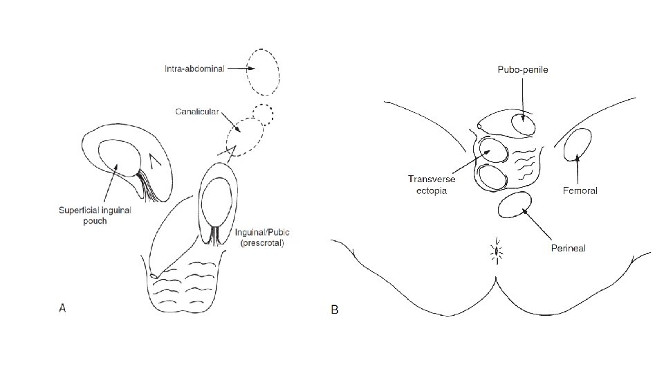
• Most undescended testes are located outside the inguinal canal because the migratory inguinoscrotal phase of testicular descent is deranged more commonly. • So, the intra-abdominal testes are relatively uncommon, occurring in 5% to 10% of cryptorchid boys. • In most cases, the undescended testis is located near the neck of the scrotum, just outside or a little lateral to the external inguinal ring, in the “superficial inguinal pouch”.
Associated syndromes • Microcephaly- Possibility of pituitary hormone or gonadotrophin deficiency • Anthrogryposis multiplex conginata- neurogenic and mechanical anomalies • Prune belly syndrome-Absence of processus vaginalis development due to prenatal urinary bladder distension. • 10% of cases of PUV • Gastroschisis and omphalocele- due to rupture of gubernaculum or poorly formed gubernaculum • Exstrophy of the bladder- poorly formed gubernaculum • Neural tube defects- due to the stimulation by sensory genitofemoral nerve is impaired
Embryology of ectopic testis • Undescended testes lying well outside the normal line of descent such as in the perineum or femoral region are rare, and their cause is unknown. • It has been suggested that this may be the result of an abnormal location of the genitofemoral nerve with consequent abnormal migration of the gubernaculum to the wrong site. • May be found in inguinal region, femoral canal, penopubic area, or even contralateral hemiscrotum.
CLASSIFICATION OF UNDESCENDED TESTES • The positions of undescended testes can be divided into those arrested in the line of normal descent and those in truly ectopic positions.
PALPABLE: • Imperfectly descended: • External inguinal ring • Superficial inguinal pouch • Gliding or mobile testis • Ectopic: • • • Lateral abdominal wall Femoral Base of penis Perineal Crossed ectopia IMPALPABLE: • Absent: • Primary failure • Vascular injury • High location: • Intra-abdominal • High intracanalicular
• A retractile testis is a normally descended testis that retracts into the inguinal canal as a result of cremaster muscle contraction. It can be manipulated down into the scrotum on examination without tension and will remain in place without causing discomfort to child. • Gliding or mobile testis descends spontaneously to the high/midscrotum but has a short processus vaginalis, such that traction on the testis causes obvious discomfort to the child. • Gliding testes are smaller in volume • Often are associated with a hypoplastic scrotum. • They show histological features of cryptorchidism and classify as undescended testes, requiring the same attention.
• The intra-abdominal testis is usually located within a few centimeters of the internal inguinal ring. • Usually identified by laparoscopy.
• A nonpalpable testis may also be absent or vanishing due to intrauterine or perinatal torsion(15 -20% of nonpalpable testis). • This condition is known as monorchia, or anorchia if both testes are absent. • Biopsy of tissue at the blind-ending gonadal vessels may reveal hemosiderin and calcification as a remnant of the previously torsed testis.
• Testes that have been documented to be in the scrotum at birth but subsequently are found to be undescended are often classified as ascending. • The situation may be due to a failure of proportional elongation of the spermatic cord, such that the testis is progressively retracted out of the scrotum by linear body growth. • More common is the iatrogenic undescended or rising testis that becomes attached to the inguinal fascia by scar tissue following an inguinal operation, e. g. for hernia or hydrocele, and is then drawn out of the scrotum with body growth.
INCIDENCE • UDT occurs in 3% of term male infants and in up to 33% of premature male infants. • The majority of testes descend within the first 9 to 12 months. • At age 1 year, the incidence of UDT is 1%, and testicular descent after 1 year is unlikely. • The overall rate of secondary testicular ascent has been reported between 2% to 45%. • 2/3 rd to 3/4 th of cases are palpable, with most being palpable within the inguinal canal or distal to the external ring.
• Right-sided undescent (53– 58%) is more common than left-sided undescent(42– 47%). • Bilateral undescended testes occur in 10– 25% of all cases of undescent.
TESTICULAR DEVELOPMENT • At term, the seminiferous tubules in the normal testis are filled with gonocytes, spermatogonia and Sertoli cells. • Leydig cells are easily identifiable within the interstitial tissue. • Accentuated growth during the second to sixth months of postnatal life is associated with a crucial surge in testosterone production and further maturation of the spermatogonia, followed by relative regression until the onset of puberty. • Spermatogonia are quantifiable as the tubular fertility index (TFI), which represents the percentage of tubules containing spermatogonia. The TFI rises after the sixth year of life, and by the eighth year spermatocytes appear. Development of a tubular lumen and Sertoli cells is followed by spermatogenesis.
Complications of crytorchidism • Temperature effect: • The scrotal testis in the human is maintained at 33°C compared with 34°C to 35°C in the inguinal region and 37°C intra-abdominally. • The physiology of the testis is well adapted to this lower temperature. • Therefore, in the undescended testis where the ambient temperature is increased, the testis undergoes progressive alteration. • Endocrine Effects: • Plasma gonadotropin and testosterone levels have been measured in infants with undescended testes, and the normal postnatal rise in plasma luteinizing hormone (LH) levels and testosterone were found to be significantly lower than normal.
• Germ Cell Development: • Germ cell deficiency in cryptorchidism was previously regarded as congenital. • It has been observed that the histology of the testes is initially normal and becomes progressively abnormal with age. • Leydig cell development is impaired in undescended testes in the first 2 to 6 months, whereas the Sertoli and germ cells appeared normal. • By the end of the second year of life, nearly 40% of undescended testes have completely lost their germ cells. • It is now well established that the transformation of neonatal gonocytes to type A spermatogonia, an early postnatal step in development of the germ cells, is deficient in infants with cryptorchidism.
• Fertility: • Fertility is lower in men with a past history of cryptorchidism. • In previous generations, it was believed that the undescended testis suffered no adverse changes until after puberty and surgical intervention was not necessary until 12 to 15 years of age. • The evidence that germ cell maturation is already abnormal after 6 months of age has led clinicians to appreciate that not only is postnatal degeneration an important issue but also that early intervention may prevent it. • Paternity rates are not deficient in unilateral cryptorchidism but, with bilateral cryptorchidism, fertility is significantly impaired.
• Malignancy: • The progressive degeneration of germ cells and dysplasia seen in cryptorchid testes is thought to be related to an increased risk of malignancy. • Relative risk for those with a history of unilateral cryptorchidism is 15 -fold or 33 -fold for bilateral undescended testes, with the risk of cancer being highest with intra-abdominal testes.
• The undescended testis and, to a lesser extent, the contralateral scrotal testis carries a greater content of dysgenetic material, which is most likely responsible for the increased risk of neoplasia. • The contralateral scrotal testis carries an increased (5%) risk of neoplastic change. • 1 in 5 tumours associated with testicular undescent arises within the contralateral scrotal testis. • Long-term follow-up is mandatory for patients with testicular undescent, particularly those who have had unilateral malignancy. • 60% of tumours developing in undescended testes are seminomas presenting at the usual young age for testicular tumours of 20– 40 years arising from abnormal gonocytes.
• Inguinal Hernia • The processus vaginalis normally obliterates after descent of the testis in the perinatal period. • Undescended testes are associated with a higher incidence of patent processus vaginalis and inguinal hernia. • A clinically evident hernia present with a cryptorchid testis is an indication for immediate intervention. • Most surgeons elect to perform a hernia repair and orchidopexy simultaneously.
• Torsion of a Cryptorchid Testis: • There is a high incidence of up to 20% for torsion in unoperated undescended testes. • The mobility of a testis within the tunica vaginalis in the superficial inguinal pouch may predispose to torsion. • Testicular-Epididymal Fusion Abnormality • Abnormal connection between the testis and the epididymis is common in cryptorchidism. • The risk of abnormal fusion is greater with testes inside the canal or the abdomen than in inguinal testes or those lying at the neck of the scrotum. • These abnormalities may be related to underlying androgen deficiency in utero.
• Failure of testicular descent, the absence of testes in the scrotum, and possible infertility cause profound psychological disturbance for both the adolescent patient and his family. • The potential impact on body image, gender identity and, hence, selfesteem and personality should not be underestimated.
DIAGNOSIS • The diagnosis and location of a UDT are determined by thorough physical examination performed in a warm room. • The patient should be examined in both a supine and a frog-legged sitting position. • The scrotum is observed for hypoplasia and examined for the presence of either testis. • In cases of monorchia, the solitary testis may be hypertrophied. • The first maneuver to locate the testis is to walk the fingers gently down the inguinal canal from the internal ring toward the scrotum, trying to push subcutaneous structures toward the scrotum. • Gentle mid-abdominal pressure may help push the testis into the inguinal canal.
• On examination, both retractile testes and low UDTs may be manipulated into the scrotum. • Once in scrotal position, the retractile testis appears to remain in place, whereas the low UDT does not. • The ipsilateral hemiscrotum is fully developed with a retractile testis, whereas it may be underdeveloped in a UDT.
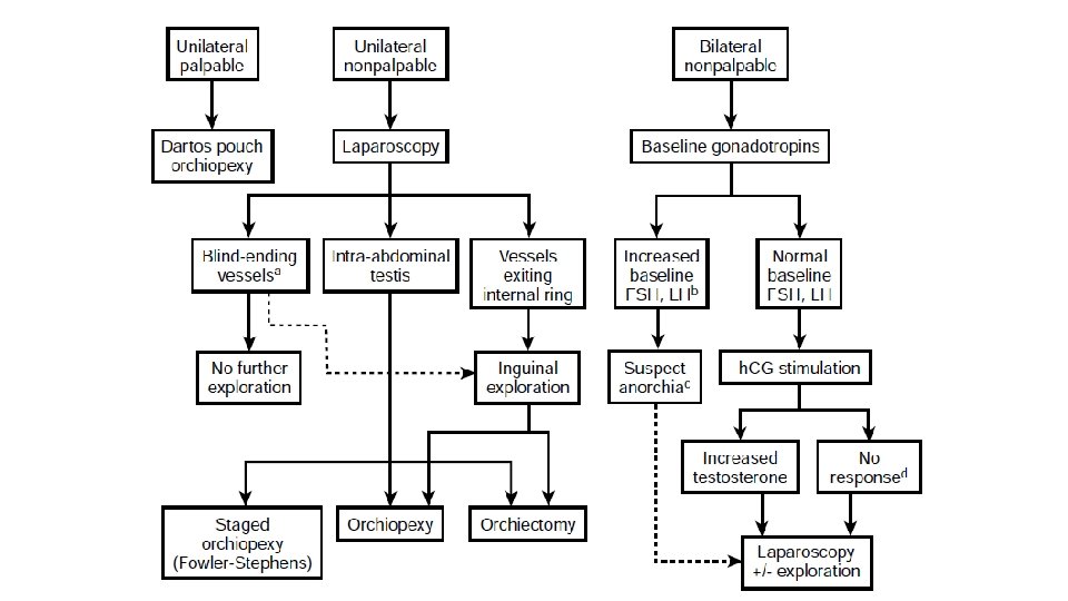
• If neither testis is palpable, anorchia, androgen insensitivity syndrome, or a chromosomal abnormality must be differentiated from bilateral UDT. • If the baseline follicle-stimulating hormone (FSH) level is elevated (three standard deviations above the mean) in a boy younger than 9 years, anorchia is likely and no further evaluation is recommended. • If baseline luteinizing hormone (LH) and FSH levels are normal and human chorionic gonadotropic (h. CG) stimulation results in an appropriate elevation of testosterone, functioning testicular tissue is likely to be present and the patient should undergo exploration. • However, if the testosterone level does not increase appropriately, nonfunctional testicular tissue may still be present and exploration should still be performed. • The h. CG stimulation test does not distinguish between normal nonpalpable testes and functioning testicular remnants.
Radiology • Radiographic imaging is rarely helpful in locating a UDT and is not recommended routinely. • In unusual situations of bilateral nonpalpable testes, MRI with gadolinium may be useful for detecting abdominal testes because testicular tissue is particularly bright on MRI.
TREATMENT: Hormone Treatment • Hormone therapy is based on the premise that the undescended testis is caused by deficiency of the hypothalamic pituitary-gonadal axis and that postnatal treatment can induce the required migration of the gubernaculum. • Therapy has been tried with testosterone, h. CG, and luteinizing hormone releasing hormone (LHRH)agonist, Buserelin. • Direct androgen therapy was abandoned many years ago because excessive doses caused precocious puberty. • Success rates for treatment range from 10% to 50%. • Children older than 4 years and those with bilateral undescended testes near the scrotal entrance or retractile testes respond most favorably to h. CG.
Surgical Treatment • Indications and Timing: • Guidelines (AAP 1996 and EAU 2012) recommend that orchiopexy in otherwise healthy males be performed by 12– 18 months of age, as the UDT is unlikely to descend after 12 months of age. • Repair may be undertaken even earlier if a symptomatic hernia is present.
• The surgical approach for UDT depends on whether the testis is palpable. • Unilateral and bilateral palpable UDT are treated the same way. • It is important to re-examine the patient after induction of anesthesia because up to 18% of nonpalpable testes may become palpable on examination under anesthesia.
• For unilateral and bilateral nonpalpable UDT, definitive management may be determined through a combination of inguinal exploration and diagnostic laparoscopy. • The nonpalpable testis is approached first through diagnostic laparoscopy and then inguinal exploration is done as needed.
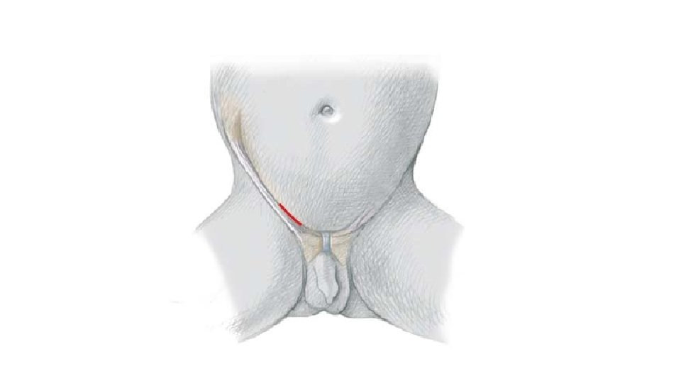
Palpable Undescended Testes • The mainstay of therapy for the palpable UDT is surgical orchiopexy with creation of a subdartos pouch. • The operation is usually performed under general anesthesia. • The incision should be made along one of Langer’s lines, over the internal ring.
• The external oblique aponeurosis is incised laterally from the external ring in the direction of its fibers, avoiding injury to the ilioinguinal nerve.
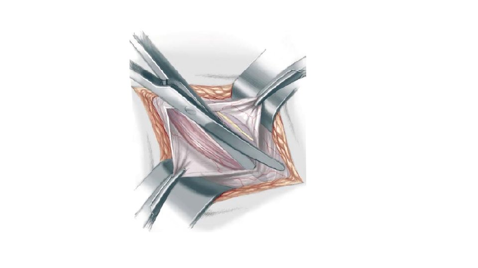
• The spermatic cord is separated from the external oblique aponeurosis and the cord and testis (usually just outside the external ring) are mobilized out of the wound by blunt dissection. • The distal gubernacular attachment is divided carefully (diathermy or scissors), avoiding any long-looping vas deferens. • A small clamp on the gubernaculum or distal tunica vaginalis allows traction on the spermatic cord. • The spermatic cord is dissected by first isolating and stripping off with blunt forceps any cremaster muscle fibres.
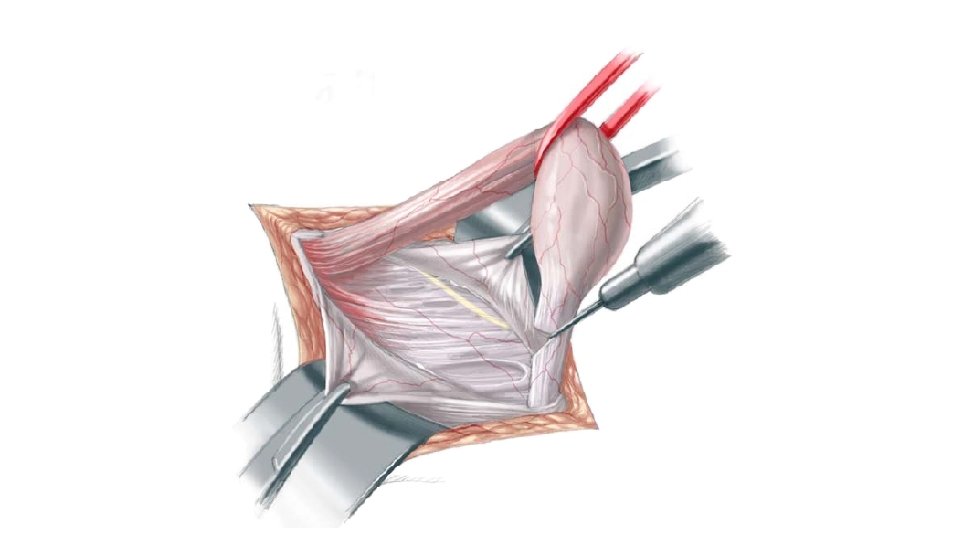
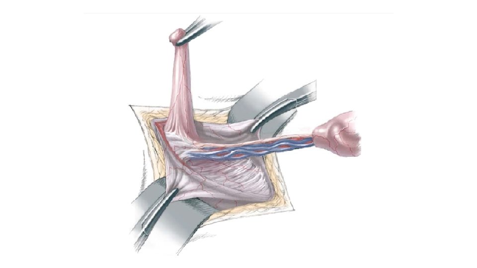
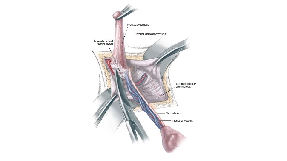
• The remnant of the processus vaginalis is usually obvious in cryptorchidism as a shiny white, translucent inelastic layer covering the vas deferens and testicular vessels. • The hernial sac is grasped with blunt forceps and the vas and vessels are carefully swept off its posterior surface en masse. • The vas deferens must be identified formally during this dissection, as it is closely adherent to the posterior surface of peritoneum. • The vas deferens and gonadal vessels are retracted away from the sac, which then can be clamped and divided.
• The processus vaginalis is twisted to exclude intraperitoneal contents and transfixed and ligated with absorbable suture. • These manoeuvre provide adequate mobilisation of the testis to provide adequate length to bring down testis to the scrotum.
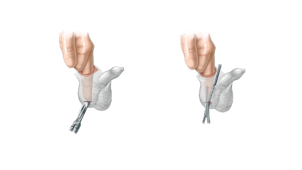
• A finger is introduced through the wound and bluntly pushed down to the scrotum. • A subcutaneous pouch is developed with scissors or small artery forceps with the finger still in place.
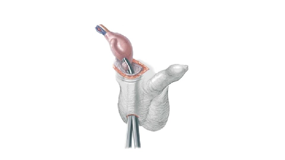
• Now the testis is brought down to the scrotum holding the gubernaculum.
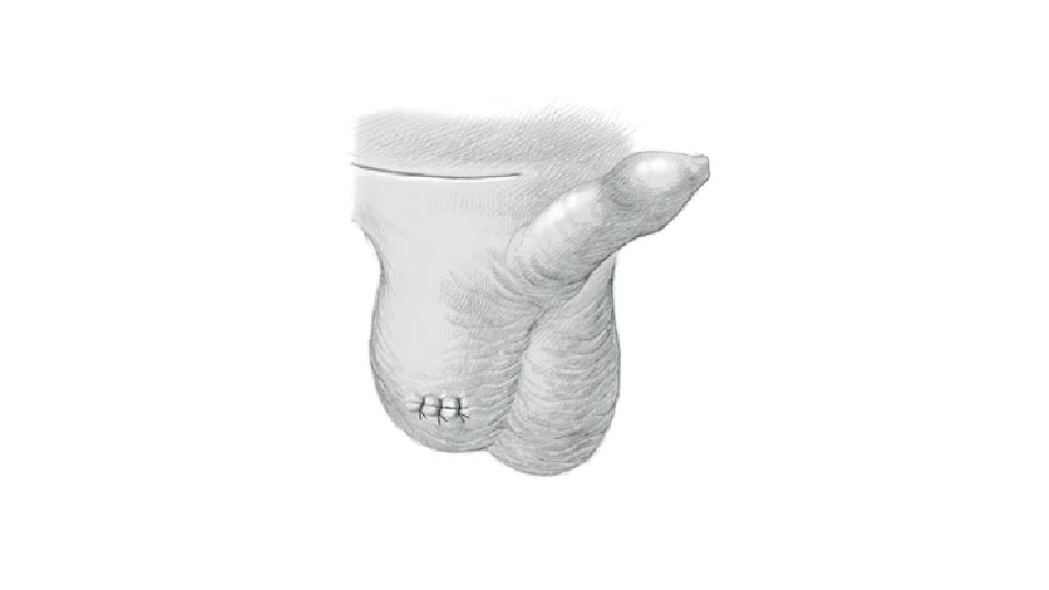
• The testis is nestled into its new subcutaneous pouch and the scrotum closed with subcuticular suture. • • The inguinal incision is closed in layers.
• The patient is seen in the outpatient clinic after a few weeks for a wound check and again several months later for testicular examination. • Final position and condition of the testis should be noted. • Although rare, complications include atrophy and retraction.
Nonpalpable Undescended Testis • The standard approach is diagnostic laparoscopy through an umbilical port.
• If the testicular vessels are seen exiting the internal ring, an inguinal incision is used to locate the testis or testicular remnant. • Orchiopexy is performed if a viable testis is found
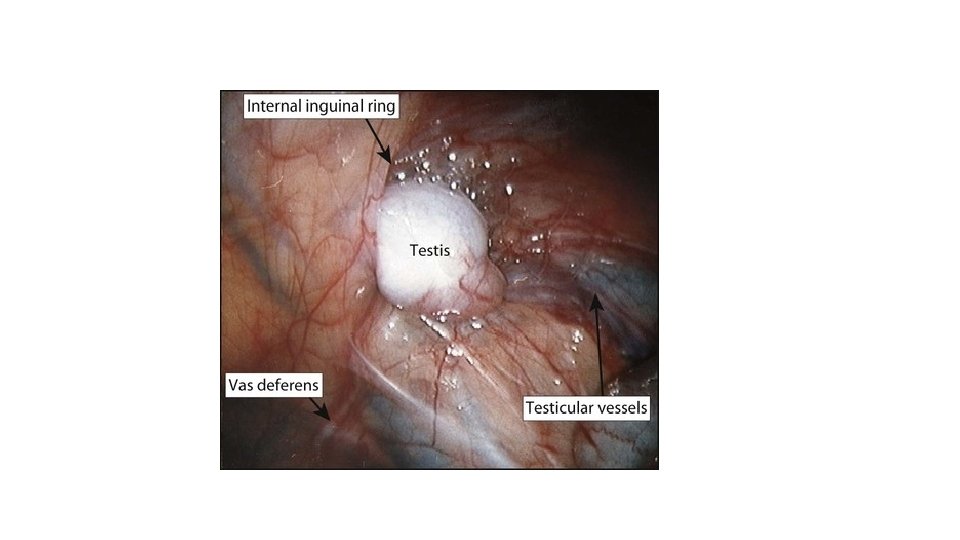
The testicular vessels and vas deferens are seen to enter the inguinal canal. There is no evidence for a patent processus vaginalis. The vessels and vas deferens appear to be of relatively normal caliber. In this situation, inguinal exploration is necessary
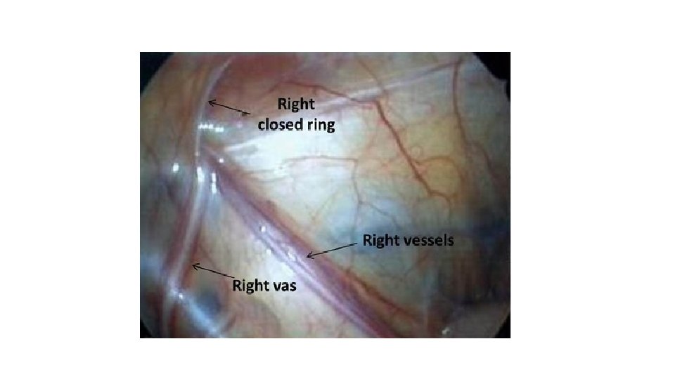
• If the vessels end blindly in the inguinal canal (vanishing testis), the tip of the vessels may be sent for pathologic examination. • Remnants of testicular tissue or hemosiderin and calcifications are indicative of probable perinatal torsion and testicular resorption.
The vas deferens and testicular vessels in this patient end blindly in the retroperitoneum. The internal ring is closed. In this very unusual situation, inguinal exploration is not necessary.
• If diagnostic laparoscopy reveals an intra-abdominal testis, several options are available. • The Fowler-Stephens orchiopexy involves ligation of the testicular vessels, which makes the testis dependent on the vasal and cremasteric arteries for viability. • After ligation of the testicular vessels, which can be done laparoscopically or by laparotomy, a delay of about 6 months is recommended before orchiopexy to allow development of collateral circulation
A) After intra-abdominal mobilization of the testis, the gubernaculum has been grasped with forceps inserted through a 10 -mm cannula that has been introduced through the scrotal incision, over the pubic tubercle and into the abdomen. The testis is then withdrawn into the cannula. Often, it is not possible to place the testis entirely into the 10 -mm port. B) The testis is delivered over the pubic tubercle and into the right hemiscrotum