ULTRASOUND NEWS November 2020 Abstract Introduction Ultrasound is

ULTRASOUND NEWS November , 2020

Abstract • Introduction • Ultrasound is the modality of choice in the evaluation of neonates and young children with suspected adrenal lesions including adrenal haemorrhage and congenital adrenal hyperplasia. It is also the initial imaging modality in children presenting with an upper abdominal mass, which may be adrenal in origin. • Topic discussion: This paper shows adrenal anatomy and demonstrates how the sonographic features change with age. It reviews the imaging features of congenital adrenal lesions, as well as benign and malignant conditions affecting the adrenal gland in childhood. • Discussion and Conclusion: Ultrasound is a useful primary imaging modality in the assessment of the adrenal gland in children. Knowledge of the changes of the adrenal gland with age is important when assessing the adrenal gland. Ultrasound is also useful for assessing abdominal masses. However, it cannot differentiate adrenal masses, therefore correlation with biochemical findings, multimodality imaging, and histology is usually required. • Keywords Adrenal gland, ultrasound, child, paediatric

Abstract • Introduction Chronic liver disease is a public health burden worldwide. Portal hypertension is a major portosystemic abnormality in chronic liver disease. This study aimed to determine the main, right, and the left portal vein diameter in patients with chronic liver disease. • Materials and methods A cross-sectional study was carried out at the Abubakar Tafawa Balewa University Teaching Hospital, Bauchi, northeastern Nigeria from December 2018 to September 2019. Ethical clearance was obtained from the institutional review board. A total of 200 subjects were recruited comprising 100 patients with chronic liver disease and 100 age-matched controls, aged 18 years and above. A transabdominal ultrasound scan was carried out measuring the main, right, and left portal vein diameter while lying supine and/or in the right anterior oblique position after overnight fasting, or 6 hours before the scan. Data analysis was done using SPSS version 22. 0. Descriptive statistics (mean, standard deviation) and Pearson’s correlation were used. • Results There were 106(53%) males and 94(47%) females, aged between 18 and 73 years with a mean age of 46. 79 ± 15. 43. The main, right, and left portal vein diameter in patients with chronic liver disease was 14. 51 ± 0. 78 mm, 6. 83 ± 0. 81 mm, and 6. 26 ± 0. 74 mm, which were higher than those of their control. The portal vein diameter positively correlated (weak) with age and respiratory phases among participants (P < 0. 05). • Conclusion This study found the main, right, and left portal vein diameter among patients with chronic liver disease to be larger than those of the controls. Ultrasonography is a reliable diagnostic tool in evaluating portosystemic pathologies. • Keywords Portal hypertension, hepatology, ultrasound

Abstract • Introduction Chronic liver disease is a public health burden worldwide. Portal hypertension is a major portosystemic abnormality in chronic liver disease. This study aimed to determine the main, right, and the left portal vein diameter in patients with chronic liver disease. • Materials and methods A cross-sectional study was carried out at the Abubakar Tafawa Balewa University Teaching Hospital, Bauchi, northeastern Nigeria from December 2018 to September 2019. Ethical clearance was obtained from the institutional review board. A total of 200 subjects were recruited comprising 100 patients with chronic liver disease and 100 age-matched controls, aged 18 years and above. A transabdominal ultrasound scan was carried out measuring the main, right, and left portal vein diameter while lying supine and/or in the right anterior oblique position after overnight fasting, or 6 hours before the scan. Data analysis was done using SPSS version 22. 0. Descriptive statistics (mean, standard deviation) and Pearson’s correlation were used. • Results There were 106(53%) males and 94(47%) females, aged between 18 and 73 years with a mean age of 46. 79 ± 15. 43. The main, right, and left portal vein diameter in patients with chronic liver disease was 14. 51 ± 0. 78 mm, 6. 83 ± 0. 81 mm, and 6. 26 ± 0. 74 mm, which were higher than those of their control. The portal vein diameter positively correlated (weak) with age and respiratory phases among participants (P < 0. 05). • Conclusion This study found the main, right, and left portal vein diameter among patients with chronic liver disease to be larger than those of the controls. Ultrasonography is a reliable diagnostic tool in evaluating portosystemic pathologies. • Keywords Portal hypertension, hepatology, ultrasound
 is a rare, benign developmental anomaly with a")
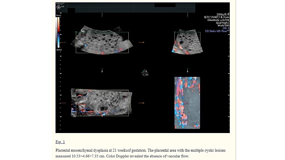
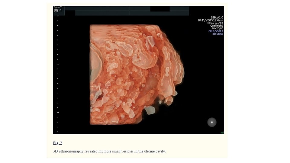
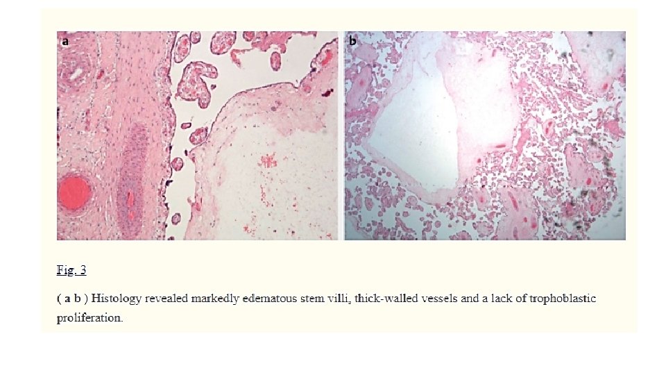
Introduction • Placental mesenchymal dysplasia (PMD) is a rare, benign developmental anomaly with a reported prevalence of 0. 02% (Arizawa and Nakayama, 2002). It is characterized by placentomegaly with multiple cystic lesions of the stem villi and vascular anomalies (Pawoo and Heller, 2014). Early detection of PMD has been described during routine prenatal ultrasound (Vaisbuch et al. , 2009). The sonographic characteristics of PMD include increased placental thickness and multiple cystic areas within the placenta with either an absence of blood flow or with low venous Doppler signals (Vaisbuch et al. , 2009). The differential diagnosis of multicystic placental lesions with the presence of a live fetus include partial molar pregnancy, multiple hematomas, chorioangioma Beckwith-Wiedemann syndrome and PMD. Chorioangiomas are well circumscribed masses within the placenta and they are characterized by the presence of a single feeding vessel with the same pulse rate as the umbilical cord (Zalel et al. , 2002). Invasive prenatal testing is required for the exclusion of partial molar pregnancy and Beckwith-Wiedemann Syndrome (Vaisbuch et al. , 2009). Definitive diagnosis of PMD is based on the pathologic examination of the placenta. Histology reveals aneurysm or dilated blood vessels that may be thrombosed. The stem villi are edematous and enlarged with thick-walled vessels, without trophoblastic proliferation (Pawoo and Heller, 2014). This case report highlights the significance of the early detection of PMD, illustrates the pitfalls in differential diagnosis and provides valuable insights regarding PMD management in a clinical setting.
 is an uncommon vascular anomaly of the placenta")
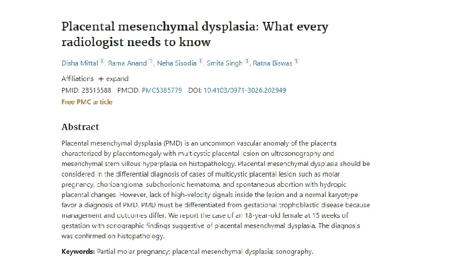
Abstract • Placental mesenchymal dysplasia (PMD) is an uncommon vascular anomaly of the placenta characterized by placentomegaly with multicystic placental lesion on ultrasonography and mesenchymal stem villous hyperplasia on histopathology. • Placental mesenchymal dysplasia should be considered in the differential diagnosis of cases of multicystic placental lesion such as molar pregnancy, chorioangioma, subchorionic hematoma, and spontaneous abortion with hydropic placental changes. However, lack of high-velocity signals inside the lesion and a normal karyotype favor a diagnosis of PMD must be differentiated from gestational trophoblastic disease because management and outcomes differ. • We report the case of an 18 -year-old female at 15 weeks of gestation with sonographic findings suggestive of placental mesenchymal dysplasia. The diagnosis was confirmed on histopathology.
- Slides: 14