
Ultrasound in Critical Obstetric Situations the Role of
Ultrasound in Critical Obstetric Situations: the Role of the Anesthesiologist Yaacov Gozal, M. D. Associate Professor of Anesthesiology Hebrew University and Hadassah Medical School Chair, Dept. of Anesthesiology Shaare Zedek Medical Center
INTRODUCTION Anesthesiologists: key role in high risk pregnancies Member of a multidisciplinary team ICU: 2 -4/1000 deliveries
INTRODUCTION Traditionally: background of previous excellent health and large physiological reserve Modern maternal characteristics: ◦ Increasing age ◦ Morbid obesity ◦ Congenital and acquired cardiac disease ◦ Assisted reproduction Diagnostic dilemmas
INTRODUCTION Emergencies: quick and accurate diagnostic tools Ultrasound: ◦ Safe and easily accessible ◦ Ease of use ◦ Connectivity and data storage ◦ Reduced size and weight
Ultrasound Routine locations: ◦ Emergency Room ◦ Delivery room ◦ Operating Room ◦ PACU ◦ ICU
Echocardiography Provides volumetric and flow data Shows the functioning heart OB patient is the ideal subject: ◦ Ant and left displacement of heart ◦ Elevated diaphragm Class I recommendation according to American, British and European guidelines

The FATE Examination Jensen MB et al, Eur J Anaesthesiol 2004; 21: 700 -707
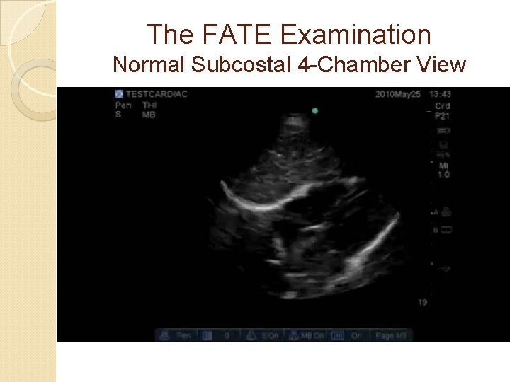
The FATE Examination Normal Subcostal 4 -Chamber View
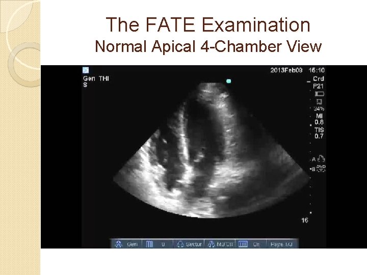
The FATE Examination Normal Apical 4 -Chamber View
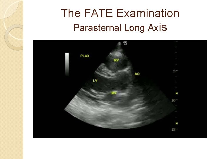
The FATE Examination Parasternal Long Axis
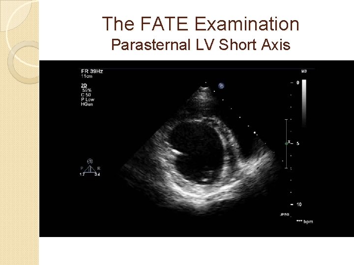
The FATE Examination Parasternal LV Short Axis
LUNG ULTRASOUND Acute respiratory failure: one of the most distressing situations Physical exam. and chest X-Ray: imperfect Need for sophisticated tests and delay management Lung U/S: standard tool in critical care
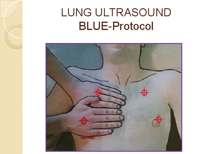
LUNG ULTRASOUND BLUE-Protocol
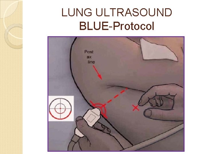
LUNG ULTRASOUND BLUE-Protocol
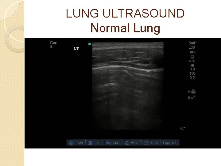
LUNG ULTRASOUND Normal Lung
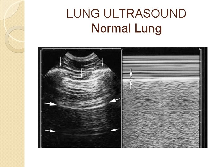
LUNG ULTRASOUND Normal Lung
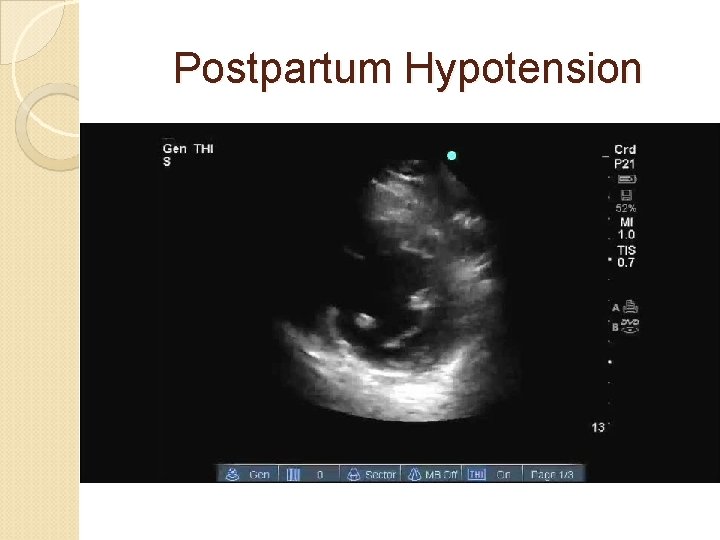
Postpartum Hypotension 29 -yr old primaparous No significant medical history Uncomplicated CS, with minimal blood loss under spinal anesthesia at 35 weeks’ gestation 6 hrs after delivery: Hypotension Tachycardia Febrile Hb: 12
Postpartum Hypotension
Postpartum Hypotension due to cardiac failure Dagnostic: postpartum cardiomyopathy No signs of IHD (ECG, chest pain, . . ) Treatment: ◦ Inotropic support ◦ Diuresis ◦ ACE inhibitors
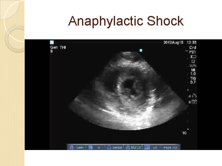
Anaphylactic Shock 35 -yr old primaparous No significant medical history Delivery suite: epidural analgesia and urinary catheter No progress CS Baby delivered: hemodynamic collapse intubation, fluids, vasopressors
Anaphylactic Shock
Anaphylactic Shock Adrenaline boluses Steroids H 1 and H 2 blockers Adrenaline continuous infusion Removal of the urine catheter
Amniotic Fluid Embolism 40 -yrs old, gravida 5, para 4 No remarkable medical history Cesarean section at 38 weeks’gestation Spinal anesthesia: hemodynamic stability After delivery, CARDIAC ARREST �Cardiac massage �Epinephrine �Intubation and ventilation �VF: cardioversion X 6
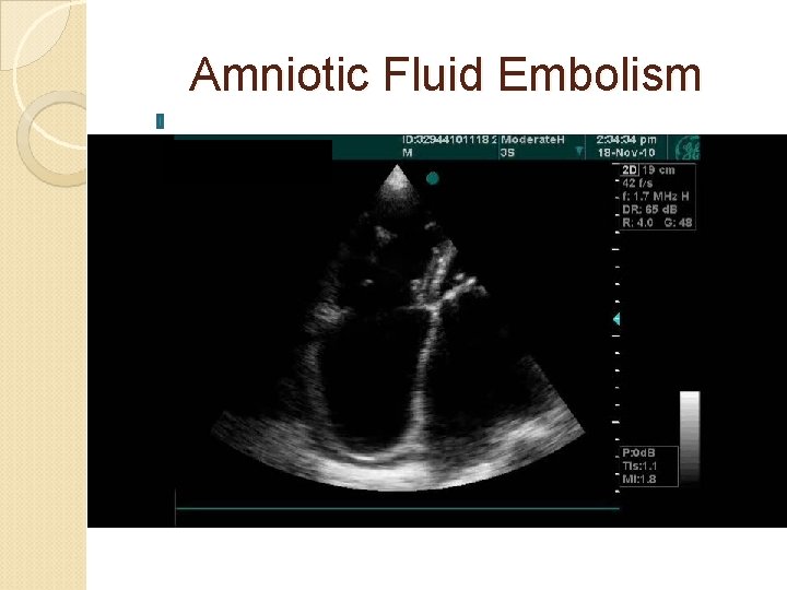
Amniotic Fluid Embolism
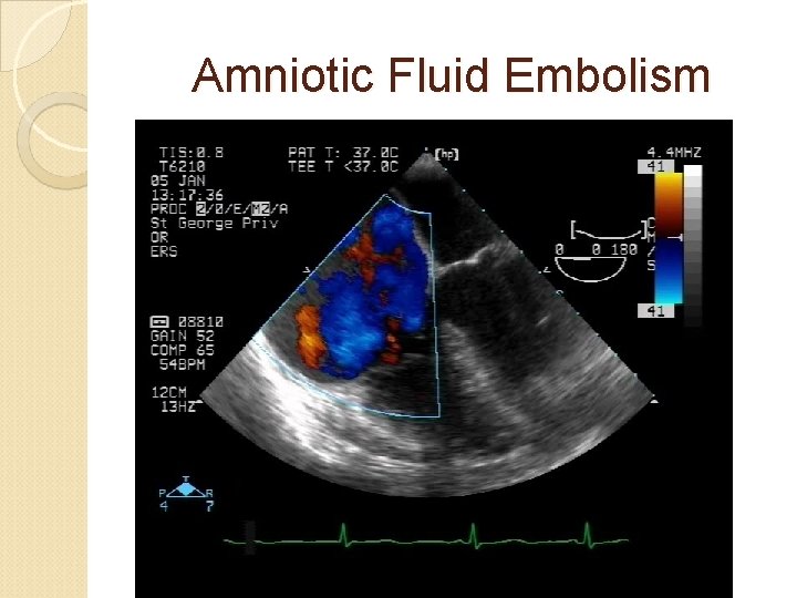
Amniotic Fluid Embolism

Amniotic Fluid Embolism DIC: TEG flat Thrombocytopenia Supportive treatment
Pulmonary Embolus 35 -yr old, Gravida 5, Para 3 Vaginal delivery at 40 weeks’gestation Postpartum hemorrhage Atonic uterus: pitocin, methergin Severe bleeding: 10 units RBC and 10 units FFP During surgery, Sa. O 2: 70% Hemodynamic instability
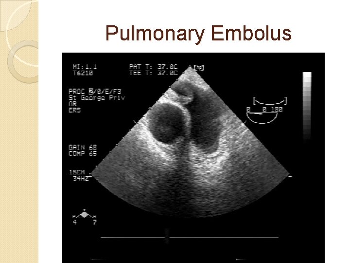
Pulmonary Embolus
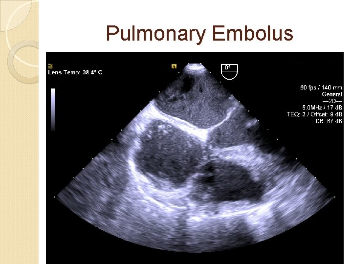
Pulmonary Embolus
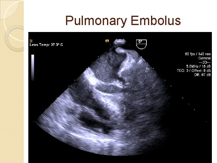
Pulmonary Embolus
LUNG ULTRASOUND Pneumothorax
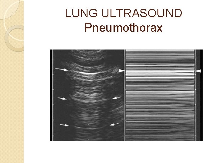
LUNG ULTRASOUND Pneumothorax
LUNG ULTRASOUND Acute Dyspnea 40 -yrs old, gravida 3, para 2 Acute respiratory distress at 31 weeks’ gestation Medical background: asthma, morbid obesity and diabetes Examination: ◦ ◦ Tachypneic Sa. O 2: 85% (RA) Bilateral wheezing Lower limb edema
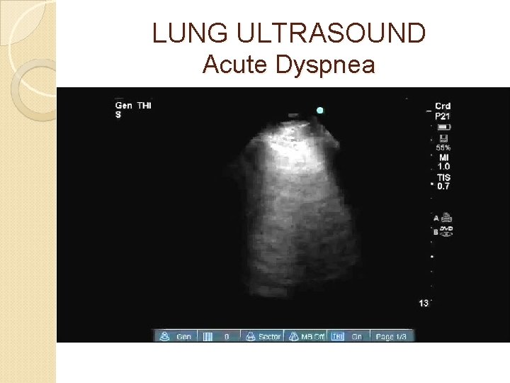
LUNG ULTRASOUND Acute Dyspnea
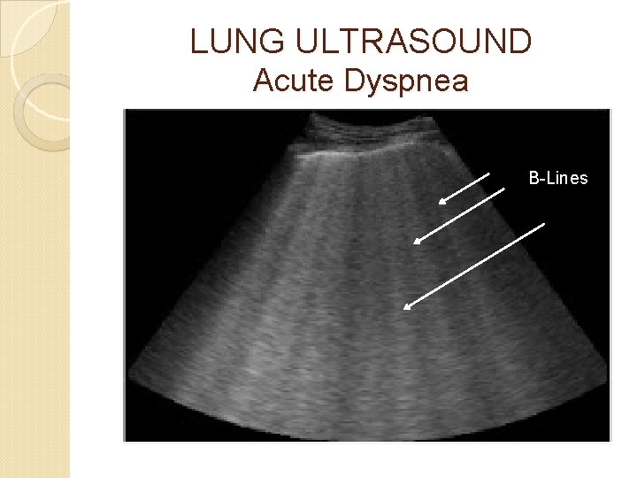
LUNG ULTRASOUND Acute Dyspnea B-Lines
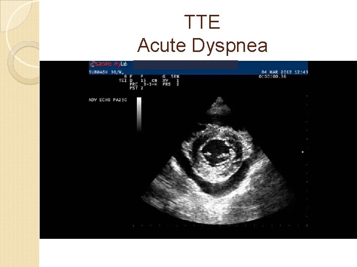
TTE Acute Dyspnea
LUNG ULTRASOUND Acute Dyspnea Diagnosis: acute pulmonary edema rather than exacerbation of asthma Management: diuretics and oxygen therapy Rapid Improvement
CONCLUSIONS Ultrasound: unique tool Diagnostic and monitoring capabilities Ultrasound = 3 rd eye of the anesthesiologist
- Slides: 38